
Capillaroscopy: Non-Invasive Microvascular Imaging and Evaluation Methods
Capillaroscopy is a non-invasive optical imaging method used to examine microvascular architecture and blood flow dynamics directly through superficial tissue layers. By combining directed illumination with specialized optical magnification, the technique allows clinicians and researchers to observe tiny capillary loops without breaking the skin barrier. Professional interpretation of these microcirculatory patterns provides valuable descriptive insight into peripheral vascular morphology.
Selecting the appropriate imaging configuration depends on whether observation takes place in outpatient clinical settings, academic research laboratories, or mobile screening environments. Factors such as optical stability, digital sensor resolution, illumination control, and mechanical stage precision determine the overall utility of each platform. Institutional buyers must carefully match instrument specifications with clinical documentation protocols and research requirements.
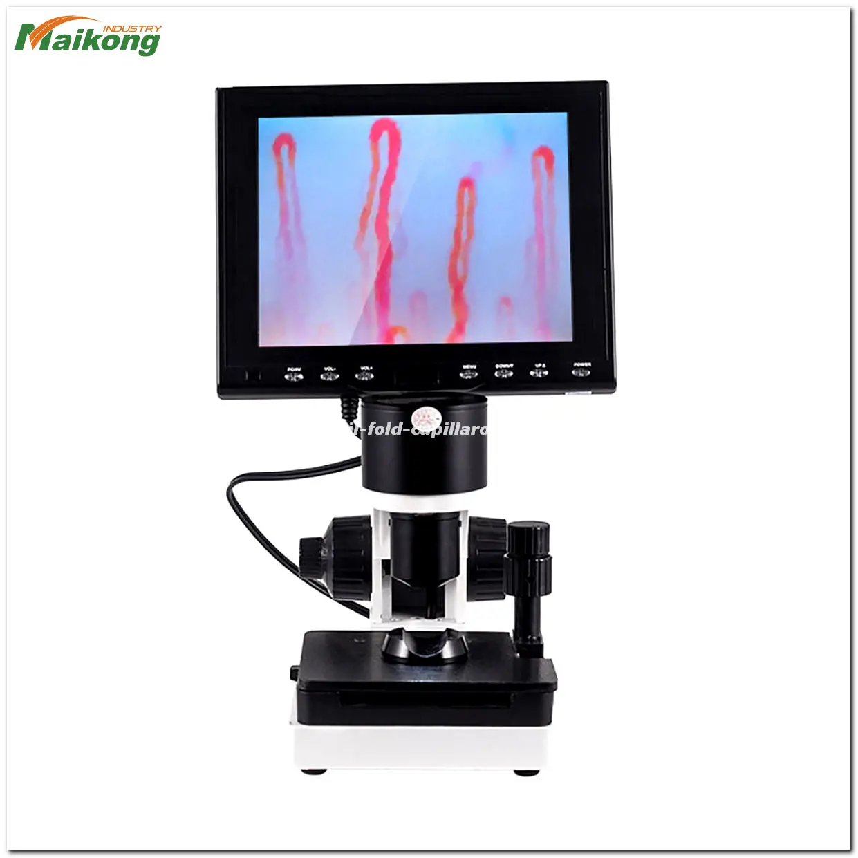
What Is Capillaroscopy?
Capillaroscopy represents an optical assessment technique designed for the in vivo visualization of human microcirculation. Historically referred to in various scientific texts as a capilliscope evaluation or simply capiloscopy, this method provides real-time, non-invasive imaging of tiny blood vessels located near the cutaneous surface. Specialized instrumentation directs focused cold light onto the target tissue while magnifying the vascular loops across ranges from optical baseline levels up to several hundred times magnification.
While the human nailfold provides the primary window for this observation, the underlying methodology applies broadly across various mucosal and cutaneous sites. In a standard nail fold capillaroscopy procedure, the capillary loops run parallel to the epidermal surface at the terminal boundary of the digit. This unique anatomical orientation allows clear lateral observation of the hairpin loop, comprising the afferent arterial limb, transition zone, and efferent venular limb.
The practice relies entirely on surface optics, immersion interfaces, and advanced image capture rather than ionizing radiation or intravenous dye injection. Modern videocapillaroscopes integrate solid-state sensors to project real-time, uncompressed visual feeds onto inspection displays. This capability allows multidisciplinary practitioners to capture static photographs, record dynamic blood flow sequences, and archive longitudinal image series for comparative morphological studies.
Observational imaging through a capilliscope provides morphological data regarding microvascular architecture. Such visual findings require qualified clinical correlation and do not by themselves establish a definitive medical diagnosis.

Microvascular loops captured under specialized magnification and uniform LED surface illumination.
How Does Capillaroscopy Work?
The operational framework of capillaroscopic imaging centers on overcoming the natural light-scattering properties of the superficial stratum corneum. Human skin scatters incident light, creating glare and opacity that obscures the underlying papillary dermis. Applying an optical coupling medium transforms the uneven skin surface into an optically coherent window through which incident light can penetrate and reflect off microvascular structures.
Once illumination penetrates the tissue, specialized achromatic lenses gather the reflected light and direct it toward an optical ocular or digital sensor. Modern platforms utilize cold-temperature LED illumination that emits minimal infrared energy, preventing localized tissue warming that could alter native blood velocity. The operator manipulates coaxial micro-positioning controls to establish a crisp focal plane across the vascular bed.
- The operator cleans the target examination site and positions the digit stably within an adjustable mechanical cradle.
- A small drop of pharmaceutical-grade mineral oil, paraffin oil, or cedarwood immersion oil is applied to the tissue to minimize light scattering.
- The technician engages the cold LED illumination source and brings the optical objective within its designated working distance.
- Coaxial coarse and fine handwheels are adjusted to locate the primary vascular row and resolve capillary definition on the display.
- The X-Y mechanical stage is mobilized laterally to document consecutive capillary fields across the full anatomical border.

Coaxial focusing mechanisms allow precise depth calibration across dynamic dermal microvascular beds.
What Microvascular Features Can Capillaroscopy Show?
Capillaroscopic observation yields qualitative and morphometric data concerning the physical layout and dynamic activity of the terminal vascular tree. Under appropriate magnification, the operator evaluates the density of the nail fold capillary distribution per millimeter, loop orientation, dimensional caliber, and the presence of morphologic variations. Healthy microcirculatory beds typically display homogeneous, upright, comb-like arrangements of hairpin loops distributed uniformly across the tissue margin.
When systemic vascular alterations occur, microvascular beds may present distinct structural variations. Practitioners document parameters including apical elongation, loop tortuosity, generalized capillary dilation, vascular architecture dropout, and microhemorrhages resulting from local extravasation. Dynamic video assessment also permits subjective observation of erythrocyte column flow, documenting continuous flow, intermittent sludging, or localized plasma gaps.
Observable Morphological Markers
- Capillary loop density and spatial distribution along the terminal margin.
- Dimensional caliber of the arterial limb, apical loop, and venous limb.
- Dynamic movement and coherence of the intravasal erythrocyte column.
- Presence of microhemorrhages, hemosiderin deposits, or localized edema.
Methodological Limitations
- Surface imaging cannot evaluate deep visceral microvascular systems.
- Severe epidermal thickening or severe tissue pigment can impede light transmission.
- Motion artifacts or tremors can temporarily blur dynamic video sequences.
- Visual morphology cannot substitute for systemic laboratory or histopathologic testing.
Evaluating cases categorized as challenging capillaroscopy requires methodical adjustment of illumination angles and oil distribution. Excessive skin thickness, recent mechanical trauma, severe contractures, or cold-induced peripheral vasoconstriction can obscure vascular visibility. Systematic operating protocols assist in obtaining clear baseline documentation even in complex observational contexts.

Digital display showing high-definition imaging of microvascular architecture with clear luminal margins.
Clinical, Research, and Educational Fields Using Capillaroscopy
The capacity to observe living human microcirculation non-invasively makes capillaroscopic equipment a valuable resource across diverse clinical disciplines and academic faculties. Specialists incorporate optical assessment into routine diagnostic workflows, translational medicine, physiology teaching, and vascular pharmacological trials. The non-destructive nature of the technique supports repeated longitudinal evaluations without discomfort to the subject.
- Rheumatology departments utilize microvascular observation to differentiate between primary microcirculatory vasospastic events and complex autoimmune connective tissue involvement.
- Dermatology practices employ surface magnification to evaluate cutaneous inflammatory patterns, psoriasis plaques, chronic wound margins, and local vascular formation.
- Vascular medicine and angiology clinics observe peripheral microcirculation to document lower-extremity insufficiency, arterial occlusive changes, and systemic microangiopathy.
- University medical laboratories and physiology schools integrate video systems into practical curricula to demonstrate hemodynamic principles and capillary flow dynamics.
- Biomedical engineering programs and pharmaceutical research teams apply standardized imaging to monitor microcirculatory response to novel therapeutic compounds.
Outpatient wellness centers and functional health practices also explore peripheral capillary observation to visually track microcirculatory response over time. In every setting, data gathered via capillary microscopy serves as supportive descriptive documentation within a broader clinical or experimental framework.
Capillaroscopy Equipment and Imaging Formats
Modern microcirculatory observation platforms encompass multiple form factors engineered to satisfy varied institutional environments. Choosing between benchtop systems, portable handheld devices, and multi-component workstations depends on physical mobility requirements, throughput demands, and the necessity for calibrated morphometric software integration.
Desktop Benchtop Systems
Integrated desktop systems combine a rigid heavy-base stand with an X-Y compound stage and dedicated LCD monitor. These workstations deliver exceptional mechanical stability and vibration isolation, making them standard fixtures in high-volume hospital clinics and university teaching laboratories.
Digital Videocapillaroscopes
Digital video platforms feature solid-state imaging sensors connected to computerized image analysis workstations. These configurations enable calibrated micrometric loop measurement, high-frame-rate hemodynamic recording, and digital patient file integration for clinical trial compliance.
Portable and Handheld Units
Lightweight portable devices offer direct USB or wireless connectivity to laptops and tablets. These flexible instruments suit multi-room clinics, mobile health screening initiatives, and bedside consultations where cart mobility is essential.
For research institutes requiring deep image capture flexibility, dedicated optical setups like a nailfold capillaroscopy microscope or a digital nailfold capillaroscopy microscope provide interchangeability between eyepieces and high-resolution camera mounts. Clinics with space constraints frequently favor a usb nailfold capillaroscopy microscope connected to existing departmental computer terminals.
Dedicated clinical suites often deploy a professional nailfold capillaroscope or an integrated nailfold capillaroscopy video system. These advanced installations feature synchronized cold illumination, a specialized nailfold capillaroscopy camera, and a comprehensive nailfold capillaroscopy imaging system designed for continuous daily workflow.

Modern microcirculatory instrumentation ranges from stationary desktop stations to portable optical devices.
Equipment Evaluation Criteria for Institutional Buyers
Procurement teams and department chairs must evaluate technical specifications against their practical operating demands before purchasing microvascular observation equipment. Verifying optical resolving power, ergonomic flexibility, and data connectivity prevents workflow bottlenecks and supports reliable diagnostic documentation.
| Feature | Specification | Procurement Relevance |
| Magnification | 200X to 600X | Sufficient magnification reveals individual capillary loops and intravascular flow motion. |
| Illumination | LED Cold Light | Prevents localized tissue heating that could artificially alter microvascular velocity. |
| Focusing | Coaxial Dual | Coarse and fine controls enable sharp focusing without disturbing the finger holder. |
| Mechanical Stage | X-Y Compound | Smooth multidirectional translation allows sequential tracking along the nailfold boundary. |
| Display Interface | LCD Monitor | Dedicated screens eliminate external computer boot-up delays during routine clinical rounds. |
| Chassis Stability | Rigid Metal Base | Heavy construction dampens benchtop vibrations that cause visual jitter at high power. |
Departmental purchasing committees must also evaluate operational training curves, decontamination protocols between examinations, and the availability of replacement components. Ensuring that a nailfold capillaroscopy device integrates smoothly into established facility cleaning routines preserves equipment longevity and maintains consistent imaging quality over extended institutional deployments.
Reference Specifications for a MAIKONG Capillaroscopy System
To assist technical committees with parameter benchmarking, the following reference specifications outline the operational architecture of a standard MAIKONG optical microcirculation workstation. These values reflect the MK-GY880 platform design and provide a baseline for comparing optical components, stage dimensions, and electrical requirements.
| Parameter | Reference Value | Technical Description |
| Brand Name | MAIKONG | Manufactured under centralized quality control protocols in GD, SZ. |
| Function | Body Health | Non-invasive observational microvascular visualization and optical inspection. |
| Amplification | 380X/500X/600X | Selectable optical amplification modules for varying magnification depth. |
| Input Power | AC 100-240V | Universal 50/60Hz alternating current input supporting international standards. |
| Output Power | DC 12V 2A | Regulated direct current output for low-voltage chassis safety. |
| Objective Lens | 5X Achromat | Achromatized optical objective offering an effective working range of 25mm. |
| Mechanical Stage | X-Y Compound | Double-layer compound mechanical platform with dual translation wheels. |
| Focus Mechanism | Coaxial Dual | Integrated coaxial coarse and fine focus wheels with tension adjustment. |
| Illumination | LED Cold Light | Internal dimmable LED cold light source with focused projection. |
| Equipment Type | Type I or B | Standardized electrical safety classification for laboratory inspection devices. |
| Chassis Power | 12V 1A DC | Direct current source driving optical sensors and internal LED illumination. |
| Built-in Camera | 80 000 pixels | Integrated solid-state CCD sensor engineered for low-noise real-time video. |
| LCD Monitor | 8/10/12 Inch | Integrated high-contrast color LCD screen options for real-time display. |
| Certification | CE Approved | Manufactured in accordance with European technical safety benchmarks. |
| Net Weight | 6KG | Solid chassis mass engineered to minimize ambient table vibration artifacts. |
| Gross Weight | 7KG | Total shipping weight including protective case and accessory layout. |
| Inner Package | Aluminum Box | Custom-fitted aluminum security box lined with dense memory foam. |
| Outer Package | Corrugated Box | Reinforced export-grade multi-layer corrugated transit carton. |
Buyers reviewing a nailfold capillaroscopy equipment price should note that exact dimensions and packaging options vary by configuration. Standard packing dimensions measure 37.5*26.5*34cm, housing the primary instrument, optical objectives, monitor, and connection cables securely within protective memory foam cutouts.
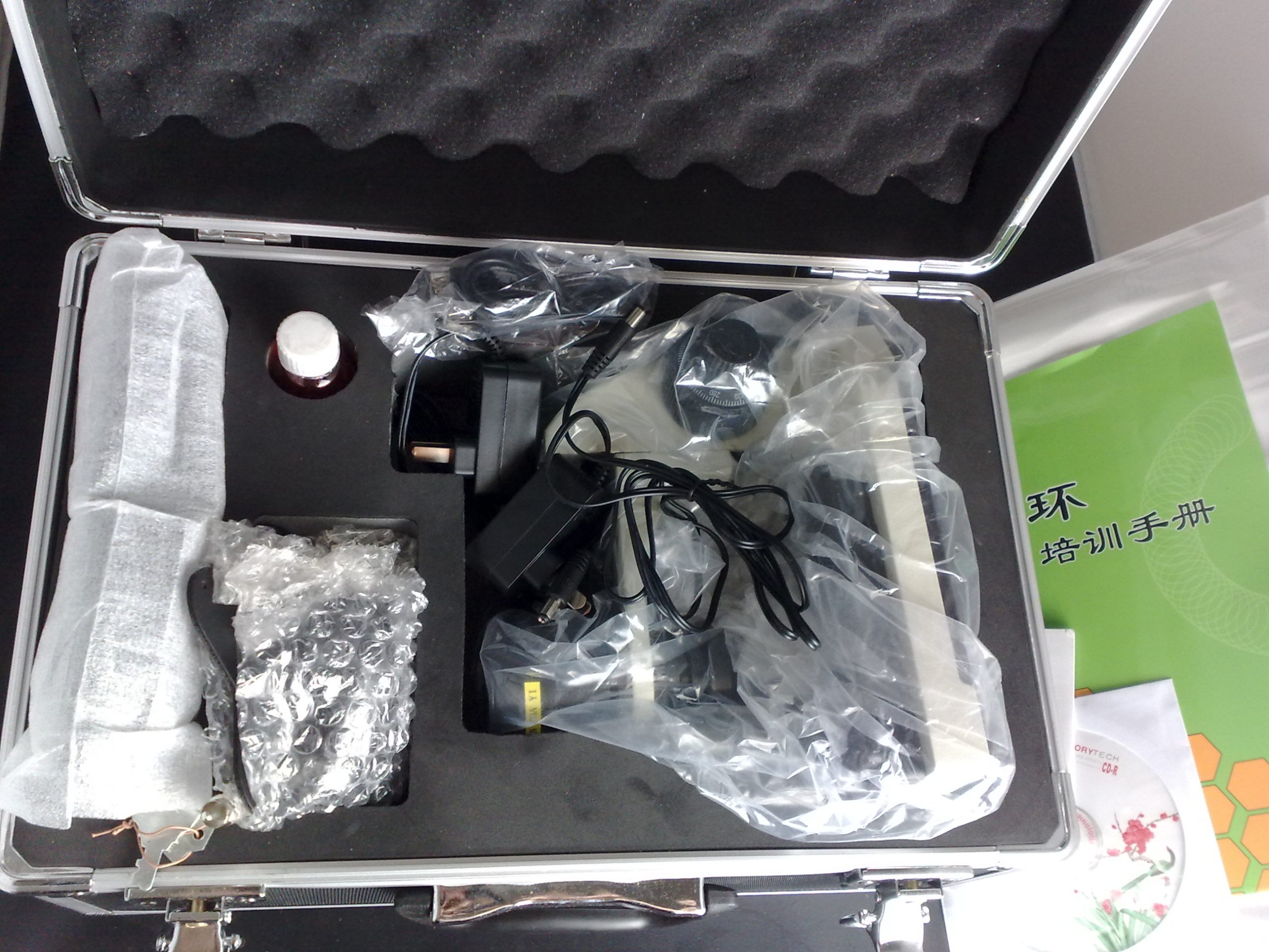
Custom aluminum security transit cases protect sensitive optical and mechanical assemblies during international freight.
Standard Package and Accessories
When deploying microvascular imaging equipment into active practice, having a complete set of validated accessories is essential for smooth installation. MAIKONG systems arrive with standardized accessory packages that allow clinical staff to begin equipment verification immediately upon unpacking.
| Component | Quantity | Operational Purpose |
| MK-GY880 Host | 1 pc | Main structural microscope base with coaxial mechanism and optical column. |
| LCD Monitor | 1 pc | Dedicated color display screen with integrated chassis mounting bracket. |
| Power Adapter | 1 pc | Universal voltage converter supplying regulated 12V direct current. |
| Cedar Oil | 1 pc | Optical immersion coupling liquid for reducing epidermal light reflection. |
| Cotton Swab | 1 pc | Lint-free applicator for precise oil distribution on the cuticle border. |
| Reference Chart | 1 pc | Morphological pattern guide for educational identification and training. |
Distributors and clinics planning an equipment Order can coordinate with our support team to request extra consumables, specialized finger clamps, or alternate immersion fluids depending on institutional protocols.
Basic Operating and Examination Considerations
Consistent environmental controls and methodical patient preparation are vital when gathering standardized microcirculatory data. Because peripheral capillary diameter and blood velocity respond dynamically to ambient temperatures, physical exertion, and emotional stress, standardized examination protocols help minimize observational variance.
Clinical protocols recommend maintaining examination rooms at a steady temperature between 22 and 24 degrees Celsius with approximately 70 percent relative humidity to minimize autonomic vasospasm during imaging.
Prior to undergoing capillary observation, subjects should remain seated comfortably for approximately 15 to 30 minutes in the acclimatized room. Their hands should be rested comfortably on the examination table at the level of the heart to prevent gravitational venous pooling or arterial hypoperfusion. Protocols also advise avoiding intense physical exercise, caffeine consumption, or smoking for at least one hour before the session.
The operator dispenses two to three drops of immersion oil—such as pharmaceutical-grade paraffin or cedarwood oil—directly onto the nailfold boundary of the non-dominant ring digit. This digit is traditionally chosen due to reduced susceptibility to occupational trauma. The finger is positioned comfortably within the finger press, and the stage handwheels are rotated until the distal capillary loops resolve clearly on the display.
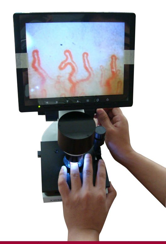
Stable finger positioning and appropriate immersion oil application are essential for clear optical observation.
Microcirculation Equipment Manufacturing and OEM Capabilities
At MAIKONG, we operate specialized production facilities in Shenzhen, GD, China, providing robust optoelectronic instruments to clinical and commercial partners worldwide. Our manufacturing infrastructure leverages an integrated regional supply chain, modern CNC machining, cleanroom optical assembly, and standardized quality auditing. We deliver high-reliability imaging instruments designed to balance high-precision performance with cost-effective capital investment.
We provide full OEM and private-label manufacturing, offering tailored chassis branding, custom casing designs, specialized firmware, and tailored packaging configurations for equipment distributors.
Learn more About us to explore our manufacturing history and browse our Factory facilities. Qualified regional distributors and healthcare supply firms are invited to connect directly through our dedicated contact portal to discuss product localization, sample evaluations, and international supply arrangements.
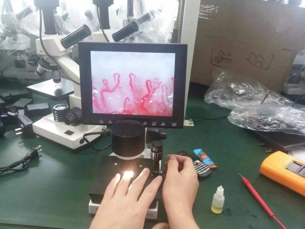
Modern precision assembly and rigorous optical alignment at our Shenzhen manufacturing facility.
Frequently Asked Questions About Capillaroscopy
What is capillaroscopy?
Capillaroscopy is a non-invasive optical examination method used to visualize living microcirculation and capillary architecture in superficial tissue beds. By using focused cold illumination and high-magnification lenses, it enables clinicians and researchers to observe vascular loops without penetrating the cutaneous surface.
Is capillaroscopy invasive?
Capillaroscopy is completely non-invasive and requires no needles, intravenous dye injections, or tissue incisions. The procedure relies entirely on harmless external light projection and surface immersion oil applied over intact skin.
What does a capillaroscopy system record?
A capillaroscopy platform records static photographs and real-time video footage of capillary loops, vessel density, and intravascular erythrocyte movement. Digital systems can also store calibrated morphometric measurements for longitudinal observational comparison.
Who may use capillaroscopy equipment?
Capillaroscopy systems are primarily operated by rheumatologists, dermatologists, vascular physicians, university researchers, and trained laboratory technicians. Independent medical practitioners and clinical investigators also utilize the equipment for physiological observation.
What is the difference between optical and digital capillaroscopy?
Optical capillaroscopy relies on traditional glass eyepieces for direct visual observation by a single operator. Digital videocapillaroscopy incorporates solid-state camera sensors and monitors, allowing multiple observers to view live video, capture digital media, and perform computerized vessel measurements.
Is capillaroscopy the same as a capillary test?
Capillaroscopy refers to the overarching optical imaging methodology and equipment system used to view microcirculation. In contrast, a nailfold capillary test describes the specific clinical examination protocol conducted on an individual subject under controlled conditions.
What should a distributor verify before importing a system?
Distributors should verify regional electrical voltage compatibility, optical resolution parameters, mechanical chassis stability, and required customs documentation. Reviewing warranty communication terms, protective freight packaging, and available user training materials is also recommended before placing bulk orders.
Can MAIKONG support OEM, private-label, or distributor projects?
MAIKONG provides extensive OEM and ODM customization services, including custom chassis colors, private logo application, specialized packaging cases, and tailored software branding. We work closely with medical distributors and institutional suppliers to fulfill both small trial orders and scheduled container shipments.
Advancing Microvascular Observation and Institutional Procurement
Capillaroscopy remains a vital, non-invasive methodology for investigating the architecture and dynamic behavior of peripheral microcirculation across clinical and research environments. Selecting the appropriate imaging instrument requires balancing optical magnification, mechanical stage stability, and digital recording capabilities to suit specific institutional workloads. By establishing reliable manufacturing partnerships and adhering to standardized observation protocols, healthcare organizations can implement effective microvascular imaging workflows.
Equip Your Facility with MAIKONG Capillaroscopy Systems
Connect with our technical team to evaluate reference specifications, request product documentation, or explore regional distributor opportunities for your market.