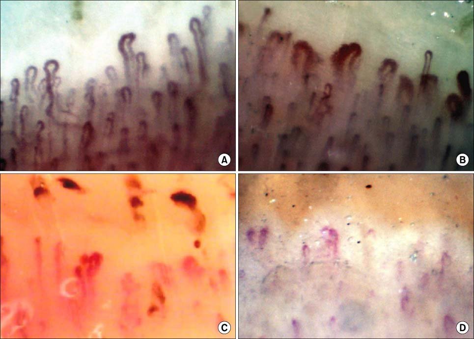
Image clarity, optical resolution, and mechanical stability determine how effectively microvascular architecture can be documented for clinical observation or laboratory research. Equipment selection depends on the required optical magnification range, sensor stability, illumination source, and mechanical stage precision across clinical and academic settings.
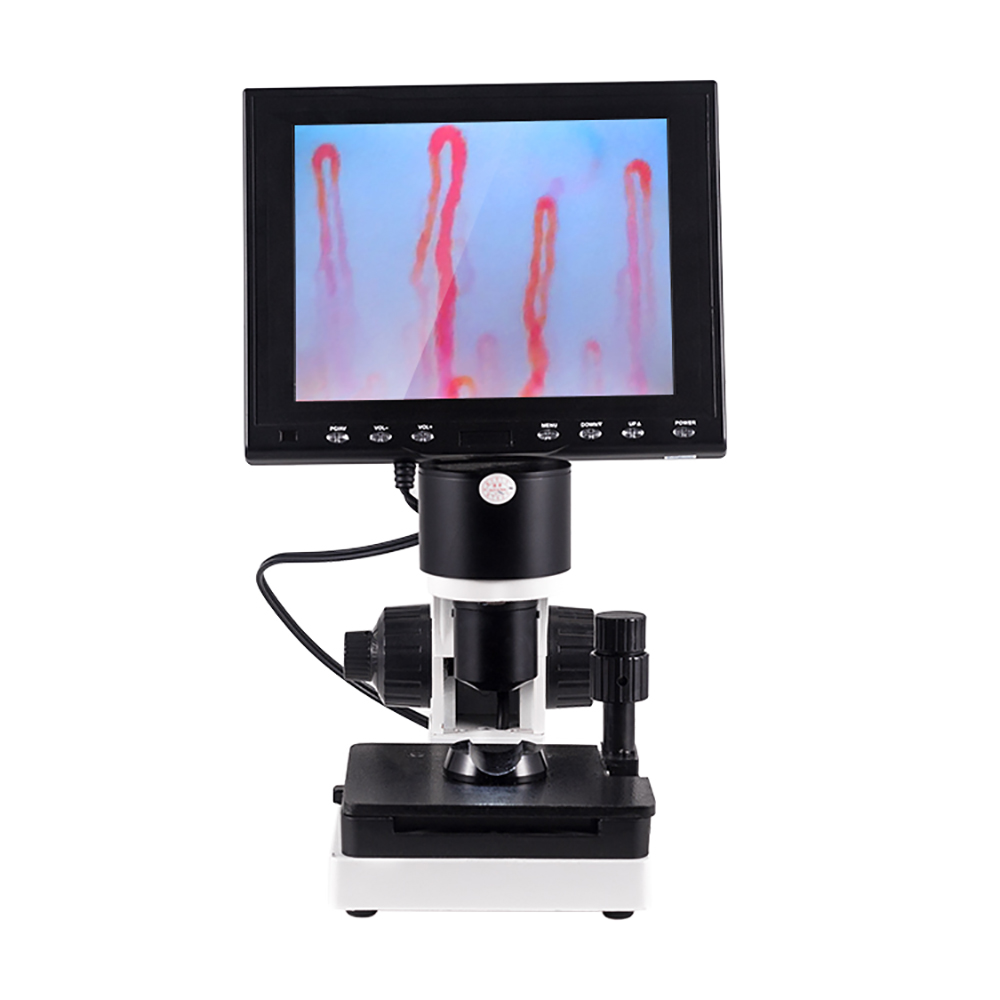
Optical and Video Components in Modern Systems
Advanced capillaroscopy platforms integrate precision optical assemblies with digital imaging sensors to capture microvascular architecture without invasive contact. High-resolution CCD or digital video sensors transfer live streams directly to attached LCD display monitors, minimizing latency during active examination.
Illumination plays a critical role in microvascular visibility because surface glare can obscure underlying dermal tissue. Dedicated LED cold-light sources project uniform illumination directly onto the nailbed target, preventing thermal discomfort while preserving structural clarity.
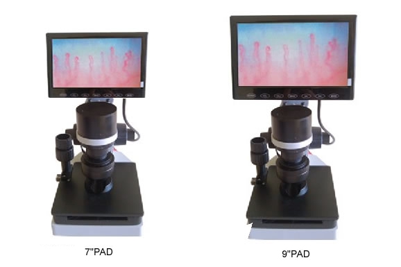
Achromatic Optical Objectives
Achromatic optical lenses reduce chromatic aberration across the field of view. The 5X objective configuration provides a 25mm effective working distance for smooth specimen alignment.
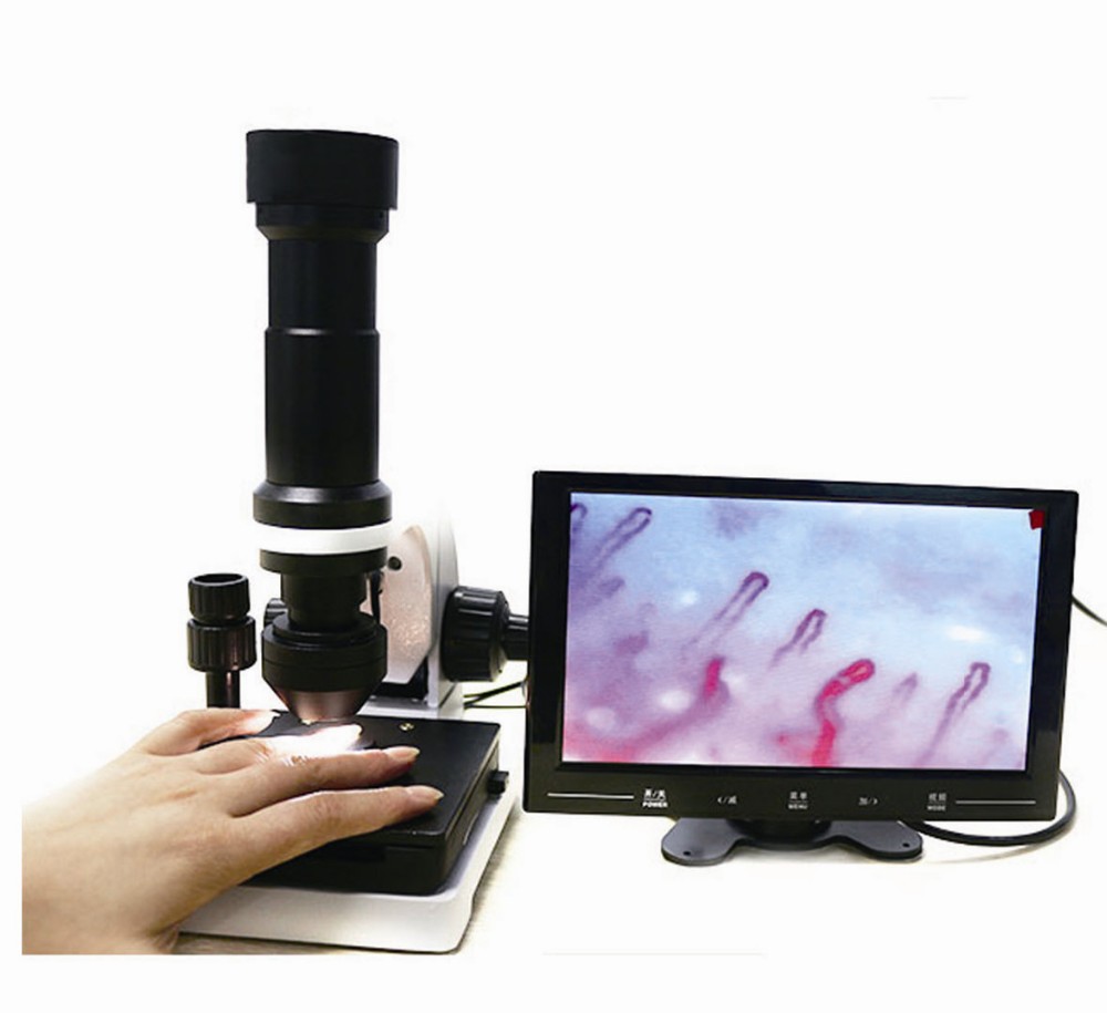
Integrated Video Sensors
High-sensitivity video sensors stream real-time microvascular dynamics to dedicated color monitors. Live output allows multi-angle observation of capillary blood flow and vascular morphology without digital artifacting.
Optical amplification configurations typically range from 240X to 380X, with extended options reaching 500X or 600X depending on research requirements. Consistent magnification allows practitioners to observe capillary loop density, caliber dilation, and microvascular flow dynamics across standardized dermal fields.
Technical Configuration and Optical Parameters
Standardized technical parameters govern hardware performance, imaging stability, and electrical compatibility across professional environments. The MK-GY880 series features balanced mechanical engineering designed for routine microvascular observation.
Review the comprehensive technical parameters below before selecting system configurations for institutional or clinical implementation.
| Parameter | Specification | Technical Context |
| Brand Name | MAIKONG | Manufacturer branding for export and distribution |
| Model Series | MK-GY880 | Base optoelectronic desktop imaging station |
| Optical Amplification | 380X or 240X | Standard optical configurations with optional 500X/600X |
| Objective Lens | 5X Achromatization | Achromatic lens with 25mm effective working range |
| Mechanical Stage | X-Y Compound Stage | Double layer mechanical platform with bilateral travel |
| Focusing Mechanism | Coaxial Controls | Coaxial coarse and fine handwheel adjustments |
| Illumination System | LED Cold Light | Built-in inner dimming to eliminate tissue heating |
| Camera Sensor | Video CCD Sensor | Built-in 80,000 pixel color dynamic video capture |
| Monitor Display | LCD Color Panel | Available in 8-inch, 9-inch, 10-inch, or 12-inch panels |
| Power Input | AC 100-240V | 50/60Hz universal adapter with DC 12V 2A output |
| Compliance | CE Approved | Standardized European conformity certification |
| Instrument Net Weight | 6KG | Solid desktop chassis preventing vibration drift |
When selecting a dedicated nailfold capillaroscopy imaging system, confirming optical specifications and display dimensions supports smooth integration into existing examination workspaces.
Included Hardware and Accessory Kits
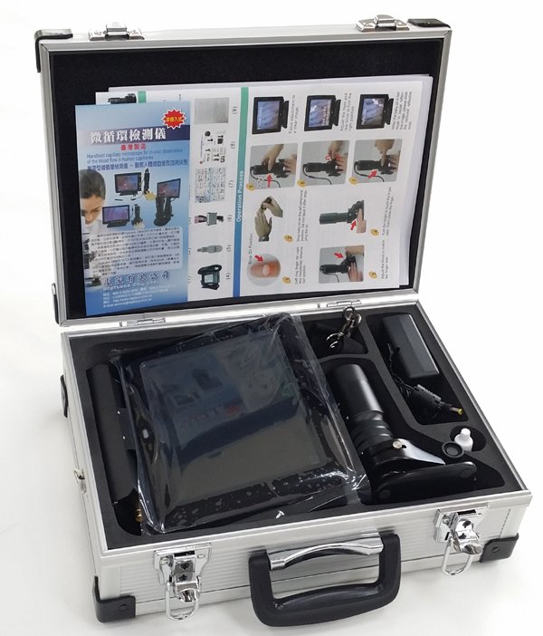
Protective packaging maintains optical alignment and mechanical calibration during international transit. Instruments ship in reinforced aluminum security cases lined with pre-cut high-density memory foam to protect precision stages and internal optical elements.
Standard accessories ship with each workstation to facilitate immediate installation and optical alignment upon arrival.
| Component | Quantity | Functional Role |
| MK-GY880 Host | 1 pc | Main optical body, base stand, and stage |
| LCD Color Monitor | 1 pc | Dedicated top-mounted display screen |
| Power Adapter | 1 pc | 100-240V universal AC to 12V DC power converter |
| Cedar Oil | 1 pc | Immersion medium for light transmission |
| Cotton Swabs | 1 pc | Clean application tool for immersion oil |
| Reference Chart | 1 pc | Capillary pattern guide and alignment aid |
| Aluminum Case | 1 pc | Combination lock transport case with foam inserts |
| Corrugated Carton | 1 pc | Heavy-duty outer shipping container |
Gross shipping weight measures approximately 7KG per single system carton, with outer packaging dimensions of 37.5 cm by 26.5 cm by 34 cm. Clinics and distributors can order replacement accessories or secondary immersion kits directly through Order coordination channels.
Mechanical Stage Controls and Focusing Mechanics
Precise finger positioning requires fine mechanical adjustments that operate independently of finger movement. The double-layer compound mechanical stage moves horizontally and longitudinally to track the microvascular bed across multiple capillary rows.
Coaxial coarse and fine handwheels control the vertical movement of the optical head. Operators rotate the outer coarse ring to bring the objective into initial proximity, followed by fine micrometer rotation to sharpen cellular microcirculation boundaries.
The coaxial focusing mechanism features adjustable tension control. Rotating the tension ring prevents unwanted focal drift while keeping movement light and responsive.
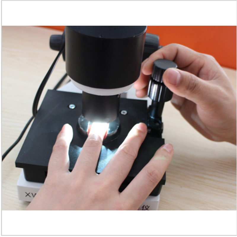
Integrated finger clamps secure the distal digit directly below the objective aperture. Stabilizing the hand reduces involuntary muscular tremor, allowing continuous observation of blood vessels and structural capillary morphology without jittering.
Setup Protocol and Live Image Acquisition
System setup follows a methodical sequence to protect the optical lens and establish clear illumination across the nailfold bed. Standardized ambient conditions help minimize physiological vasoconstriction before evaluation begins.
Operators follow standard assembly and optical focusing steps during routine operation:
- Mount the LCD monitor onto the host chassis bracket and connect the video and power cables to the base ports.
- Connect the AC power supply to a grounded electrical outlet and turn on the monitor power button.
- Place the subject in a seated position with the hand supported at heart level on the examination surface.
- Apply a drop of cedar oil or paraffin oil to the distal cuticle margin of the left fourth digit to improve light transmission and reduce epidermal scatter.
- Slide the prepared finger into the stage clamp beneath the objective lens aperture.
- Rotate the coarse handwheel until initial epidermal vessels appear on the display screen.
- Adjust the fine focus wheel and stage translation dials to center and sharpen the target capillary loops.
Under appropriate operational conditions, locating the initial microvascular row takes seconds once finger placement and immersion oil application are complete.
Observation Contexts in Clinical and Laboratory Settings
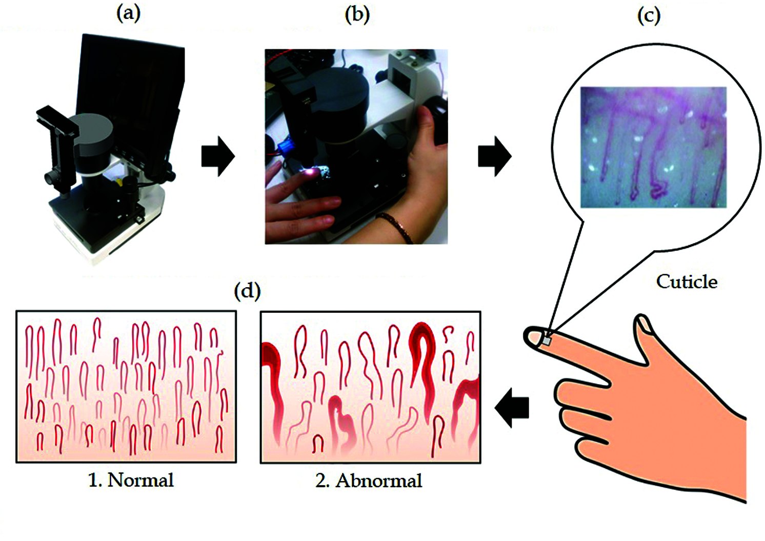
Visualizing microvascular architecture assists clinical specialists in examining vascular responses across dermal tissues. Non-invasive observation provides visual insight into capillary density, flow anomalies, and morphological changes without requiring skin biopsies.
Rheumatology and vascular medicine specialists frequently use capillaroscopic equipment to evaluate patients exhibiting Raynaud phenomenon or structural changes associated with systemic sclerosis and connective tissue diseases. Academic institutions incorporate high-magnification video platforms into physiology curricula to demonstrate peripheral hemodynamics directly.
- Rheumatology evaluations observing capillary dilation, avascular zones, and microvascular hemorrhages.
- Vascular medicine research examining peripheral arterial occlusive disorders and localized circulation.
- Dermatology assessments tracking microvascular loops in cutaneous inflammatory processes.
- Academic microcirculation laboratories conducting biomedical imaging and physiological flow studies.
- Cosmetic science investigations evaluating cutaneous capillary network responses to topical formulations.
Clinics expanding their diagnostic capabilities can review nailfold capillaroscopy equipment options to select systems tailored to high-volume outpatient evaluations.
Imaging Modalities and Optical Configurations
Healthcare facilities and research centers utilize varying optical devices for microvascular evaluation, ranging from simple optical magnifiers to integrated video consoles. Understanding the structural differences helps buyers match hardware capabilities to organizational needs.
Compare console-based video systems with handheld optical tools across primary operational factors:
Integrated Video Systems
- Integrated LCD color monitor displays live capillary blood flow simultaneously for multi-viewer assessment.
- Two-layer compound mechanical stage holds the finger firmly to reduce motion artifact.
- High-magnification optics from 240X to 380X provide detailed visualization of capillary morphology.
- Standardized benchtop chassis built with stable aluminum and steel components.
Handheld Optical Devices
- Compact lightweight footprint suitable for portable screening and quick bedside assessments.
- Manual hand stabilization without compound mechanical stage positioning.
- Lower magnification levels focusing on broader vascular patterns rather than single capillary loops.
- Battery-powered or direct USB connection dependent on external laptop hardware.
For specialized clinics requiring dynamic loop tracking, a stationary capillaroscopy imaging system offers superior mechanical stability during prolonged visual examinations.
Equipment Evaluation Checklist for Purchasers
Procurement teams and distributors must weigh hardware reliability, optical specifications, and logistical support prior to placing equipment orders. Systematic evaluation prevents hardware incompatibilities across clinical networks.
Assess these critical hardware criteria during technical procurement evaluations:
Verify that stage mechanics allow fine micrometric translation across both X and Y axes, ensuring stable field tracking during continuous observation.
Check the optical working distance and magnification levels required for your facility’s protocols. Confirm that the LED illumination produces uniform cold lighting without hotspots that could create reflective flare across immersion oils.
Evaluate video output interfaces to ensure compatibility with institutional recording setups. Verify shipping case specifications, power supply certifications, and availability of replacement parts before finalizing distributor agreements.
Operational Limitations and Practical Handling Factors
Capillaroscopic imaging is fundamentally operator-dependent and requires practical training to obtain reproducible fields of view. Skin pigmentation, thick calluses, and ambient room temperatures can directly affect epidermal transparency and peripheral perfusion.
Maintained room temperatures between 22 and 24 degrees Celsius help minimize cold-induced vasoconstriction during examination. Patients should rest comfortably prior to evaluation, avoiding strenuous physical exercise or recent nicotine exposure that alters peripheral vascular tone.
Capillaroscopy imaging systems provide visual observation of microvascular structures and do not provide automated diagnostic determinations. Clinical interpretations must be conducted by qualified medical professionals according to established clinical criteria.
Objective lenses must be cleaned gently with optical swabs after every examination to prevent oil accumulation on the achromatic front element. Storing the system inside its protective aluminum case shields sensitive video sensors and mechanical gears from dust contamination.
OEM Production and MAIKONG Supply Capabilities
MAIKONG operates advanced optoelectronic manufacturing facilities based in SZ, GD, China, delivering dependable imaging systems to clinical and research distributors worldwide. Our supply chain supports standard device lines as well as custom OEM configurations tailored to institutional specifications.
Partners benefit from flexible private-label branding, customized monitor dimensions, specialized optical magnification options, and comprehensive warranty support. We work closely with international medical wholesalers to support dependable procurement timelines and strict optical quality control.
Establish Your Distribution Partnership
Connect with our engineering and procurement team to discuss wholesale pricing, custom OEM hardware specifications, or U.S. regional distribution rights.
Frequently Asked Questions About Capillaroscopy Systems
What is a nailfold capillaroscopy imaging system?
A nailfold capillaroscopy imaging system is a specialized optoelectronic microscope equipped with cold LED illumination and video sensors. It enables non-invasive visual observation of peripheral capillary microcirculation at the nailbed.
Why is immersion oil applied to the nailfold during examination?
Immersion oil, such as cedar oil or paraffin oil, increases skin transparency by reducing surface light scattering. This step allows light to penetrate the epidermal stratum corneum and reveal underlying capillary loops clearly.
Can the system diagnose vascular or autoimmune diseases automatically?
No, the system does not provide automated diagnostic results or replace clinical assessments. It provides high-magnification optical imagery that qualified medical specialists interpret alongside clinical findings.
What magnification levels are available on the MK-GY880 system?
The MK-GY880 platform provides optical magnification configurations of 240X and 380X as standard options. Extended specifications with 500X or 600X amplification can be requested for advanced research applications.
What display screen sizes can be configured with the station?
The integrated LCD color monitor is available in 8-inch, 9-inch, 10-inch, and 12-inch dimensions. Wholesalers and clinical procurement teams can specify their preferred screen size during order configuration.
How are MAIKONG capillaroscopy systems packaged for international transport?
Each instrument is packed inside a secure aluminum password case fitted with custom memory foam. The aluminum case is then placed inside a heavy-duty corrugated shipping box to protect optical components.
Advancing Microvascular Observation Standards
Selecting a high-precision nail fold capillaroscopy imaging system provides clinics and laboratories with the optical stability needed for comprehensive microvascular observation. Integrating coaxial focusing, cold LED illumination, and dynamic video capture supports reliable scientific evaluation across academic and specialty healthcare environments.