Nailfold Capillaroscopy Video System: Technical Selection and Setup Guide
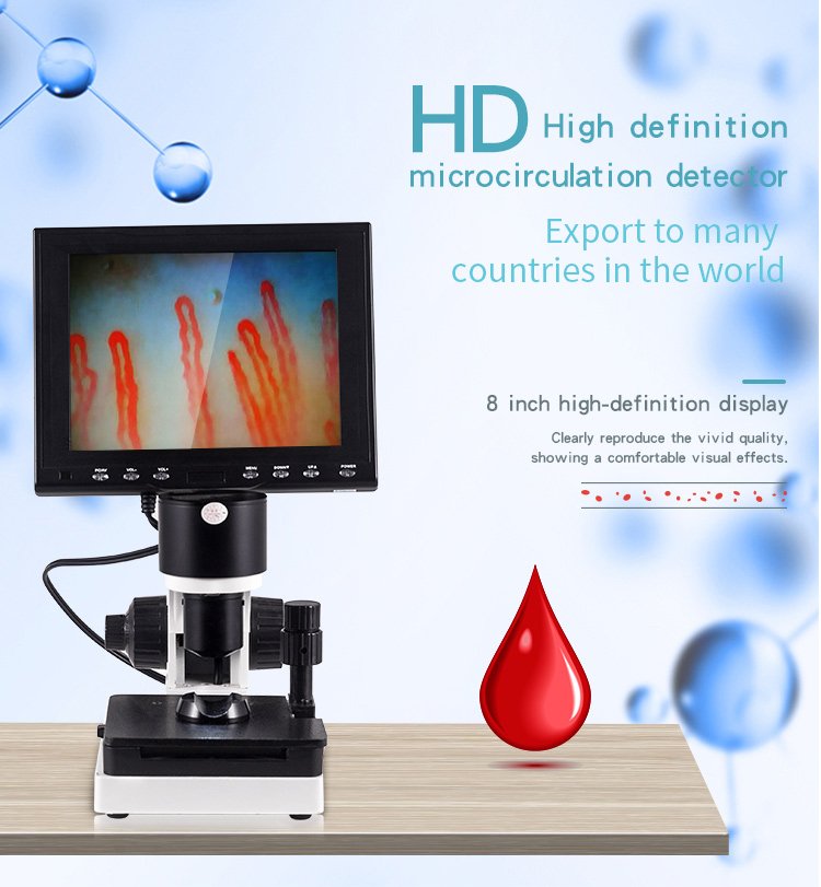
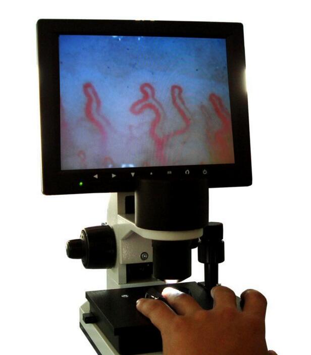
A nailfold capillaroscopy video system is a specialized optoelectronic workstation designed for non-invasive microcirculation examination. It combines high-magnification optical lenses, target-directed LED cold illumination, and high-resolution video sensors to project live microvascular blood flow directly onto a dedicated display screen.
Selecting the appropriate setup depends on required optical magnification, camera sensor configurations, mechanical stage stability, and specific documentation needs in specialty clinics or research laboratories. Careful alignment of hardware features with daily workflow demands maintains steady imaging performance across routine assessments.
What Is a Nailfold Capillaroscopy Video System?
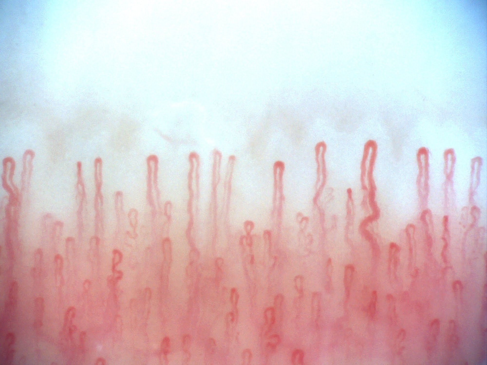
A nailfold capillaroscopy video system integrates an optical magnification column with dynamic video streaming technology to inspect the dermal microvascular beds of the human periungual region. By positioning the digit beneath an achromatic objective, the instrument exposes the horizontal orientation of nail fold capillary architectures without surgical puncture or tissue disruption.
Unlike simple magnifying loupes, a nail fold capillaroscopy video system converts captured optical light into electronic video streams via built-in charge-coupled device (CCD) or digital sensors. This architecture permits multiple observers to analyze dynamic erythrocyte transit, capillary morphology, and perivascular fields simultaneously on an external color monitor.
During standard nail fold capillaroscopy examinations, immersion fluids such as cedar oil or paraffin oil reduce surface light refraction on the keratinized cuticle. The resulting high-contrast image clearly reveals apical loops, vascular distribution patterns, and surrounding dermal tissue morphology.
The primary advantage of a dedicated video capillaroscopy system is the capability to translate real-time capillary hemodynamics into clear, shareable video documentation without relying exclusively on ocular eyepieces.
How the MAIKONG Video System Operates
The MAIKONG system utilizes an integrated optoelectronic architecture engineered to simplify microvascular inspections while preserving image stability. Coordinated optical, electronic, and mechanical assemblies support clear observation across consecutive assessments.
Optical and Lighting Subsystem
An achromatic 5X objective delivers crisp magnification across an effective working range of 25mm. Built-in LED cold light projection focuses clean, heat-free illumination directly onto the nailfold boundary to prevent tissue overheating.
Video Capture and Display
The imaging module incorporates an imported CCD sensor architecture that channels distortion-free video feeds directly to a color LCD monitor. Operators can assess vascular flow characteristics live without noticeable transmission lag.
Mechanical Finger Stage
An X-Y double-layer compound mechanical stage secures the finger holder during inspection. Coaxial coarse and fine handwheels allow operators to isolate capillaries rapidly and sharpen focal planes with minimal physical drift.
For facilities evaluating upgrades, the system pairs smoothly with digital processing platforms, bridging conventional optics and modern digital nailfold capillaroscopy microscope workflows for institutional data retention.
Key Features for Clinical and Research Workstations
Specialized healthcare departments require imaging platforms that deliver consistent image quality during repetitive daily evaluations. The MAIKONG video capillaroscopy system provides mechanical and optical features tailored to the rigorous demands of microvascular observation.
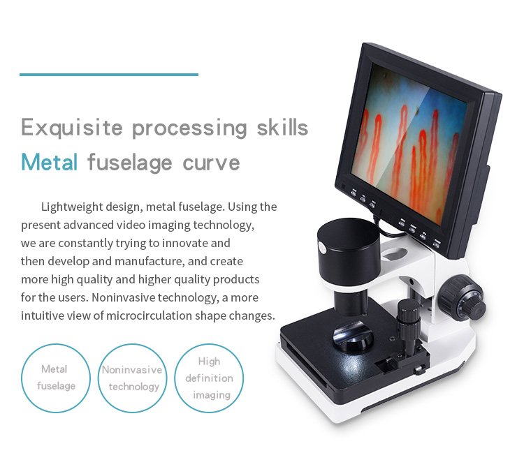
Precision coaxial handwheels allow smooth vertical translation for acute focal alignment.
Selecting an instrument equipped with dedicated mechanical fixtures significantly minimizes physical vibrations. Handheld alternatives often struggle with fine hand tremors, whereas a stationary stage stabilizes the field of view during detailed nail capillary microscopy evaluations.
Institutions transitioning from portable viewers to a professional nailfold capillaroscope benefit from balanced operational ergonomics and rugged industrial hardware designed for high-throughput screening sessions.
Workstation Advantages
- Non-invasive assessment preserves physiological vascular flow during observation.
- Coaxial coarse and fine mechanical adjustments isolate capillary loops swiftly.
- Integrated LED cold light eliminates focal thermal distortion in delicate tissue.
- Live video display supports collective case discussions and student instruction.
Operational Considerations
- Requires stable desktop space and reliable mains electrical power supply.
- Proper image contrast requires correct immersion oil application.
- Image quality remains dependent on environmental room temperature stability.
Technical Specifications and Operational Parameters
Hardware parameters determine visual clarity, magnification thresholds, and system compatibility across institutional networks. The following parameters reflect the standard engineering build of the MAIKONG MK-GY880 series workstation.
| Component | Standard Value | Technical Specifications |
| Brand Name | MAIKONG | Manufactured under stringent quality management protocols in SZ, GD. |
| Product Model | MK-GY880 Host | Specialized optoelectronic nailfold microcirculation video examination workstation. |
| Magnification | 380X or 240X | Optional optical magnification tiers include 380X, 500X, or 600X upon order specification. |
| Objective Lens | 5X Achromatic | Delivers an effective working distance of 25mm for broad field inspection. |
| Stage System | X-Y Mechanical | Double-layer compound mechanical stage with independent lateral and longitudinal controls. |
| Focusing Module | Coaxial Controls | Ergonomic coaxial coarse and fine handwheels for precise focal plane tracking. |
| Light Source | LED Cold Light | Internal dimming module projects targeted, non-heating illumination on the target. |
| Camera Sensor | CCD Module | Integrated 80,000-pixel video sensor delivering real-time analog video signal feed. |
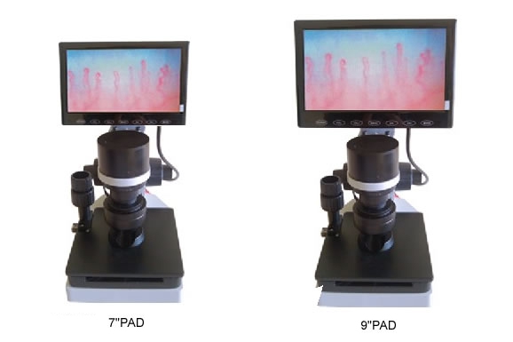
| Display Monitor | Color LCD | Available in 8-inch, 9-inch, 10-inch, or 12-inch configurations based on station setup. |
| Power Input | 100-240V AC | Universal 50/60Hz AC input converting to stable DC 12V 2A operating power. |
| Device Weight | 6KG Net Weight | Total gross packaged weight equals 7KG with protective hard flight case. |
| Certification | CE Approved | Meets applicable international safety directives for laboratory optoelectronic equipment. |
Buyers integrating this workstation alongside existing nailfold capillaroscopy equipment or evaluating a usb nailfold capillaroscopy microscope should verify custom display resolutions with our engineering team prior to shipment.
Packaging Specifications and Standard Inclusions
To prevent misalignment of sensitive optical paths during international transit, each MAIKONG workstation ships in a dedicated reinforced enclosure. Memory foam padding isolates mechanical assemblies from impact shocks.
| Item Included | Quantity | Packaging Details |
| MK-GY880 Host | 1 Piece | Precision microscope core housed in molded anti-vibration memory foam. |
| LCD Monitor | 1 Piece | Integrated color viewing monitor with adjustable mounting bracket. |
| Power Adapter | 1 Piece | Universal switching power unit with region-appropriate electrical cable. |
| Cedar Oil | 1 Bottle | Immersion fluid designed to optimize light transmission and reduce surface reflection. |
| Cotton Swabs | 1 Pack | Clean application tools for targeted immersion fluid distribution on cuticles. |
| Reference Chart | 1 Piece | Laminated microvascular pattern visual guide for baseline alignment. |
| Inner Enclosure | 1 Case | Lockable aluminum briefcase with combination latch for secure storage. |
| Outer Carton | 1 Carton | Heavy-duty corrugated box measuring 37.5 by 26.5 by 34 centimeters. |
For clinic networks seeking streamlined procurement, review the formal Order schedule or explore bulk production options directly through our modern Factory facility in SZ, GD.
Confirm Your Custom Workstation Package
Review standard configurations, verify monitor sizes, and coordinate custom distributor packaging directly with our export logistics team.
Step-by-Step System Setup and Preparation Protocol
Obtaining reliable, low-noise microvascular imagery requires systematic device preparation alongside controlled environmental conditions. Following a standard setup procedure minimizes operational delays and safeguards delicate optical assemblies.
- Mount the color LCD screen onto the upper bracket of the MK-GY880 mechanical host and secure all retaining screws.
- Attach the dedicated video signal cable and DC power cord to the corresponding ports on the base assembly.
- Connect the power adapter to an AC outlet, observe the active power indicator, and depress the monitor switch to activate the screen.
- Instruct the subject to rest comfortably at a table, positioning the examination hand level with the heart in a room maintained at 22 to 24 degrees Celsius.
- Apply two drops of cedar oil or medical paraffin oil across the periungual fold of the designated finger to optimize optical refraction.
- Slide the prepared finger into the stage bracket beneath the achromatic objective lens.
- Turn the coarse handwheel until vascular structures become discernible, then rotate the fine handwheel to establish sharp focus.
- Shift the X-Y mechanical stage controls to position the initial row of capillary loops centrally within the monitor frame.
Standardizing testing workflows supports repeatable results when conducting a routine nailfold capillary test across diverse research cohorts using a stabilized nailfold capillaroscopy device.
Hardware and Procurement Evaluation Criteria
Selecting an institutional video system requires balancing imaging clarity, hardware durability, and post-purchase technical support. Evaluating these factors beforehand prevents unexpected compatibility issues in clinical departments.
Optical Resolution and Magnification
Verify whether your clinical protocol demands 240X, 380X, or higher optional tiers like 600X. High-grade achromatic optics eliminate spherical aberrations, maintaining sharp capillary outlines across the entire visual field.
Display Integration and Signal Flow
Assess whether your facility operates standalone viewing stations or requires external archiving. Confirm monitor dimensions, signal format compatibility, and digital capture cards if integrating with centralized electronic filing archives.
Institutions auditing commercial options should review our published nailfold capillaroscopy equipment price guidance or discuss high-definition sensor customization with an optional dedicated nailfold capillaroscopy camera.
Professional Applications in Clinical and Laboratory Settings
A video-based capillaroscopy platform provides substantial documentation utility across numerous clinical specializations, academic departments, and microvascular research divisions. Live digital visualization enables thorough morphological evaluations without invasive intervention.
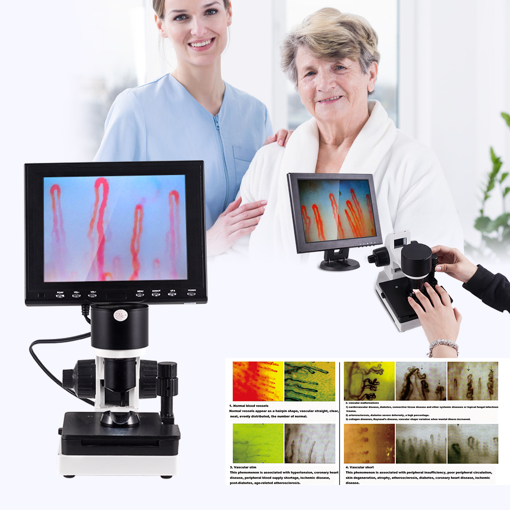
Video streaming platforms facilitate simultaneous collaborative review in specialized clinical environments.
In academic rheumatology and vascular medicine, documentation platforms assist investigators in assessing structural vascular changes. The video feed helps track capillary density, abnormal loop enlargement, and microhemorrhages over consecutive longitudinal visits.
- Rheumatology departments investigating peripheral vascular vasospasm and structural alterations associated with nailfold capillaroscopy scleroderma research protocols.
- Vascular medicine clinics documenting peripheral circulatory dynamics, acrocyanosis, and microvascular patterns in patients with Raynaud phenomenon.
- Dermatology practices monitoring periungual tissue integrity, chronic inflammatory dermatoses, and local wound healing vascular beds.
- Academic research laboratories tracking systemic microvascular adaptation and capillary density changes across diverse clinical study cohorts.
- Medical training institutions demonstrating real-time hemodynamics, capillary loops, and subpapillary venular plexuses to students using nail videocapillaroscopy workflows.
Operational Limitations and Examination Standards
While an advanced video capillaroscopy workstation provides remarkable visual access to the microcirculation, it represents an observational imaging instrument rather than an automated diagnostic tool. Qualified medical personnel must interpret all observed vascular architectures within the broader context of complete clinical findings.
Physiological variables significantly influence peripheral blood flow. Exposure to cold ambient air, emotional distress, recent tobacco usage, or intense physical exertion can induce transient capillary constriction that obscures baseline morphology.
Proper operator technique is essential. Applying excessive downward pressure on the finger rest can compress fragile superficial capillaries, falsely mimicking avascular fields or diminished capillary density. Standardizing room conditions, patient rest periods, and delicate stage manipulation helps prevent observational artifacts.
A video capillaroscopy platform does not independently diagnose disease or replace comprehensive clinical judgment; qualified healthcare professionals must conduct all image interpretations following standardized institutional protocols.
Facilities seeking intermediate magnification screening before detailed video evaluation may review optical alternatives such as a nailfold videocapillaroscope or a portable nailfold capillaroscopy magnifier.
Frequently Asked Questions
What is a nailfold capillaroscopy video system?
A nailfold capillaroscopy video system is an optoelectronic instrument combining microscopic objective lenses, cold illumination, and high-definition video sensors to display live microcirculation video on an integrated monitor. It provides non-invasive visual access to superficial capillary beds without surgical disruption.
What magnification options are available on the MK-GY880?
Standard configurations feature 240X or 380X optical magnification. Optional higher-magnification modules, including 500X and 600X configurations, are available upon request during custom order specification.
Does the workstation include an integrated display screen?
Yes, standard packages include a dedicated color LCD monitor mounted directly to the host frame. Buyers can select 8-inch, 9-inch, 10-inch, or 12-inch display formats depending on workstation space requirements.
What accessories are included in the standard flight case?
Each case contains the MK-GY880 host, color LCD screen, regulated power adapter, cedar oil, application swabs, and a reference pattern chart. The complete workstation arrives cushioned inside an aluminum combination case.
Can MAIKONG support OEM, private-label, or distributor orders?
MAIKONG provides comprehensive OEM and private-label manufacturing from our facility in SZ, GD. We assist medical equipment distributors with custom branding, hardware modifications, tailored packaging, and flexible wholesale pricing structures.
What should buyers verify before completing an order?
Buyers should confirm preferred optical magnification, display monitor size, plug type, and any external video recording requirements. Clarifying these specifications in advance streamlines custom factory configuration and international freight delivery.
Inquire About Workstations and Distribution Opportunities
Integrating a dependable video capillaroscopy workstation elevates microvascular observation capabilities across hospital departments, vascular clinics, and research laboratories. MAIKONG delivers robust optoelectronic hardware supported by extensive manufacturing expertise in SZ, GD.
Request a Formal Quote or Explore Dealership Terms
Contact our medical export division to request detailed technical datasheets, verify custom equipment configurations, or discuss becoming an authorized regional distributor.