
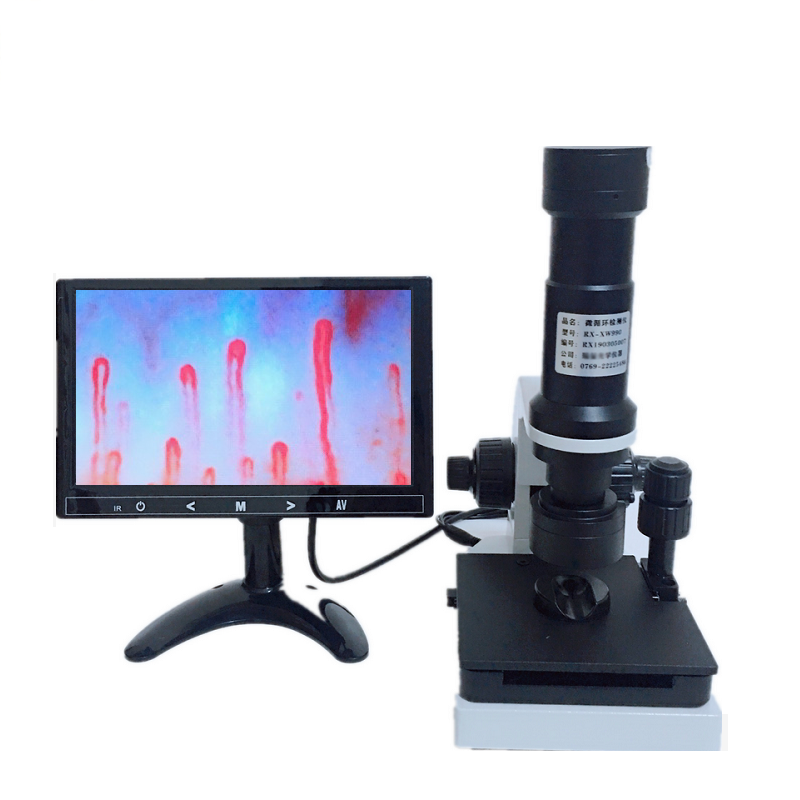
A nail fold capillary is a microscopic terminal blood vessel loop located in the subungual cutaneous tissue at the base of the fingernail. These terminal loops run parallel to the skin surface in the nailbed cuticle margin, providing an accessible site for direct optical visualization of human microcirculation. Examining these microvascular structures allows researchers and clinicians to evaluate capillary morphology, vessel architecture, and red blood cell flow dynamics without invasive surgical intervention.
Optical magnification quality, illumination stability, mechanical stage precision, and digital sensor resolution determine how effectively these microvessels can be inspected. Selecting the proper equipment configuration depends on the demands of the examination environment, varying between mobile assessment settings and stationary clinical research workstations.
What Is a Nail Fold Capillary?
The nail fold capillary network forms the distal boundary of the systemic microcirculation. In most areas of human skin, dermal microvessels ascend perpendicular to the epidermal layer, revealing only the tips of capillary loops when viewed from above. In the periungual region, the dermal papillae orient horizontally along the terminal nail fold, allowing an optical observer to inspect the entire hairpin-shaped vascular loop from the arterial limb to the venous limb.
Each standard loop consists of an ascending afferent arterial branch, an apical turning point, and a descending efferent venous limb draining into the subpapillary venular plexus. Under steady illumination and appropriate optical magnification, erythrocytes travel through these microvascular loops in single file, displaying real-time rheological movement. This structural alignment makes the nail fold the primary anatomical site for vital microvascular microscopy in physiological and biomechanical research.
The horizontal orientation of the nailbed dermal papillae allows direct visualization of the entire capillary loop length, rather than a cross-sectional point, making it suitable for non-invasive morphological measurement.
Why Nail Fold Capillary Observation Is Useful for Professional Work
Direct evaluation of the nail fold vasculature provides quantitative insight into systemic peripheral microcirculation. Because microvascular alterations often precede gross anatomical changes in connective tissues and peripheral vascular beds, observing capillary patterns aids clinical investigations and academic studies. Microvascular researchers inspect these zones to assess vascular integrity, endothelial responsiveness, and regional blood perfusion.
Observation of nailfold capillaries serves distinct observational and operational purposes across multiple professional settings:
- Rheumatology and microvascular research facilities evaluate capillary morphology, loop density, and structural irregularities in autoimmune conditions.
- Dermatology clinics examine cuticular capillary integrity, tissue perfusion, and localized inflammatory reactions near the distal digits.
- Academic medical universities and physiological teaching laboratories demonstrate capillary hemodynamics, leukocyte adhesion, and vascular rheology in living tissue.
- Vascular assessment clinics and rehabilitation centers monitor peripheral blood distribution and post-therapy vascular responsiveness over extended periods.
- Integrative wellness centers and functional health practices evaluate baseline digital circulation patterns as part of holistic vascular screenings.
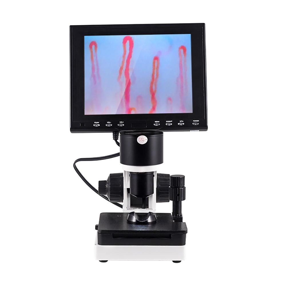
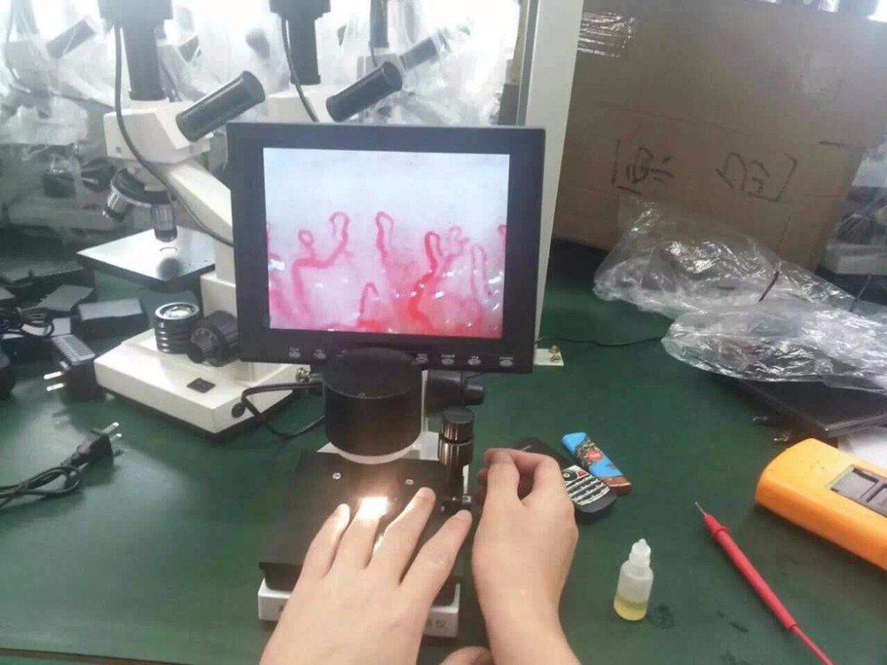
Dedicated finger positioning stages maintain steady alignment during dynamic microcirculation examination.
How a Nail Fold Capillary Microscope Works
A dedicated nail capillary microscopy system combines an optical microscope core, high-efficiency illumination, and a specialized stage assembly. The skin surface is naturally opaque due to light scattering in the stratum corneum. Applying a drop of medical-grade immersion medium, such as paraffin oil or cedar oil, aligns refractive indices, reduces surface scattering, and renders the uppermost epidermal layer translucent.
Light from an integrated cold LED array directs an incident epi-illumination beam onto the cuticle margin at a calibrated angle. Optical lenses with achromatic correction magnify the vascular field, directing the focused image either into an eyepiece or directly onto an integrated CCD or CMOS sensor. Coaxial coarse and fine adjustment mechanisms allow the operator to navigate focal planes smoothly across microscopic depths without disturbing the resting digit.
Refractive index matching between the skin and the optical objective minimizes backscatter, allowing epi-illumination to penetrate the stratum corneum and reveal moving erythrocyte columns.
Modern optoelectronic systems digitize this optical stream, transmitting live video feeds to dedicated monitors or computational software suites. These digital signals capture erythrocyte flow velocity, vessel diameter variations, and loop counts, supporting structured image archiving for longitudinal evaluation.
Equipment Types for Nail Fold Capillary Observation
Selecting the proper nailfold capillaroscopy equipment depends on workflow volume, mobility needs, and data management preferences. Systems range from compact hand-held scanners to integrated stationary workstations with compound mechanical stages.
Handheld and Portable Devices
Handheld scanners feature integrated LED illumination rings and lightweight bodies suited for bedside evaluations, multi-room clinics, and field screenings. These units prioritize rapid deployment, though they require steady operator handling during high-magnification captures.
Stationary Video Workstations
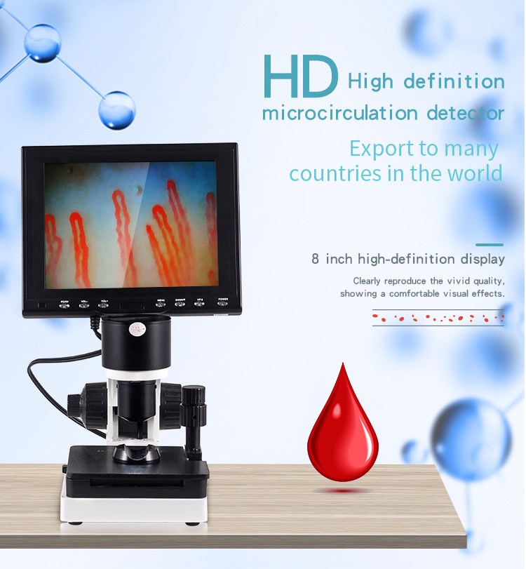
Stationary video systems mount high-performance optics and compound X-Y mechanical stages on stable desktop frames. These setups deliver steady focus and micrometric navigation, making them ideal for high-throughput clinical research and documentation.
USB-interfaced systems connect directly to external laptops or desktop workstations, utilizing host software for image capture, measurement calibration, and database storage. Standalone units with integrated LCD screens operate independently without requiring external computer drivers, simplifying setup in busy treatment environments.
A Practical Observation Workflow for Microvascular Assessment
Consistent observation outcomes depend on structured preparation of both the testing environment and the mechanical equipment. Standardizing the observation workflow reduces movement artifacts and stabilizes peripheral vascular tone. Operators should note that the following steps describe equipment operation procedures rather than diagnostic or therapeutic protocols.
- Stabilize the examination room at a steady ambient temperature, generally between 22 and 24 degrees Celsius, with relative humidity near seventy percent to prevent cold-induced peripheral vasoconstriction.
- Seat the subject comfortably and allow a rest period of fifteen to thirty minutes, keeping the hand supported on the examination surface at heart level.
- Place one to two drops of immersion medium, such as clear cedar oil or medical paraffin oil, directly onto the cuticle of the selected digit to improve optical transmission.
- Position the oiled digit securely inside the finger bracket under the microscope objective, gently tightening the finger rest without compressing local digital blood flow.
- Turn on the cold LED light source and rotate the coaxial coarse adjustment handwheel until the surface of the skin becomes discernible on the display.
- Rotate the fine adjustment handwheel to bring individual vascular loops into sharp focus, using the X-Y mechanical stage controls to sweep horizontally along the distal capillary row.
- Capture static images or dynamic video recordings for documentation, archiving data in accordance with facility record-keeping procedures.
Maintaining gentle finger contact prevents involuntary tremor from displacing the focal plane. Operators should follow standard sanitation protocols between subjects by wiping stage contacts with approved surface disinfectants.
Magnification, Lighting, Camera, and Display Specifications
Optical design directly influences image fidelity when viewing submillimeter microvessels. While entry-level magnifiers operate between 50X and 100X, specialized systems routinely employ 240X, 380X, or higher total optical amplification. Balancing optical resolution with sensor pixel density prevents digital interpolation artifacts that could obscure delicate capillary loops.
Optical Magnification Core
- True optical zoom preserves structural detail without pixelation.
- Achromatic 5X objective lenses minimize chromatic aberration along vessel edges.
- Generates clear delineation of apical loops and descending limbs.
- Requires precise micrometric focusing due to shallow depth of field.
Digital Sensor and Display
- High-sensitivity CCD or CMOS chips translate optical light to electronic video.
- Integrated LCD monitors provide real-time dynamic streaming for multiple observers.
- Cold LED lighting arrays prevent localized tissue heating during prolonged inspection.
- Camera rotation controls allow realignment of image orientation on screen.
Selecting an appropriate nailfold capillaroscopy camera requires evaluating dynamic range and sensitivity under low-light conditions. High-grade image sensors deliver stable video streams without jitter, helping researchers trace red blood cell velocity accurately.
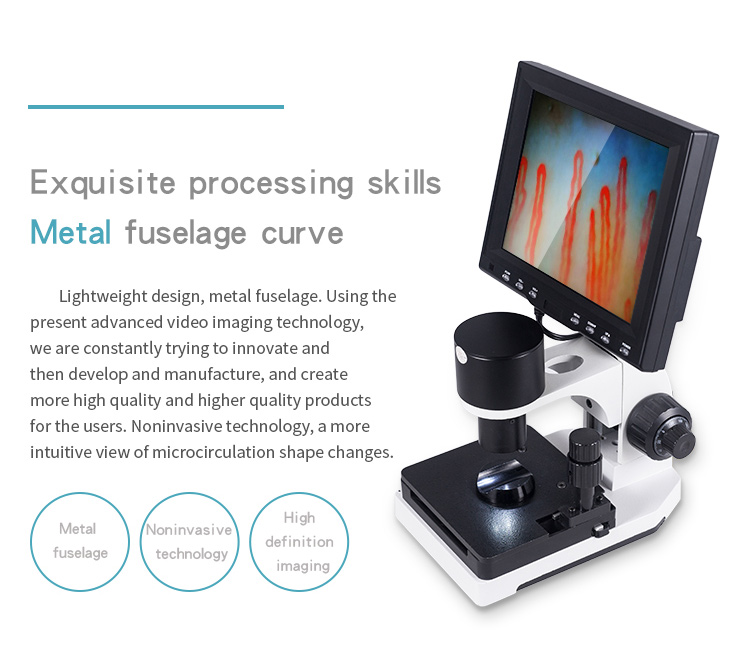
High-resolution monitors display dynamic erythrocyte flow in real time for clinical instruction and review.
Technical Specifications for a MAIKONG Nail Fold Capillary System
MAIKONG builds optoelectronic observation instruments designed for high mechanical stability and clear microvascular imaging. Our MK-GY880 series integrates precision coaxial focusing mechanics, a double-layer mechanical stage, and integrated cold lighting for high-throughput operational environments.
| Specification | Parameter Standard | Operational Scope | Technical Details |
| Brand Name | MAIKONG | System series | Manufactured by MAIKONG under specialized quality control workflows. |
| Amplification | 380X or 240X | Standard optical | Delivers calibrated magnification for individual capillary loop visualization. |
| Optional Zoom | 380X 500X 600X | Factory option | Configurable optical magnifications available upon technical order confirmation. |
| Power Input | AC 100-240V | Universal line | Operates across 50Hz and 60Hz global electrical systems. |
| Power Output | DC 12V 2A | Low voltage | Provides stable direct current to optical sensors and illumination modules. |
| Objective Lens | 5X 25mm range | Optical front | Features achromatization design to maintain sharpness across the entire field. |
| Mechanical Stage | Double layer | X-Y compound | Permits smooth micrometric transverse and longitudinal travel of digit rest. |
| Focus Mechanism | Coaxial design | Coarse and fine | Allows rapid rough acquisition followed by micro-level focal tuning. |
| Internal Dimming | LED cold light | Epi-illumination | Directs heat-free concentrated light directly onto the target skin site. |
| Protection Type | Type I or B | Safety class | Built to standard electrical equipment safety guidelines. |
| System Camera | 80000 pixels | Internal sensor | Integrated imaging chip delivers real-time live video streaming. |
| Display Screen | Color LCD | 8 to 12 inch | Available in 8-inch, 9-inch, 10-inch, or 12-inch sizes. |
| Certification | CE approved | Select models | Applicable only to specifically certified factory configurations. |
| System Weight | Net 6KG | Chassis weight | Solid weighted base prevents desk vibrations during magnification. |
| Gross Weight | Gross 7KG | Packaged weight | Total weight including heavy-duty aluminum transport case. |
| Case Dimensions | 37.5×26.5x34cm | Package volume | Compact footprint for practical transport and secure clinic storage. |
Each professional nailfold capillaroscope system from MAIKONG undergoes multi-step factory inspection to verify optical centering and mechanical stability before shipment.
Packing Accessories and Included Components
MAIKONG packages each observation station with precision-cut memory foam within an aluminum hard-shell case to protect sensitive optoelectronic elements during transit. Buyers should review packing lists on individual proforma invoices, as component contents may vary depending on customized procurement specifications.
| Component | Quantity | Package Role | Included Details |
| MAIKONG Host | 1 pc | Main hardware | Core optoelectronic unit containing stage, objective, and coaxial controls. |
| Color Monitor | 1 pc | Display screen | Mounts securely to the upper frame for dynamic observation. |
| Power Adapter | 1 pc | Power supply | Regulated direct-current transformer compatible with regional mains. |
| Cedar Oil | 1 pc | Immersion liquid | Optical transmission fluid used to reduce skin light scattering. |
| Cotton Swabs | 1 pack | Application tool | Lint-free applicators for spreading immersion medium across nailbeds. |
| Reference Chart | 1 pc | Visual reference | Comparative diagram for anatomical orientation during training. |
| Inner Package | 1 pc | Protective case | Aluminum hard-shell security case equipped with customized memory foam. |
| Outer Package | 1 pc | Shipping carton | Reinforced corrugated carton built to withstand international freight. |
Facilities requiring a nailfold capillaroscopy portable device configuration can store all essential components inside the lockable aluminum case for safe transport between examination rooms.

Reinforced aluminum cases protect optoelectronic components and glass objectives during shipping and storage.
What U.S. B2B Buyers Should Check Before Ordering
Procurement teams, clinical department directors, and commercial distributors should evaluate several technical and operational factors before purchasing microcirculation instrumentation. Assessing these details beforehand helps align equipment capabilities with research protocols or clinic workflows.
Verify whether your software setup requires a standalone monitor or direct computer interface capture cards, as operating system drivers and image archiving pipelines vary between models.
When selecting a digital nailfold capillaroscopy microscope, buyers should review the following technical parameters:
- Optical resolution versus digital zoom: Ensure the baseline optical lenses provide true magnification of at least 240X to avoid relying on blurred digital scaling.
- Stage stability: Verify that the X-Y mechanical stage provides sufficient gear resistance to prevent drift while moving across consecutive fingernails.
- Thermal footprint: Confirm that the system uses LED cold illumination rather than halogen bulbs to avoid heating the subject’s finger and altering blood flow dynamics.
- Supply chain replacement parts: Confirm that the manufacturer maintains spare objectives, power adapters, and display panels for long-term operational maintenance.
- Packaging specifications: Ensure that transit cases include impact-resistant custom foam inserts suitable for regional or international transit.
MAIKONG Supply, OEM, Private-Label, and Distributor Support
MAIKONG operates advanced optoelectronic manufacturing facilities based in SZ, GD, China, serving healthcare equipment wholesalers, clinical researchers, and OEM brand partners worldwide. Our production lines combine precision optical machining, cleanroom optical assembly, and rigorous electronic testing to deliver dependable microvascular observation hardware.
We provide comprehensive OEM and private-label customization options for American medical device distributors and corporate brands. Our engineering team accommodates customized exterior chassis colors, silk-screened brand logos, tailored packaging graphics, and specialized user documentation. Distributors can order sample units for evaluation before committing to volume manufacturing runs, with flexible production scheduling to meet commercial distribution targets.
Expand Your Microvascular Equipment Distribution
Connect with our technical supply division to review wholesale pricing schedules, private-label branding options, and factory export configurations.
Frequently Asked Questions
What is a nail fold capillary?
A nail fold capillary is a terminal microvascular loop located in the cuticular skin border of the human fingernail. Because these vessels lie parallel to the epidermal surface, they can be visualized directly under optical magnification without surgical procedures.
Why is immersion oil required during nail fold observation?
Immersion oil reduces light scattering across the irregular stratum corneum of the skin. Matching the refractive index between the surface skin layers and the optical lens allows incident LED light to pass through, creating a clear optical view of the capillary bed.
How does optical magnification differ from digital magnification?
Optical magnification relies on physical glass lenses to enlarge the image without loss of detail or fidelity. Digital magnification enlarges existing pixels through software interpolation, which can cause image pixelation if the optical core lacks sufficient resolution.
Can MAIKONG systems operate on standard U.S. electrical voltages?
Yes, MAIKONG observation systems feature universal power adapters compatible with 100V to 240V alternating current at 50Hz and 60Hz. They connect to standard North American power outlets using appropriate low-voltage direct-current adapters.
How long does an operator need to locate the first capillary loop?
Operators familiar with the coaxial focus knobs and X-Y stage controls typically locate the first distal capillary row within seconds after placing the oiled digit. Proper resting and ambient temperature controls help streamline this positioning process.
What customization options does MAIKONG offer for OEM partners?
MAIKONG provides custom chassis silk-screening, custom startup software logos, specialized monitor sizes, and customized flight case branding. Wholesale partners can discuss specific technical and packaging requirements directly with our engineering team.
Advancing Microvascular Observation Technology
Evaluating the nail fold capillary architecture provides essential physiological insight for clinical researchers, academic educators, and vascular specialists. Selecting an optoelectronic system with robust coaxial focusing, achromatic lenses, and stable LED cold illumination supports reliable documentation of peripheral microcirculation. Commercial distributors, research institutions, and healthcare purchasing teams can contact MAIKONG to discuss wholesale configurations, OEM partnerships, and supply arrangements.
Contact MAIKONG Supply and Distribution
Reach out directly to discuss system configurations, distributor pricing, and private-label production schedules.