
Nailfold Capillaroscopy Magnifier for Microvascular Observation
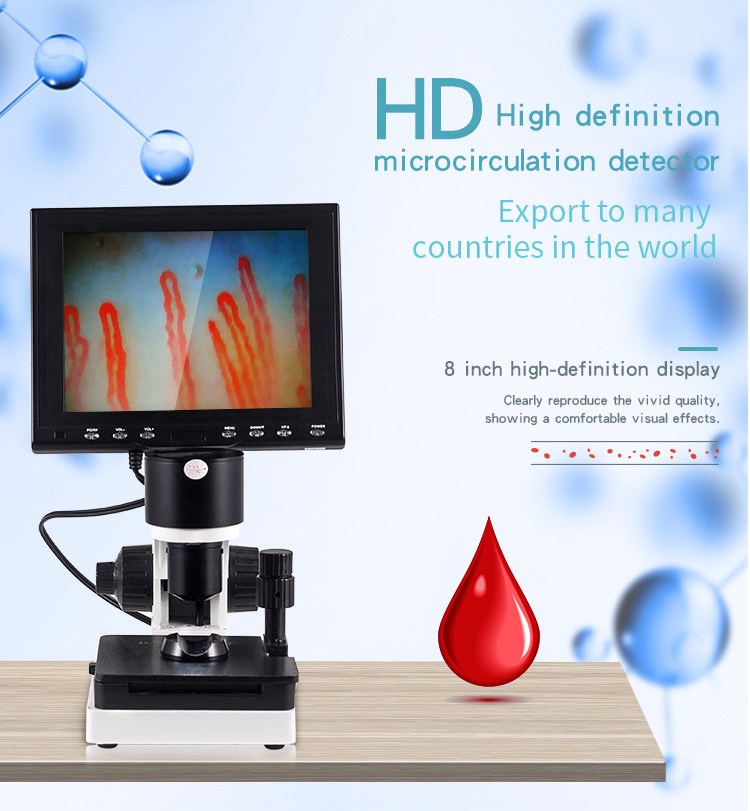
A nailfold capillaroscopy magnifier provides direct optical enlargement to observe periungual microcirculation, capillary loop morphology, and localized red blood cell movement. Dedicated optical magnifiers allow clinicians and microcirculation researchers to examine dermal capillary beds non-invasively through clear focal alignment without requiring cumbersome benchtop setups.
Selecting the appropriate capillary observation format depends on practical clinical throughput, field of view preferences, and documentation requirements. Compact handheld units deliver rapid bedside viewing, while integrated console systems provide stabilized platforms for detailed research assessment.
What Is a Nailfold Capillaroscopy Magnifier?
A capillaroscopy magnifier is a targeted visual instrument engineered specifically to inspect the terminal vascular networks situated beneath the proximal nail fold cuticle. Unlike general dermatoscopes or magnifying loupes, these units incorporate targeted focal planes and perpendicular illumination geometries that penetrate superficial skin scattering when paired with immersion fluid.
By positioning the digit beneath the objective lens, an operator can view capillary arrangement, luminal diameters, and vascular architecture across the distal row. The optical design maintains high chromatic correction, preventing edge distortion during prolonged microscopic assessment of microcirculatory pathways.
Visual observation with an optical magnifier serves research and clinical screening workflows and does not substitute for independent professional medical judgment or formal disease classification.
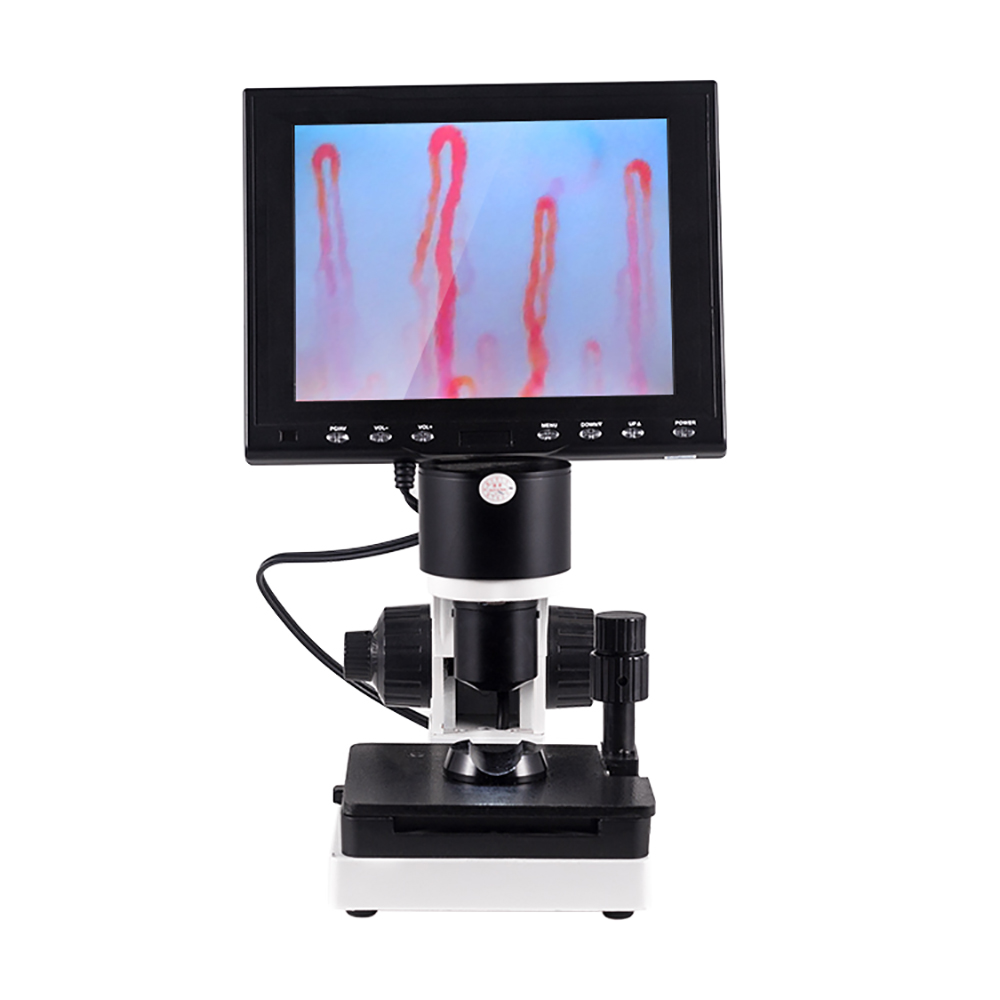
Dedicated optical alignment allows unobstructed observation across the proximal nail fold margin.
Key Features to Evaluate Before Purchasing
Evaluating microvascular viewing instruments requires examining optical performance alongside daily handling mechanics. Optical clarity remains closely tied to lighting management, working distance, and mechanical stage stability during delicate positioning.
- Optical Magnification: Nominal magnification options typically range from 240X up to 380X or higher, offering sufficient resolution to assess capillary loop dimensions without excessive field narrowing.
- Glare-Free Illumination: Integrated LED cold-light sources prevent tissue warming while directing targeted beams directly across the nail plate to highlight intravascular flow.
- Stable Finger Fixtures: Ergonomic rests and mechanical clamps steady the digit against involuntary tremor, preserving focus during real-time observation.
- Coaxial Focus Adjustment: Independent coarse and fine focus handwheels allow swift initial height adjustment followed by smooth focal tracking.
- Immersion Compatibility: Contact plates and lens cones must safely support common transmission mediums such as cedar oil, paraffin oil, or mineral oil to eliminate reflective air pockets.
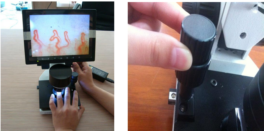
Dual-axis coaxial focusing enables rapid image acquisition across delicate vascular fields.
Magnifier-Style Viewer vs. Digital Microscope System
Determining whether to acquire a handheld nail fold capillary viewer or a console nailfold capillaroscopy microscope depends on practical reporting workflows. Direct magnifiers offer rapid deployment for quick visual screening, whereas larger systems integrate digital sensors and video monitors for archival documentation.
Facilities focused primarily on immediate visual surveys benefit from the small footprint of portable optical viewers. In contrast, academic departments and vascular specialty practices frequently implement an integrated nailfold capillaroscopy camera or complete nailfold capillaroscopy video system to export reproducible image series for research files.
Optical Magnifier Viewer
- Compact footprint suited for flexible room-to-room transit.
- Immediate setup without requiring computer operating system drivers.
- Focuses entirely on live direct visualization.
- Minimal cable management and lower benchtop footprint.
Digital Microscope Station
- Integrated video sensor exports footage to dedicated monitors.
- Mechanical dual-axis mechanical stage stabilizes horizontal finger movement.
- Facilitates group review and detailed laboratory image capture.
- Accommodates interchangeable objectives and digital storage accessories.
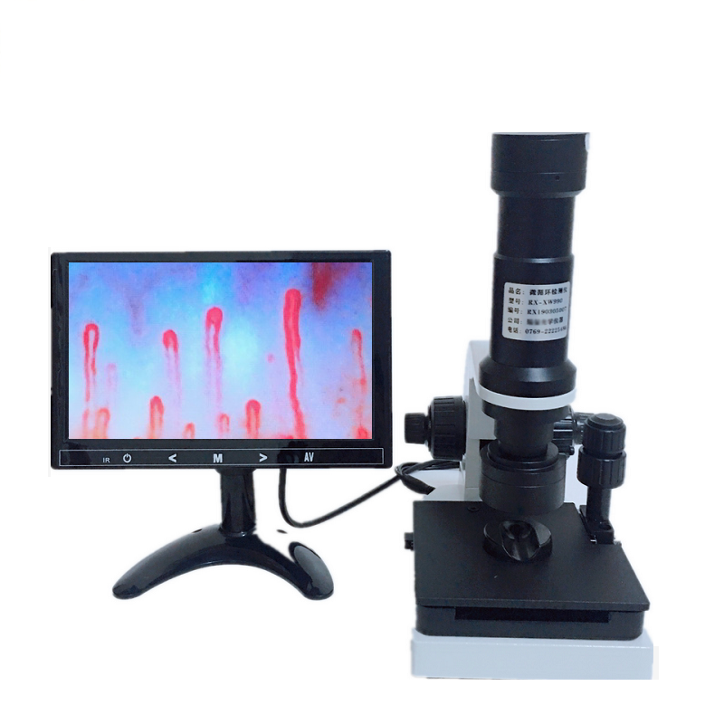
Console units expand direct magnification into multi-user video display workflows.
Professional and Commercial Environments
Microvascular visualization tools support multidisciplinary evaluation in routine outpatient clinics as well as advanced academic laboratories. Selecting an instrument format tailored to operational pace streamlines daily microcirculation evaluations.
Specialty Practices
Rheumatology and vascular medicine clinics utilize optical magnification units for secondary structural assessment of peripheral capillaries during comprehensive vascular health exams.
Research Labs
University physiology laboratories and biomedical engineering programs deploy stable magnifiers to monitor peripheral blood flow behavior under varied experimental variables.
Equipment Distributors
Commercial suppliers and regional wholesalers stock portable viewers and complete console stations to satisfy diverse institutional requests across regional healthcare systems.

Adaptable optical viewing configurations serve specialized diagnostic centers and university research programs alike.
Example Technical Specifications
When selecting specialized microcirculation hardware, reviewing optical, physical, and electronic parameters helps match equipment capabilities with clinic workflow demands. The specifications below represent an advanced MAIKONG MK-series optoelectronic workstation with integrated display and coaxial mechanics.
| Parameter | Specification | Operating Detail |
| Packing Dimfnsing | 37.5*26.5*34cm | Secure dimensions for transit |
| Amplification | 380X or 240X | High optical magnification |
| Input | AC 100-240V | Universal AC 50/60Hz supply |
| Output | DC12V, 2A | Stable low-voltage output |
| Objective | 5X | Effective range 25mm |
| Objective type | Achromatization | Corrected optical lens elements |
| Mechanical stage | X-Y compound | Double layer compound stage |
| Focusing mechanism | Coaxial focus | Coarse and fine adjustments |
| Inner dimming | LED cold light | Targeted projection without heat |
| Type | I or B | Standardized equipment build |
| Power | 12V, 1A | Dedicated DC power source |
| Built-in camera | 80 000 pixels | Integrated sensor feed |
| LCD monitor | 8 or 9 inch | Direct color LCD screen |
| Certificate | CE approved | Standardized export clearance |
| Net weight | 6KG | Heavy stabilized bench base |
| Gross weight | 7KG | Total boxed shipping weight |
| Inner package | Aluminum box | Locking box with memory foam |
| Out package | Corrugated box | Reinforced exterior carton |
Additional custom configurations through MAIKONG can provide optional amplification levels of 380X, 500X, or 600X, paired with 8-inch, 10-inch, or 12-inch Color LCD monitors. Handheld or portable models may incorporate ultra-lightweight housings, including specialized lamp designs weighing approximately 250 g, depending on the specific setup selected.
Example Packaging and Included Accessories
Every commercial optical order ships with essential accessories necessary for immediate operational setup and optical maintenance. Secure internal boxing protects precision glass elements against shock during domestic transport or inter-facility transfer.
| Spare parts | Quantity | Component Function |
| GY880 host | 1pc | Main optical chassis with stage |
| LCD monitor | 1pc | Dedicated display screen unit |
| power adapter | 1pc | Regulated wall power supply |
| cedar oil | 1pc | Optical interface contact medium |
| cotton swab | 1pc | Lens and finger preparation tool |
| chart | 1pc | Reference morphology display sheet |
Exact accessory inclusions vary according to ordered quantities and custom OEM packaging requirements. Facilities may request supplemental sample bottles, specialized mounting adapters, or custom-fitted travel cases during consultation.
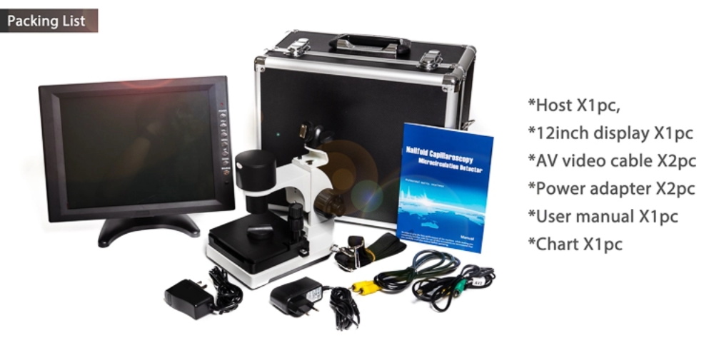
Protective aluminum cases safeguard optical assemblies during shipping and transit between clinic facilities.
Production and Supply Capabilities
MAIKONG operates advanced manufacturing lines in SZ, GD, delivering high-precision optoelectronic viewing systems for international distribution. By maintaining stringent assembly tolerances, the production team delivers cost-effective supply solutions for private-label brands and clinical facilities.
Wholesale partners receive responsive technical coordination, structured warranty fulfillment, and comprehensive original equipment manufacturing (OEM) options. Direct factory engagement enables flexible adjustments across screen dimensions, lens magnification assemblies, and exterior brand markings.
Production Advantages
- Direct manufacturing oversight in SZ, GD ensures high mechanical consistency.
- Custom OEM and private-label options for regional distributors.
- CE approved manufacturing standards supporting international clinical export.
Procurement Considerations
- Customized magnification barrels require advance component lead times.
- Specific display sizes must be verified based on production batch schedules.
Establish Regional Wholesale Distribution
Clinics, laboratories, and regional healthcare suppliers can coordinate bulk shipments and explore custom-branded distribution agreements directly with MAIKONG production directors.
Frequently Asked Questions
What level of magnification is needed for observing nailfold capillaries?
A magnification range of 200X to 380X is generally suitable for evaluating capillary loop arrangement, morphology, and blood flow velocity. Advanced configurations offering 500X or 600X magnification provide expanded optical views for specialized laboratory inspection.
How does an LED cold-light source benefit capillary viewing?
Cold-light LED illumination projects bright, targeted light onto the skin margin without causing uncomfortable thermal expansion or vascular vasodilation. This directed projection minimizes surface glare while keeping the target region comfortable during extended visual exams.
Why is oil applied to the nail fold prior to observation?
Applying immersion oil, such as cedar oil or paraffin oil, smooths irregular skin textures and matches optical refractive indexes. This application reduces light scatter at the epidermal boundary, allowing clearer visualization of subsurface microcirculation.
What is the difference between a standalone viewer and a videocapillaroscope?
A standalone nail fold capillaroscopy magnifier is designed for direct optical observation through an eyepiece or integrated lens. In contrast, a videocapillaroscopy system incorporates high-resolution digital cameras and computer software to record, measure, and archive vascular images over time.
Can MAIKONG customize equipment specifications for OEM partners?
MAIKONG provides comprehensive OEM and private-label customization, including custom branding, tailored packaging boxes, and specific LCD display dimensions. Wholesale buyers can confirm hardware options and optical configurations directly with our manufacturing department.
Procurement Consultation and Wholesale Inquiries
Selecting the right nailfold capillary viewer requires balancing optical enlargement, mechanical stage stability, and organizational workflow needs. Connecting directly with manufacturing representatives facilitates accurate model matching, sample evaluation, and wholesale planning for your clinical or commercial enterprise.
Contact MAIKONG for Specifications and Quotations
Inquire today to discuss wholesale orders, custom OEM branding, or distributorship opportunities across your local market.