
Nail Capillary Microscopy: Clinical Principles and Microvascular Observation
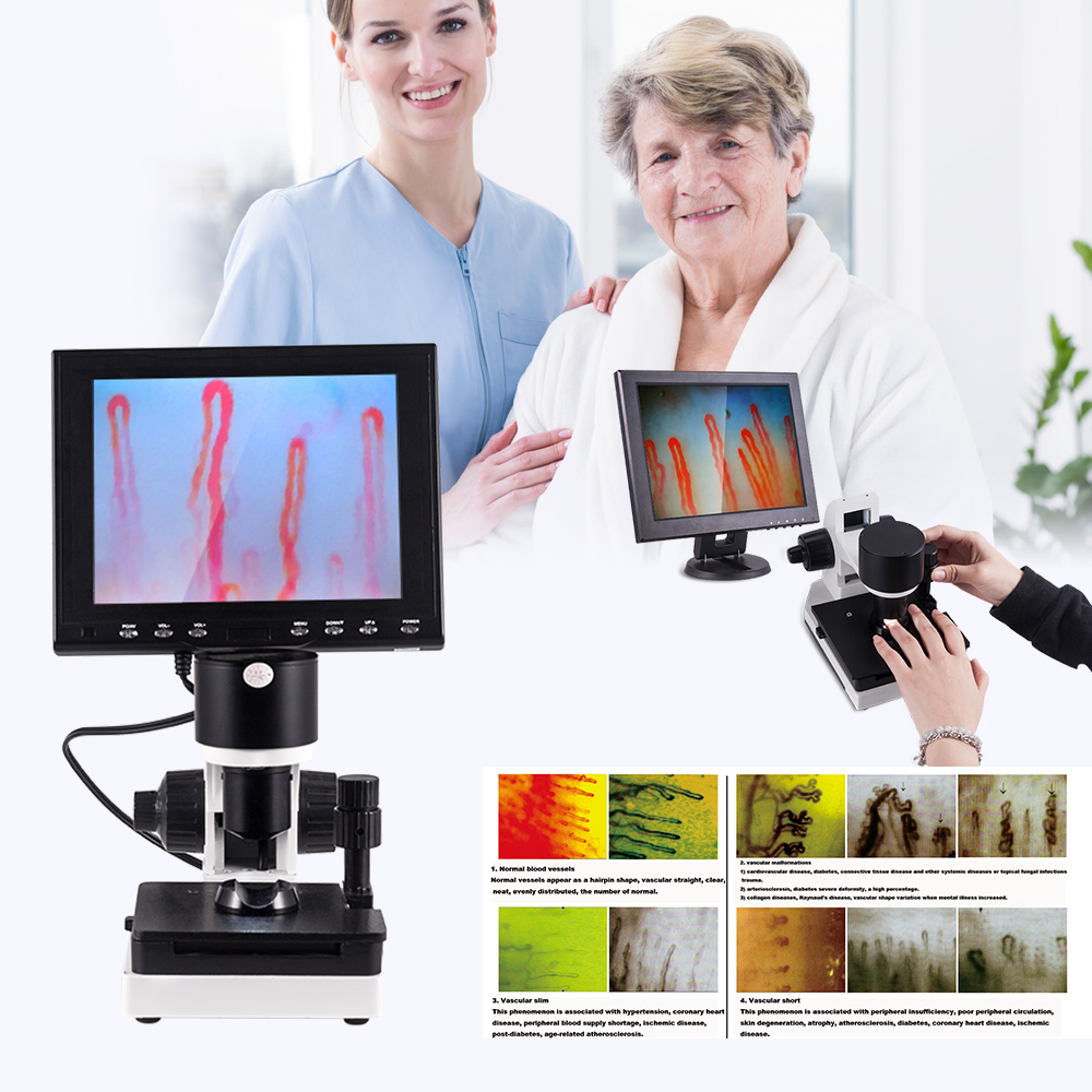
Nail capillary microscopy is a non-invasive optical technique that visualizes the terminal microvascular architecture of the periungual nailfold skin under direct illumination. The examination displays the structural arrangement of capillary loops, baseline red blood cell columns, and local vessel morphology without disrupting cutaneous tissue. Clinicians and microcirculation researchers utilize this optical assessment to evaluate microvascular integrity directly at the dermal-epidermal boundary.
Microvascular clarity depends on accurate optical alignment, refractive index matching using immersion oil, and stable magnification stages. Proper baseline environmental control and rigorous patient preparation determine the diagnostic utility and reproducibility of every recorded capillary field.
Understanding Nail Capillary Microscopy and Terminology
Nailfold microscopy evaluates the horizontal terminal vascular loops that run parallel to the epidermal surface in the finger nailfold. In most anatomical regions, dermal capillaries orient perpendicularly to the skin surface, displaying only tiny red dots under superficial magnification. At the distal nailfold, the angulation of the dermal papillae flattens the capillary loops, allowing complete longitudinal viewing of the arterial limb, hairpin apical turn, and venous limb.
While standard dermoscopy and low-power loupes provide wide-angle surveys, higher-magnification microscopy provides granular visual data on individual loop dimensions and cellular column movement. The technique integrates into wider clinical frameworks alongside nail fold capillaroscopy examinations to document quantitative and qualitative structural changes over time.
Direct Nailfold Microscopy
- Evaluates capillary morphology at magnifications between 200X and 600X.
- Permits dimensional measurement of apical loops and limb diameters.
- Requires precise coaxial focusing and stable mechanical stage control.
Wide-Field Surface Inspection
- Operates at lower magnifications between 10X and 40X.
- Surveys general loop distribution across multiple adjacent fingers rapidly.
- Offers lower optical resolution of individual capillary wall borders.
Modern microvascular observation relies on uniform descriptive terms rather than variable visual descriptions. Precise distinction between overall field density, local vessel architectural distortion, and extravasated red blood cells establishes a reliable baseline for cross-specialty clinical dialogue.
Direct optical inspection reveals the longitudinal layout of distal capillary loops alongside the periungual boundary.
Optical Principles and Imaging Architecture
A specialized digital nailfold capillaroscopy microscope combines focused optical components, solid-state image sensors, and calibrated illumination to penetrate the stratum corneum. Keratinized skin scatters incident light extensively, which prevents deep tissue visibility under standard illumination angles. Integrating targeted cold LED illumination directly around the objective eliminates glare and reduces cutaneous thermal expansion during lengthy assessments.
A dedicated nailfold capillaroscopy camera with high-sensitivity charge-coupled device (CCD) architecture captures continuous dynamic video frames from the focal plane. Achromatic objective systems correct spherical and chromatic aberrations across the visual field, yielding crisp margin resolution of minute red blood cell aggregates inside narrow lumen walls.
Cold LED Illumination
Built-in cold LED systems supply directed light without heating sensitive finger tissue. Stable temperature prevents unwanted local vasodilation during observation.
Achromatic Optics
Precision 5X achromatic objectives maintain color balance across twenty-five millimeters of working range, eliminating color fringing at capillary margins.
Coaxial Stage Controls
Dual-knob coarse and fine mechanics allow precise focal tracking across uneven nail cuticles with smooth vertical micrometer adjustments.
Smooth double-layer compound mechanical stages permit progressive scanning along the entire distal row of each finger. Coaxial controls prevent erratic movements, maintaining unbroken digital tracking while recording microvascular fields.
Microvascular Morphological Features and Clinical Observation
Observation of the nail fold capillary network focuses on specific structural characteristics across the distal perimeter. Healthy capillaries show a homogenous distribution, resembling neat hairpins or inverted U-shaped structures arranged in even, parallel rows. Distinct deviations in loop shape, size, or spatial frequency signal microvascular disturbance that requires careful documentation.
When assessing patients evaluated for connective tissue conditions, clinicians note the presence of microhemorrhages, dilated loops, and avascular areas. In systemic autoimmune conditions, such as those assessed during a nailfold capillaroscopy scleroderma evaluation, characteristic changes progress from initial giant capillary expansion to widespread capillary loss and neoangiogenic ramification.
Capillary density typically ranges between seven and twelve loops per millimeter along the distal row. A significant drop in loop density indicates structural microvascular damage, which should be assessed alongside detailed laboratory findings.
Microvascular hemorrhages indicate capillary wall disruption, appearing as focal brown or red deposits separated from the active vascular lumen. Severe morphological anomalies present as bushy, branched, or extremely twisted capillary formations resulting from disheveled endothelial repair processes.

High-magnification digital display highlighting loop arrangement, baseline spacing, and microvascular clarity.
Step-by-Step Examination Protocol
Performing a reliable nailfold capillary test demands a repeatable, standardized process to avoid image artifacts. The clinician must position the patient comfortably with the examined extremity supported firmly at heart level on the examination stage. Sudden movements or improper finger pressure on the mechanical stage can cause artificial ischemia and blur the recorded capillary loops.
Applying an optical interface fluid bridges the refractive index difference between the epidermal keratin layer and the objective lens. Paraffin oil or cedar oil fills microscopic surface irregularities, allowing light to enter the subepidermal capillary plexus directly.
- Clean the distal nailfold with a soft swab to clear particulate debris without rubbing or agitating the cuticle.
- Place two drops of cedar oil or medical-grade paraffin oil along the margin of the ring finger nailfold.
- Position the finger securely within the mechanical finger seat directly below the objective aperture.
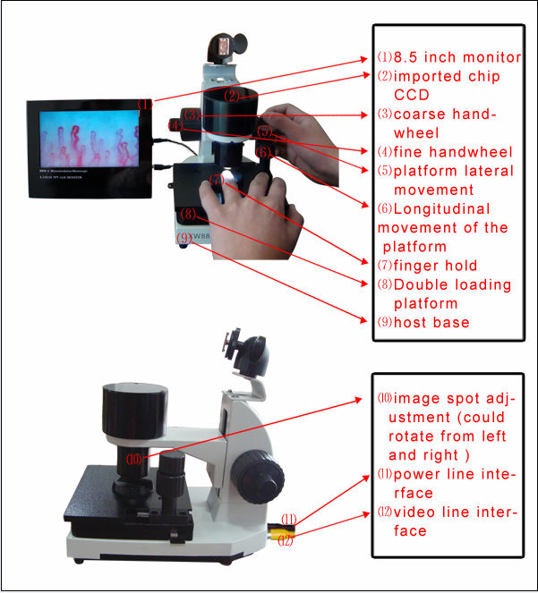
- Adjust the coarse handwheel until the initial tissue silhouette comes into preliminary focal view on the display.
- Rotate the coaxial fine adjustment handwheel to sharpen the vascular margins and resolve red blood cell columns.
- Traverse the entire distal capillary row using the lateral and longitudinal stage controls to examine every sector.
The fourth finger of the non-dominant hand offers optimal skin transparency due to reduced physical trauma and thinner cuticle tissue. When necessary, inspect digits two through five systematically to obtain a representative assessment across the hands.
Standardized Environmental and Patient Preparation Conditions
External room conditions and transient physiological states alter peripheral microvascular tone significantly. The human microcirculation responds immediately to cold exposure, emotional stress, and systemic chemical stimulants through dramatic precapillary arteriolar vasoconstriction. Uncontrolled testing settings can lead to misinterpreting transient functional vessel closure as structural capillary loss.
Adherence to standardized preparation protocols minimizes physiological variance and supports reproducible visual data across multiple sessions. Maintaining static ambient conditions isolates chronic architectural remodeling from acute hemodynamic fluctuations.
Perform examinations in a temperature-controlled room held strictly between 22 and 24 degrees Celsius, with relative humidity maintained at approximately 70 percent. The patient must remain seated at rest for fifteen to thirty minutes before recording any images.
Patients must refrain from smoking, consuming caffeine, or eating large meals for one hour prior to capillary assessment. In addition, patients should avoid strenuous physical exertion and hand washing with cold water during the immediate sixty minutes before evaluation.
Cardiovascular medications, vasoactive supplements, and the physiological phases of the menstrual cycle in female patients also influence microcirculatory blood flow. The supervising physician must interpret all observed findings alongside complete pharmacological profiles and systemic clinical records.
Consistent room temperature and resting patient position provide stable physiological conditions for capillary tracking.
Clinical Research and Observational Scope
Academic institutions and biomedical departments apply high-resolution nail videocapillaroscopy to study systemic endothelial behavior across multiple clinical settings. Because the nailfold offers a direct non-invasive window into living human microvessels, researchers investigate structural responses to pharmaceutical agents, environmental pressures, and systemic inflammation. Quantitative metrics derived from visual recordings help track long-term microvascular remodeling.
Comprehensive video systems, including the nailfold capillaroscopy video system, support objective frame-by-frame measurement of red blood cell velocities and vascular caliber changes. These observations support investigations in vascular medicine, dermatology, metabolic pathology, and physical rehabilitation science.
Rheumatology & Autoimmunity
Observation of microvascular patterns helps differentiate primary Raynaud phenomenon from systemic connective tissue diseases with microvascular involvement.
Dermatology & Angiology
Evaluation of cutaneous microangiopathy, psoriasis margins, peripheral venous stasis, and localized tissue remodeling patterns.
Metabolic & Endocrine Studies
Tracking structural microvascular changes and flow irregularities associated with diabetic microangiopathy and metabolic syndrome progression.
Specialized research applications also explore localized skin microcirculation on the tongue, earlobes, and lips to compare vascular dynamics across anatomical beds. Furthermore, biomedical materials teams assess microcirculatory responses to thermal, far-infrared, and conductive surface stimuli under controlled laboratory conditions.
Methodological Strengths and Practical Limitations
Utilizing a professional nailfold capillaroscope delivers immediate real-time visualization of living microvascular loops without ionizing radiation, venipuncture, or contrast media. Compact configurations, such as a handheld nailfold capillaroscope, provide practical versatility for multi-room clinics and bed-side assessments. High-definition optical lenses resolve loop morphology quickly while preserving patient comfort.
Despite these clear operational benefits, the technique has inherent constraints that demand rigorous standardization and experienced clinical interpretation. Nailfold capillary examination remains an observational modality that visualizes anatomy and local blood columns rather than delivering automated disease diagnoses.
Advantages
- Completely non-invasive with zero tissue disruption or radiation exposure.
- Permits immediate live video recording and side-by-side comparative tracking.
- Rapid image localization within seconds when using stable mechanical staging.
- Well suited for longitudinal monitoring of microvascular morphology.
Limitations
- Highly dependent on room temperature and patient pre-test resting stability.
- Requires deliberate operator training to distinguish true pathology from microtrauma.
- Cuticular hyperkeratosis or severe skin pigmentation can impede optical penetration.
- Cannot independently diagnose systemic diseases without broader clinical context.
Accurate interpretation requires distinguishing systemic microvascular patterns from localized nailfold microtrauma, which often causes isolated hemorrhages in manual laborers or nail biters. Clinicians must synthesize microscopy records with serology, immunological markers, and physical assessments to arrive at responsible diagnostic conclusions.
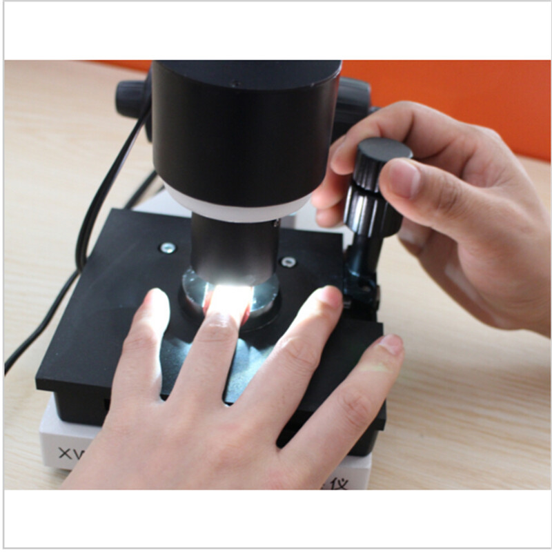
Careful operator evaluation separates true architectural microvascular patterns from localized cuticular microtrauma.
Technical Specifications and System Configurations
Selecting reliable nailfold capillaroscopy equipment requires matching optical amplification, stage stability, and sensor resolution to institutional requirements. Dedicated clinical workstations demand sturdy coaxial focus gears and double-layer compound mechanical stages to prevent drift during high-power observation. Incorporating an achromatic optical train preserves dimensional fidelity across variable field diameters.
A specialized nailfold capillaroscopy microscope designed for professional observation maintains low optical distortion across its full working distance. Below are representative technical specifications for the MK-series microcirculation platforms configured for research laboratories and specialty clinics.
| Parameter | Specification | Operational Detail |
| Brand Name | MAIKONG | Original manufacturing standards |
| Model Series | MK-Series | All factory models prefix with MK- |
| Optical Magnification | 380X 500X 600X | Configurable optical zoom options |
| Input Power | AC 100-240V | Universal 50/60Hz international compatibility |
| DC Output | DC 12V 2A | Stable direct-current operation |
| Objective System | 5X Achromatic | Effective 25mm working distance |
| Stage System | X-Y Double Layer | Smooth coaxial compound mechanical stage |
| Focusing Mount | Coaxial Coarse-Fine | Fine-pitch vertical travel control |
| Illumination | LED Cold Light | Internal projection without tissue heating |
| Safety Type | Type I or B | Standard electrical medical enclosure |
| Camera Sensor | 80000 Pixels CCD | High-sensitivity dynamic imaging sensor |
| Display Panel | 8 10 12 Inch | Dedicated high-contrast color LCD monitors |
| Compliance | CE Approved | European conformity certification standard |
| Chassis Weight | 6KG Net 7KG Gross | Rigid vibration-dampening metal alloy body |
Earlier standard configurations with 240X or 380X magnification and 8-inch or 9-inch integrated color LCD displays remain supported across clinical environments. Facility procurement teams can confirm custom optical configurations and specific hardware attachments directly with the factory before final assembly.
Standard Packaging and Accessory Allocation
Safe transit of delicate optoelectronic equipment requires reinforced shipping packaging. Each complete station includes fitted high-density memory foam enclosures set within a locked aluminum case, which sits inside a multi-wall corrugated master carton. This double-layer protective arrangement prevents misalignment of the optical prism and CCD sensor during air or ground transit.
Alternative hardware designs, such as a compact USB nailfold capillaroscopy microscope or an optical nailfold capillaroscopy magnifier, utilize lightweight cases tailored for mobile screening kits. Standard clinical orders ship complete with all essential operational supplies needed for immediate assembly.
| Spare Parts | Quantity | Component Description |
| GY880 Host | 1 pc | Main optical stage and microscope assembly |
| LCD Monitor | 1 pc | Dedicated high-resolution color display |
| Power Adapter | 1 pc | Regulated 12V direct-current power supply |
| Cedar Oil | 1 pc | Optical immersion fluid for light transmission |
| Cotton Swab | 1 pc | Lint-free applicators for cuticle preparation |
| Chart | 1 pc | Reference morphological classification guide |
Carton dimensions for complete crated units measure 37.5 by 26.5 by 34 centimeters with a total shipping weight of seven kilograms. Procurement teams should verify local electrical plug configurations and monitor screen preferences when submitting equipment requisitions.
Equipment Selection Criteria for Clinical and Research Facilities
Selecting the right nailfold capillaroscopy device requires balancing optical resolving power against practical workflow needs. Academic medical centers often demand high-magnification systems with integrated digital storage and measurement software. In contrast, outpatient clinics often prioritize ergonomic setups with quick-focus stages for rapid routine screening.
Integrating a modern nailfold capillaroscopy imaging system allows clinical departments to archive digital video clips, track longitudinal vascular alterations, and share standardized records across hospital information networks. Storing calibrated image files directly supports multi-center collaborative research and clinical documentation.
Evaluate optical lens glass quality rather than relying solely on digital interpolation. High true optical magnification preserves edge sharpness and fine structural detail that digital zooming cannot reproduce.
Key technical criteria include checking stage travel smoothness, ensuring LED light uniformity without thermal transfer, and verifying driver stability for software integrations. Establishing standardized imaging procedures early protects data integrity across longitudinal clinical trials.
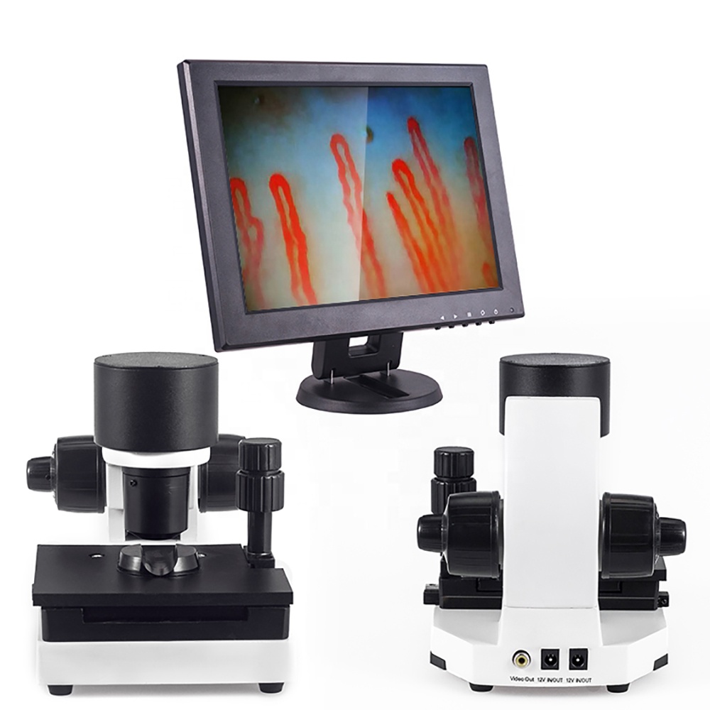
Integrated digital imaging systems support reliable image capture, objective calibration, and longitudinal study storage.
MAIKONG Manufacturing Standards and Supply Chain
We produce the MK-series microcirculation instrumentation at our specialized manufacturing facility in SZ, GD. Leveraging established Chinese high-technology supply chains, our factory incorporates precision optical machining, cleanroom optical assembly, and thorough multi-point optoelectronic quality verification. This manufacturing structure delivers reliable optoelectronic equipment while maintaining cost-effective production economics.
As an established nailfold capillaroscopy manufacturer, MAIKONG provides comprehensive OEM customization, private labeling, and tailored hardware configurations for commercial distributors and research facilities. Our engineering teams support custom casing finishes, software branding, tailored objective selections, and bespoke transport cases.
- Advanced production facilities located within the technology manufacturing corridor of SZ, GD.
- Rigorous optoelectronic alignment testing for every mechanical stage, objective lens, and CCD assembly.
- Flexible OEM production supporting custom monitor integrations, branded housing, and private-label packaging.
- Direct factory logistics coordination supplying medical device distributors and academic institutions globally.
Inquiries regarding international distribution partnerships, commercial agency agreements, and bulk wholesale logistics are coordinated directly through our export services office.
Frequently Asked Questions About Nail Capillary Microscopy
What does nail capillary microscopy show?
The examination visualizes distal periungual capillary loop morphology, capillary density per millimeter, vascular dilation, microhemorrhages, and relative red blood cell column dynamics. It displays the structural architecture of peripheral terminal microvessels directly under optical magnification.
Is nail capillary microscopy an invasive procedure?
No, the procedure is completely non-invasive and painless. It relies on superficial optical light reflection through the stratum corneum without piercing the skin, administering radiation, or extracting tissue samples.
How long does image acquisition take per patient?
Initial vascular loop localization takes only a few seconds per finger once the stage is positioned. A thorough bilateral assessment across all ten digits typically requires fifteen to twenty minutes of examination time.
Why is oil required on the nailfold during examination?
A few drops of cedar oil or paraffin oil match refractive indices between the lens and the uneven keratin surface. This reduces surface light scattering and clarifies subepidermal capillary visualization.
What is the difference between optical magnification and digital magnification?
Optical magnification uses precision glass lenses to enlarge the true optical image without degrading resolution. Digital magnification merely enlarges recorded pixel data electronically, which often introduces blur and pixelation.
Is a portable capillaroscopy device suitable for clinical screening?
A nailfold capillaroscopy portable device offers rapid bed-side and community mobility, making it practical for preliminary visual screenings. Dedicated benchtop systems remain preferable when ultra-stable micrometer tracking and reproducible research metrics are required.
What should a distributor clarify before purchasing capillaroscopy equipment?
Buyers should confirm sensor resolution, true optical objective power, monitor display options, power supply standards, and the current nailfold capillaroscopy equipment price. In addition, clarify packaging cases, warranty terms, and OEM labeling availability with the manufacturer.
Does a capillary microscope diagnose medical conditions by itself?
No, the microscope is strictly an optical observation instrument and does not formulate automated medical diagnoses. Qualified medical professionals must evaluate all observed vascular patterns alongside comprehensive laboratory and clinical findings.
Summary of Microvascular Examination Standards
Standardized nail capillary microscopy offers clear, non-invasive optical insight into terminal human microcirculation for research and clinical assessment. Consistent examination environments, careful optical index matching, and stable stage hardware protect data reproducibility across longitudinal evaluations. Experienced clinical correlation remains essential to place microvascular observations into sound diagnostic context.
Establish a Distribution Partnership or Procure Laboratory Systems
Connect with our technical supply team in SZ, GD to discuss clinical equipment configurations, OEM brand customization, and regional distribution agreements.