
Dilated Nailfold Capillaries in Microvascular Assessment
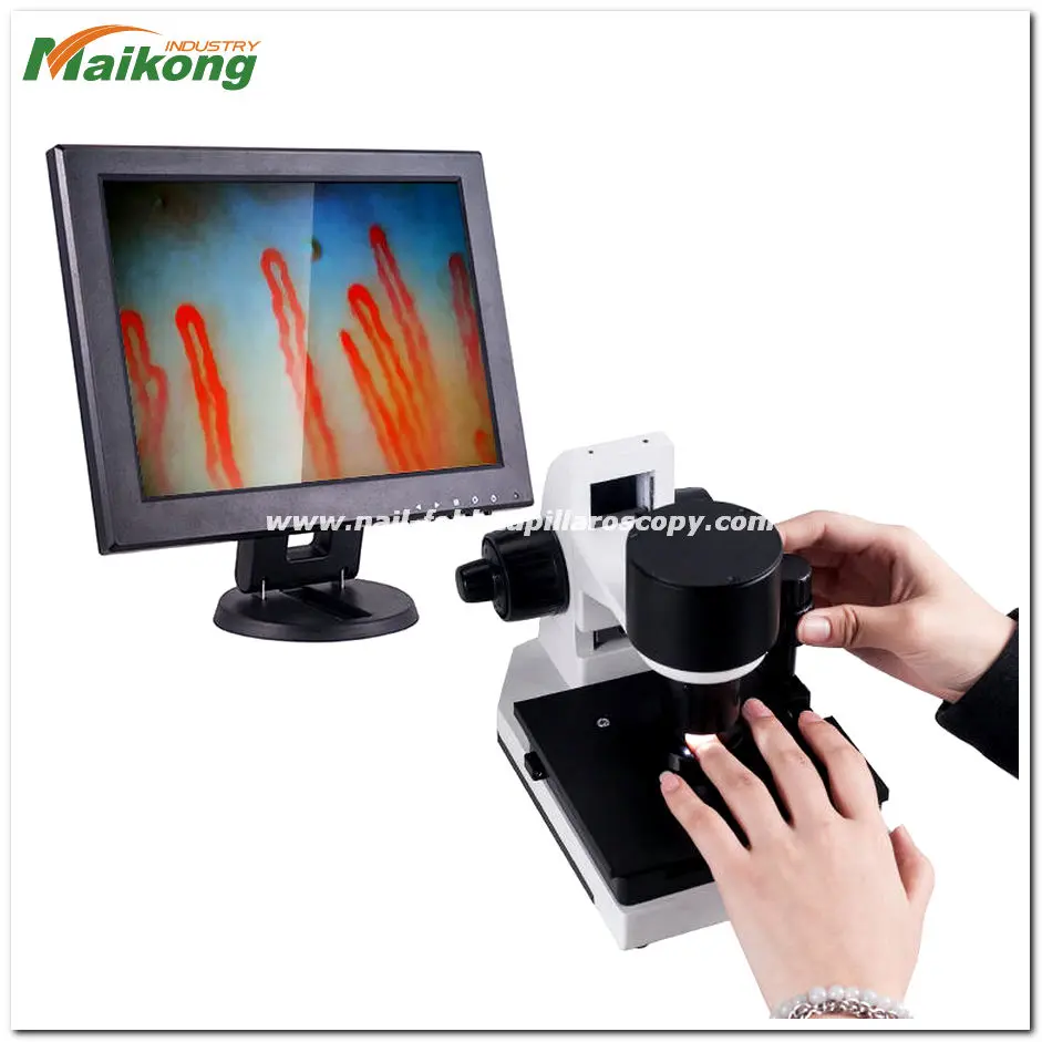
Dilated nailfold capillaries are microvascular loops in the distal periungual margin that exhibit abnormal structural widening compared to healthy hairpin loops. These enlarged vessels reflect microvascular alterations that appear frequently during high-magnification dermatological or rheumatological inspection. In visual microcirculation exams, distinguishing these widening vessels from normal physiological variants helps clinicians determine whether additional diagnostic workups are warranted.
Determining the clinical relevance of enlarged vessels depends on overall capillary architecture, loop distribution across digits, and standardized examination conditions. A solitary wider loop may represent local microtrauma or minor variations in blood flow, while widespread apical widening across multiple fingers often points toward systemic microvascular remodeling.
What Dilated Nailfold Capillaries Look Like Under Magnification
Under optical magnification, healthy nailfold capillaries display a clean, parallel arrangement resembling inverted U-shapes or hairpins. Dilated nailfold capillaries deviate from this uniform layout through noticeable expansion of the afferent arterial limb, efferent venular limb, or apical turning loop. The vessel walls often look prominent, showing darker or more congested columns of moving red blood cells.
Enlargement can occur symmetrically across both limbs or concentrate primarily at the apex of the loop. When assessing these vessels, observers look closely at whether the widening maintains a smooth lumen or presents irregular, sac-like contours along the vascular tree. Irregular contours often signify more extensive vascular remodeling compared to smooth, uniform enlargement.
Apical dilation refers specifically to widening at the crest of the capillary curve, whereas homogeneous dilation involves continuous enlargement from the ascending limb through the apical loop and descending limb.
Observers also assess adjacent tissue features when inspecting dilated nailfold capillary loops during digital examination. Surrounding edema, local extravasation, or microhemorrhages often cluster near widened vessels. Spotting these coexisting visual changes helps characterize the local vascular territory before recording final morphological notes.

Defining Capillary Loop Dimensions and Dilation Criteria
Quantitative measurement establishes clear boundaries between physiological vessel diameters and true capillary enlargement. In typical clinical assessments, clinicians evaluate the apical diameter as well as the caliber of the arterial and venous branches. A normal apical diameter generally remains slender, whereas abnormal dilation produces vessel calibers that distinctly exceed baseline thresholds.
Researchers commonly separate enlarged loops into moderate dilation and extreme ectasia, often described as giant capillaries. While both categories represent widened microvessels, giant loops show marked, bulbous swelling that dwarfs surrounding normal loops. Recognizing these caliber differences allows structured documentation across successive clinical examinations.
| Loop Type | Caliber Profile | Visual Architecture | Clinical Observation Value |
| Normal Loop | Slender caliber | Uniform hairpin | Reflects preserved dermal microvascular integrity |
| Dilated Loop | Widened limb | Smooth expansion | Indicates early or localized microvascular alteration |
| Giant Loop | Bulbous caliber | Marked ectasia | Signals prominent microangiopathy requiring systemic evaluation |
When measuring a nail fold capillary, standardized caliper tools built into digital imaging software provide objective dimensional data. These measurements focus strictly on the distal vascular row nearest the nail plate. Assessing deeper, non-terminal plexus layers can introduce optical confusion because horizontal subpapillary networks naturally carry larger calibers.
Standardizing Examination Conditions for Microvascular Inspection
Environmental factors and patient preparation directly alter peripheral blood vessel tone and visual clarity. Exposure to cold temperatures induces physiological vasoconstriction, which can mask true vessel dilation or give false impressions of reduced flow. Establishing stable testing rooms helps maintain accurate peripheral microcirculation dynamics during visual exams.
Standardized workflows minimize observational variability between different evaluation sessions across clinics and research laboratories. Applying non-irritating immersion media to the periungual cuticles transparentizes the keratinized epidermal layer, eliminating surface glare and diffuse reflection. The procedure should always follow a consistent sequence to maintain repeatable results.
- Keep the examination room at a steady ambient temperature while allowing the subject to acclimatize comfortably prior to imaging.
- Seat the individual with hands supported at approximate heart level to avoid hydrostatic venous distension or ischemia.
- Place two drops of immersion oil, such as cedar oil or pharmaceutical paraffin oil, over the distal nailfold cuticles.
- Position the target digit carefully on the mechanical finger platform beneath the optical objective lens.
- Focus the illumination and optical stage systematically across the distal capillary row from the second to the fifth digit bilaterally.
Systematic workflows reduce external distortion and clarify loop morphology when performing a comprehensive nail fold capillaroscopy assessment. Avoiding physical stress, caffeine consumption, and acute nicotine exposure prior to the session further protects the baseline vascular caliber from temporary external fluctuations.
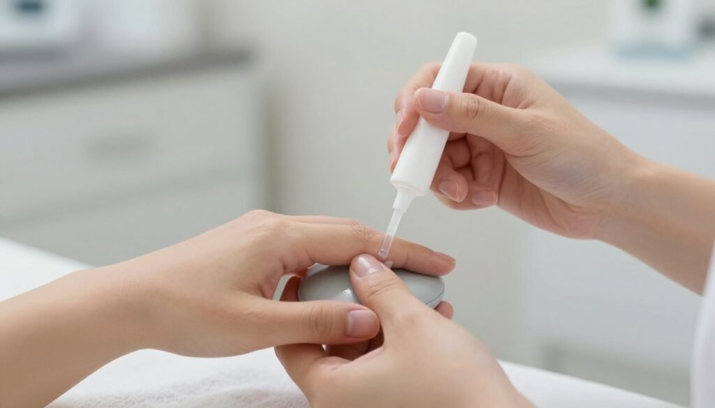
Optical Magnification and Positioning Workflow
Precise visualization of subtle microvascular widening requires stable optical alignment and suitable focal depth. Using dedicated equipment such as a nailfold capillaroscopy microscope allows operators to locate the distal capillary row smoothly. The interaction between mechanical stability, light angle, and optical resolving power dictates image fidelity.
Achieving crisp views of periungual loops involves managing several optical parameters simultaneously during the assessment. Coaxial adjustment controls deliver smooth movement when switching focus between superficial cuticle contours and deeper capillary columns. Stable platform controls keep the target vessel centered without vibration artifacts.
Coaxial Focusing
Smooth coarse and fine adjustment wheels let operators pinpoint the exact capillary depth without sudden optical overshoot.
X-Y Mechanical Stage
Compound coordinate movement shifts the observation platform steadily along the entire periungual margin across each finger.
Cold LED Illumination
Targeted non-thermal lighting penetrates epidermal layers without generating local tissue warming that could alter vessel tone.
Integrating high-definition optical lenses provides clear separation between the red blood cell columns and perivascular spaces. Operators avoid applying direct downward pressure against the finger plate, because compressive force blanches delicate capillaries and distorts baseline loop dimensions.
Differentiating Primary Raynaud Phenomenon from Connective Tissue Diseases
A primary clinical purpose of identifying enlarged periungual loops is distinguishing benign peripheral vasospasm from evolving autoimmune microangiopathy. Primary Raynaud phenomenon typically presents with preserved capillary architecture, uniform loop density, and normal apical diameters. In contrast, secondary Raynaud symptoms often display pronounced microvascular remodeling.
When dilated loops appear alongside extensive capillary dropouts, clinicians recognize a hallmark transition toward an early systemic pattern. Comparing architectural findings across several fingers clarifies whether the observed vasospasm remains functional or stems from structural endotheliopathy. Evaluating these distinctions requires comprehensive visual documentation.
Primary Vasospastic Signs
- Regular, hairpin-shaped loop architecture
- Preserved capillary density without avascular zones
- Absence of giant or irregularly enlarged loops
- Minimal or absent extravasate microhemorrhages
Secondary Microangiopathy Signs
- Irregular or widespread loop enlargement
- Presence of characteristic giant capillaries
- Noticeable capillary loss with emerging avascular spaces
- Recurrent microhemorrhages along the distal row
Observing a distinctive nailfold capillaroscopy scleroderma pattern provides essential descriptive clues that guide further serological testing. In this context, dilated loops often represent the earliest detectable structural sign of systemic microvascular involvement, preceding overt tissue fibrotic changes.

Clinical Conditions Associated with Microvascular Alterations
Capillary dilation rarely occurs in total isolation when caused by underlying systemic rheumatological disorders. Conditions affecting endothelial health, immune tolerance, and connective tissue synthesis frequently manifest visible alterations at the distal nail fold. Recognizing these clinical contexts assists multidisciplinary evaluation teams.
Enlarged capillary structures emerge across several recognized rheumatic and autoimmune disorders during specialized clinical screening. While visual inspection cannot substitute for comprehensive clinical evaluation, it highlights microvascular vulnerability across specific conditions.
- Systemic sclerosis, where progressive endothelial swelling leads to giant loops, widespread capillary dropouts, and architectural disorganization.
- Dermatomyositis, which frequently features enlarged loops alongside branched, bushy arborized capillaries and prominent hemorrhagic spots.
- Mixed connective tissue disease, displaying varying degrees of vascular dilation intertwined with non-specific microvascular tortuosity.
- Systemic lupus erythematosus, occasionally presenting with elongated or dilated loops, tortuous hairpins, and prominent subpapillary plexus visibility.
Performing a structured nailfold capillary test enables practitioners to map these microvascular changes systematically. Documenting the distribution of enlarged loops over time provides objective descriptive data regarding local microcirculation patterns.
Morphological Variations Across the Distal Capillary Row
Interpreting nailfold microcirculation requires focusing specifically on the terminal loops running parallel to the skin surface along the cuticle margin. Deeper horizontal vascular networks lie underneath, but the terminal row provides the clearest visual marker of peripheral perfusion. Pathological dilation predominantly manifests within these distal-most structures.
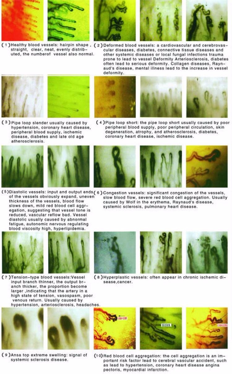
In addition to simple lumen widening, clinicians observe various abnormal shapes that develop during progressive endothelial remodeling. Understanding these morphological variations assists examiners in differentiating non-specific tortuosity from genuine microangiopathy.
Microhemorrhages frequently occur immediately adjacent to significantly enlarged capillary loops, appearing as discrete red-brown hemosiderin deposits that migrate outward with epidermal nail growth.
When capillary damage progresses, the distal row may display irregular clusters of enlarged loops surrounded by empty areas lacking perfusion. Combining morphological observations with specialized nail capillary microscopy techniques helps investigators record loop densities accurately per millimeter of tissue.

Image Acquisition Challenges and Artifact Management
Obtaining reliable, diagnostic-grade recordings of nailfold microvessels requires overcoming common physical and physiological artifacts. Subungual microtrauma, calloused cuticle skin, and accidental hand movement during recording can obscure capillary morphology or simulate vascular pathology. Examiners must systematically differentiate true biological changes from technical artifacts.
Careful optical management and steady positioning support consistent image collection across diverse clinical subjects. Evaluating the advantages and limitations of digital imaging workflows highlights essential best practices for technical operators.
Advantages of Digital Capture
- Facilitates objective measurement of vessel calibers via software reticles
- Enables direct side-by-side comparison across repeated patient visits
- Allows rapid review across full video dynamic capture sequences
Technical Challenges
- Excessive lens pressure causes artificial loop compression and blanching
- Microtrauma from aggressive manicures can mimic microhemorrhages
- Heavy epidermal pigmentation requires precise light angle adjustments
Modern nail videocapillaroscopy configurations overcome several positioning limitations by combining sensitive video sensors with stable mechanical stages. Operators can observe real-time red blood cell transit within enlarged loops while eliminating hand-tremor artifacts.
Critical Limitations of Capillaroscopic Observation
Visual observation of dilated periungual capillaries serves as an observational assessment tool rather than a standalone diagnostic test. An enlarged microvascular loop confirms the presence of morphological alteration, but it cannot independently establish a specific disease diagnosis. Comprehensive diagnosis always requires integrating patient history, physical examination, and serological findings.
Microvascular dimensions also vary naturally between individuals based on occupational hand use, environmental exposure, and constitutional factors. Manual laborers or individuals exposed to repetitive mechanical vibration frequently present isolated enlarged or tortuous capillaries without underlying systemic disease.
Capillaroscopic findings represent descriptive observational data; they do not establish independent clinical diagnoses and must always be evaluated alongside comprehensive laboratory tests and clinical exams.
Examiners utilizing a nailfold videocapillaroscope must document findings using cautious, descriptive terminology. Characterizing vessel width, capillary density, and architectural regularity without jumping to clinical conclusions maintains professional assessment integrity.
Technical Considerations for Clinical and Research Imaging Systems
Selecting appropriate visualization equipment depends on whether the intended workflow prioritizes high-throughput clinical screening, detailed academic microcirculation research, or mobile examinations. Different hardware designs offer distinct balances between portability, magnification power, and software measurement sophistication. Understanding these differences assists clinical directors and procurement managers.
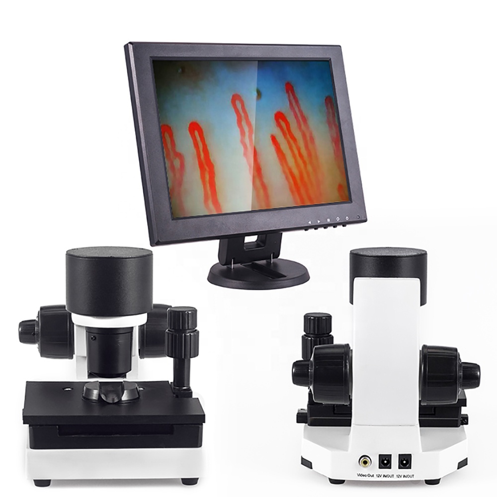
Modern clinical centers utilize varied formats of nailfold capillaroscopy equipment to support specialized vascular evaluations. Exploring primary configuration styles clarifies which setup fits specific facility requirements.
Benchtop Digital Video Workstations
Equipped with dedicated coaxial stands, mechanical finger platforms, and high-definition monitors to deliver stable optical clarity for rheumatology clinics.
Integrated Portable Capillaroscopy Units
Self-contained systems housing integrated displays and protective carrying enclosures, ideal for multi-room outpatient facilities and bedside evaluations.
Key technical criteria when reviewing imaging platforms include cold illumination stability, fine focus precision, and smooth stage positioning. Reliable optoelectronic construction allows researchers to record consistent vessel metrics without mechanical drift during prolonged assessment sessions.
MAIKONG MK-GY880 Capillaroscopy System Architecture
At MAIKONG, based in SZ, GD, China, we manufacture advanced medical optoelectronic instruments developed specifically for non-invasive microcirculation observation. Our MK-GY880 series integrates high-magnification optical pathways, coaxial fine focusing mechanisms, and dedicated cold LED illumination. As a professional nailfold capillaroscopy manufacturer, we support international clinical facilities, university research laboratories, and OEM distributors worldwide.
Our manufacturing protocols utilize precision optical components designed to deliver clear dynamic imaging of periungual capillary architecture. The detailed technical specifications for the MK-GY880 system demonstrate our commitment to reliable microvascular observation hardware.
| Parameter | Specification | Functional Context |
| Brand Name | MAIKONG | Original manufacturer identification |
| Amplification | 380X or 240X | Standard optical observation magnifications |
| Optional Power | 380X/500X/600X | Optional objective lens configurations |
| Electrical Input | AC 100-240V | Global 50/60Hz power grid compatibility |
| Electrical Output | DC 12V 2A | Regulated direct-current system supply |
| Objective Lens | 5X Achromatization | Effective 25mm distortion-free range |
| Mechanical Stage | X-Y Double Layer | Compound mechanical platform movement |
| Focusing Mechanism | Coaxial Adjustment | Combined coarse and micro focus control |
| Inner Dimming | LED Cold Light | Targeted non-thermal illumination |
| Protection Type | Type I or B | Electrical safety isolation grade |
| Built-in Camera | 80000 Pixels | Direct video sensor stream to screen |
| Display Monitor | 8 to 12 Inch | Color LCD screen options available |
| Certification | CE Approved | Verified manufacturing compliance standards |
| Instrument Weight | 6KG Net 7KG Gross | Sturdy desktop operational footprint |
Each MK-GY880 system ships in robust transport packaging engineered to protect optical alignment during international transit. Our standard packaging configurations provide secure storage for clinical deployment and research operations.
| Item | Quantity | Specification Details |
| GY880 Host | 1pc | Central optoelectronic microscope base |
| LCD Monitor | 1pc | Dedicated high-contrast color display unit |
| Power Adapter | 1pc | Standard DC regulated power supply module |
| Cedar Oil | 1pc | Optical immersion contact medium |
| Cotton Swab | 1pc | Application tool for cuticle immersion fluid |
| Vessel Chart | 1pc | Reference morphological comparison chart |
| Inner Case | 1pc | Aluminum lock box with memory foam insert |
| Outer Carton | 1pc | Reinforced corrugated shipping container |
| Case Dimension | 37.5×26.5x34cm | Standard outer shipping package dimensions |
Frequently Asked Questions About Dilated Nailfold Capillaries
What defines dilated nailfold capillaries during an examination?
Dilated nailfold capillaries are microvascular loops in the distal nailfold that show significant widening across the apical curve or limbs compared to standard hairpin vessels. They represent structural microvascular changes that warrant systematic observation and clinical correlation.
Does finding dilated nailfold capillaries confirm an autoimmune disease?
No, finding dilated capillaries does not confirm an autoimmune disease by itself. While widened loops appear frequently in systemic sclerosis and related connective tissue disorders, they represent descriptive visual findings that require correlation with physical symptoms and laboratory tests.
How do examiners distinguish dilated loops from giant capillaries?
Examiners distinguish dilated loops from giant capillaries by measuring apical caliber and assessing overall vessel enlargement. Giant capillaries display extreme, bulbous ectasia several times larger than typical dilated loops, often signaling advanced microvascular remodeling.
Why is immersion oil applied before capillary observation?
Immersion oil is applied to reduce epidermal light scattering and make the keratinized skin layers translucent. This step allows cold light to penetrate cleanly down to the distal capillary row without surface glare.
Can manual labor or cuticle trauma cause capillary dilation?
Yes, repetitive occupational trauma, cosmetic cuticle injury, or local physical impacts can cause localized capillary widening or microhemorrhages. Examiners typically evaluate all eight fingers to determine whether changes are isolated to one trauma site or distributed systemically.
Summary of Microvascular Examination Considerations
Dilated nailfold capillaries provide valuable visual evidence of peripheral microvascular remodeling that assists qualified clinicians in evaluating connective tissue disorders. Accurate assessment relies on standardized examination conditions, objective caliber measurement, and disciplined clinical correlation. Equipping healthcare facilities and research laboratories with stable optical systems supports consistent microvascular observation over time.
Partner with MAIKONG for Capillaroscopy Equipment
Qualified medical equipment distributors, clinical procurement teams, and academic research departments seeking reliable capillaroscopy microscopes can discuss customization, OEM options, and local supply partnerships directly with our manufacturing headquarters.