
Capillaroscopy and Thermography: Principles of Microvascular Assessment
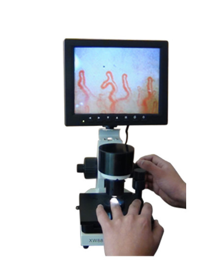
Peripheral microvascular evaluation relies on specialized non-invasive modalities to evaluate capillary architecture and cutaneous perfusion. Combining optical capillaroscopy and thermography allows clinical researchers and vascular specialists to examine physical vessel structures alongside functional surface temperature patterns. While capillaroscopy provides high-magnification visualization of individual capillary loops, thermography maps regional infrared radiation reflecting underlying cutaneous blood flow.
Selecting the appropriate instrumentation depends on whether the clinical objective requires morphological measurement of microvessel dimensions or broader thermal response analysis. Modalities differ in sensor technology, ambient environmental sensitivity, preparation protocols, and analytical parameters. Understanding how both techniques function independently and complementarily supports standardized evaluation across academic, clinical, and laboratory settings.
Optical Morphology vs. Surface Temperature Assessment
Direct capillary imaging and infrared thermal mapping address fundamentally distinct physiological domains. Capillaroscopy magnifies the superficial capillary beds of the skin, capturing direct microscopic images of vessel distribution, loop shapes, and intraluminal erythrocytes. In contrast, infrared thermography records emitted long-wave infrared radiation from the skin surface, converting heat emissions into calibrated temperature distribution maps.
The structural detail provided by optical magnification visualizes microvessel density, apical loop diameter, and abnormal morphological alterations. Thermographic data provide insight into vasomotor tone and autonomic temperature regulation across broader skin territories. When utilized in research or specialized vascular evaluation, these two approaches provide separate structural and functional data points rather than interchangeable findings.
Optical Capillaroscopy Focus
- Direct visualization of individual capillary loops at magnifications up to 380X or 600X.
- Morphological measurement of loop width, apex shape, microhemorrhages, and vessel density.
- Localized field of view restricted to the periungual nailfold or specific mucosal areas.
Infrared Thermography Focus
- Surface measurement of radiant infrared skin emissions without tissue contact.
- Assessment of functional vasomotor regulation, thermal symmetry, and rewarming rates.
- Wide field of view encompassing entire digits, hands, or broad anatomical zones.
Evaluating peripheral vascular conditions through both lenses gives a more comprehensive overview of peripheral status. An anatomical disruption captured under magnification does not necessarily mirror the real-time thermal gradient seen on a whole-hand thermogram. Utilizing capillaroscopy and thermography together establishes a clear boundary between physical vessel integrity and autonomic perfusion responses.
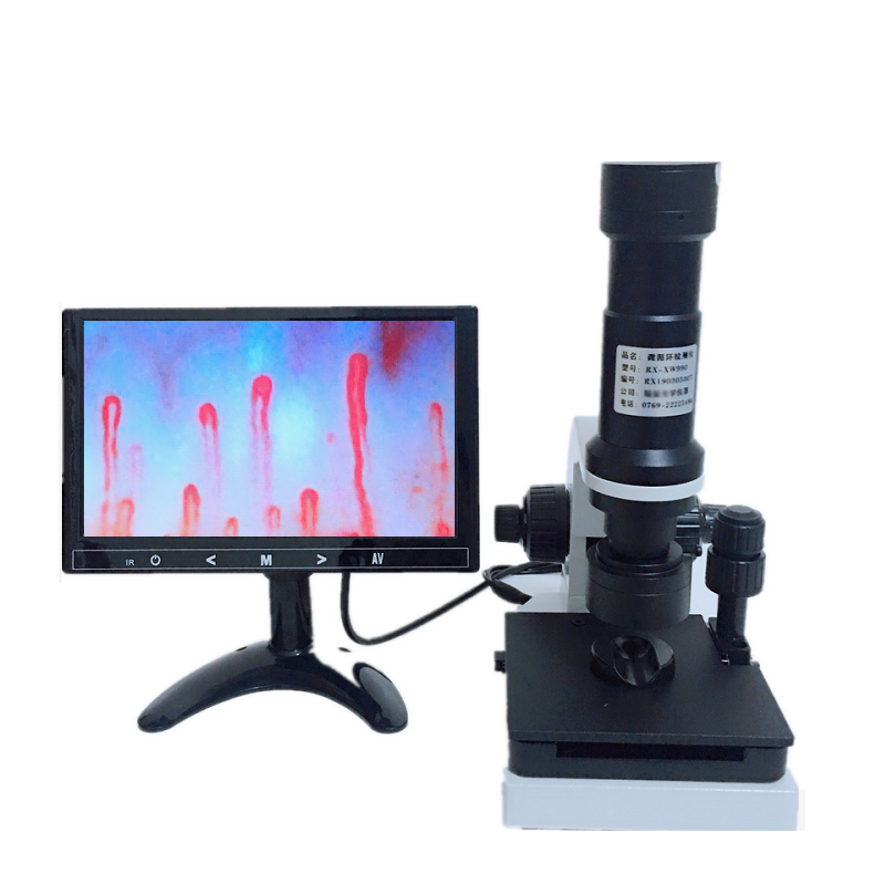
Dedicated microvascular assessment stations allow controlled structural imaging and environmental monitoring.
Principles and Diagnostic Role of Nailfold Capillaroscopy
Nailfold capillaroscopy serves as an established optical technique for assessing the microcirculation in the distal nailbed. Because capillary loops at the periungual border run parallel to the skin surface rather than perpendicular, light microscopy reveals the entire vascular loop from arteriolar limb to venular limb. Clinicians and researchers apply transparent immersion oils to reduce epidermal light reflection, allowing detailed visualization through specialized nailfold capillaroscopy equipment.
Systematic observation focuses on identifying structural hallmarks such as capillary dilation, giant loops, loop architecture derangement, and microvascular hemorrhages. When conducting a structured nailfold capillary test, practitioners assess multiple fingers to construct a representative profile of the patient’s peripheral capillary system. Pathological disruptions of the nail fold capillary network frequently reflect systemic alterations that cannot be detected by macrovascular evaluation methods.
- Capillary Loop Density: Counting active visible capillary loops per linear millimeter along the distal margin.
- Dimension Quantification: Measuring the diameter of the afferent limb, transition zone, and efferent limb.
- Morphological Regularity: Detecting architectural deviations such as tortuosity, ramification, or bush-like arborization.
- Microvascular Extravasation: Identifying pericapillary hemosiderin deposits resulting from microhemorrhages.
- Avascular Fields: Documenting localized zones characterized by the complete loss or absence of capillary loops.
Advanced implementations utilize digital sensors to store still frames and live video feeds for quantitative measurement. High-resolution nail capillary microscopy provides an objective baseline for tracking morphological progression or stability over time. The structural clarity obtained under immersion lighting remains exclusive to optical microscopy and cannot be reproduced by thermal sensors.
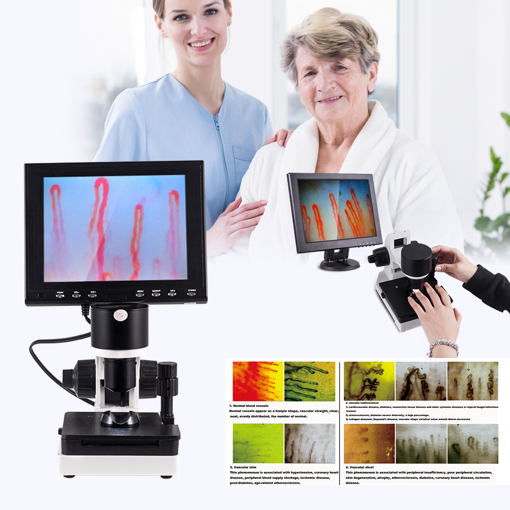
Precise optical positioning over the nailfold border provides direct line of sight to horizontal capillary loops.
Principles and Thermal Imaging Dynamics in Vasomotor Evaluation
Infrared thermography evaluates microvascular integrity through non-contact detection of heat patterns emitted by the skin. Cutaneous temperature is directly mediated by subcutaneous and dermal blood perfusion, which is controlled by autonomic vascular smooth muscle activity. Thermal cameras measure infrared radiation in the long-wave spectrum, rendering digital color-coded thermograms that display precise thermal gradients across distal extremities.
Unlike morphological microscopy, thermographic assessments excel at observing dynamic responses to controlled physiological challenges. Vasomotor provocation protocols, such as standardized cold-immersion or controlled temperature recovery tests, evaluate how rapidly peripheral blood flow rebounds following vasoconstriction. Thermograms record spatial variations across individual fingers, allowing clinicians to observe asymmetric thermal distribution or delayed rewarming intervals across adjacent digits.
Infrared thermography reflects cutaneous perfusion driven by arteriovenous anastomoses and dermal plexuses, capturing physiological vascular reactivity rather than capillary structural damage.
Because thermal cameras detect radiant energy across an entire hand simultaneously, they provide broad contextual perspective during systemic examinations. Thermal deficits often correspond to localized vasoconstriction, microvascular shunting, or regional autonomic asymmetry. However, thermographic output remains an indirect proxy for perfusion, necessitating careful interpretation alongside environmental controls and anatomical assessment.
Functional vascular evaluation protocols demand standardized spatial setups and calibrated digital display monitors.
Technical and Functional Comparison of Both Modalities
Choosing between or combining optical capillaroscopy and thermal imaging requires a thorough evaluation of technical attributes. While digital microscopes demand strict optical coupling and fine focus adjustment, thermographic sensors require optical calibration and controlled ambient reflection. The operational parameters of each method define their analytical boundaries in research laboratories and outpatient clinics.
Digital capillaroscopy using a dedicated nailfold videocapillaroscope delivers spatial resolution down to micrometers, isolating single microvessels. Thermographic imaging provides spatial resolution at the millimeter scale while delivering thermal sensitivity capable of resolving fractions of a degree Celsius. The following matrix illustrates the foundational operational distinctions between these instrumentation types.
| Feature | Capillaroscopy | Thermography | Analytical Significance |
| Primary Target | Capillary loops | Skin temperature | Structural vessel architecture versus functional thermal distribution |
| Contact Mode | Optical immersion | Non-contact | Microscopy requires skin oiling while infrared capture occurs at a distance |
| Spatial Scale | Micrometer level | Millimeter level | Microscopic single loops compared to whole-digit thermal gradients |
| Field of View | Narrow periungual | Broad extremity | Focused single nailfold margin versus multi-digit thermal symmetry |
| Dynamic Range | Structural shapes | Thermal changes | Direct morphology versus time-resolved rewarming kinetics |
| Key Variable | Loop dimensions | Thermal gradients | Morphological metrics versus regional temperature variations |
Neither instrument replaces the physiological data provided by the other. While a digital microscope reveals avascular patches or capillary microhemorrhages, it does not record thermal symmetry across all digits. Conversely, thermal cameras detect cold digital patterns but cannot determine whether the low temperature stems from structural vessel loss or reversible vasospasm.
Multimodal Microvascular Assessment in Clinical Research
Integrating optical capillaroscopy with thermal imaging creates a robust multimodal testing protocol for vascular medicine and rheumatological research. Connective tissue evaluations frequently benefit from distinguishing primary vasospastic events from organic microangiopathies. In conditions characterized by secondary Raynaud phenomenon, identifying morphological changes in capillary loops helps differentiate structural damage from functional dysregulation.
In observational studies of systemic autoimmune diseases, such as nailfold capillaroscopy scleroderma patterns, optical imaging identifies early, active, or late microvascular architecture damage. Meanwhile, infrared thermography tracks whether digital surface temperatures rewarm appropriately following cold provocation. Evaluating both data sets helps researchers understand whether impaired thermoregulation correlates directly with capillary density loss.
Multimodal research protocols combine structural microvessel scoring via optical microscopy with objective rewarming curves from thermal imaging to assess vascular status comprehensively.
Standardized workflows often perform infrared thermography first to prevent the skin-to-probe contact and immersion oils used during microscopy from altering cutaneous temperature readings. Once thermal baselines and provocation tests conclude, practitioners clean and prepare digits for detailed nail videocapillaroscopy. This orderly sequence preserves data integrity for both analytical modalities.

Sequential examination setups allow clinicians to capture thermal data before applying immersion media for microscopy.
Controlled Examination Conditions and Operational Factors
Both optical capillaroscopy and infrared thermal mapping are sensitive to external environmental factors and subject physiology. Precise assessment demands rigorous control of the examination room to avoid ambient artifacts that distort vascular tone or image resolution. Variations in ambient room climate, air drafts, and subject activity can significantly alter microcirculatory blood flow.
For consistent capillaroscopy results, operating environments should maintain a steady ambient temperature of 22 to 24 degrees Celsius and relative humidity near 70 percent. The subject should remain seated with hands positioned at heart level to normalize hydrostatic venous pressure. Stabilizing patients for 15 to 30 minutes in the examination room prior to measurement reduces physiological fluctuations caused by external transit.
- Activity Restrictions: Subjects should avoid strenuous physical exercise for one hour prior to undergoing evaluation.
- Vasomotor Influences: Operators must document recent tobacco exposure, hand washing, caffeine, or food intake prior to testing.
- Medication Auditing: Clinicians should record concurrent vasoactive pharmacotherapy without instructing patients to alter prescribed regimens without supervision.
- Immersion Medium: Applying two to three drops of cedar oil or paraffin oil to the nailfold minimizes epidermal light refraction and scattering.
- Chronobiological Timing: Conducting follow-up evaluations at the same time of day minimizes the impact of diurnal vasomotor cycles.
Adhering to strict stabilization protocols allows laboratories to collect reproducible vascular data. Because cutaneous capillary perfusion responds rapidly to emotional, thermal, and mechanical stressors, standardizing pre-test procedures remains essential. Operator consistency during finger positioning and focus adjustment further ensures dependable image acquisition.
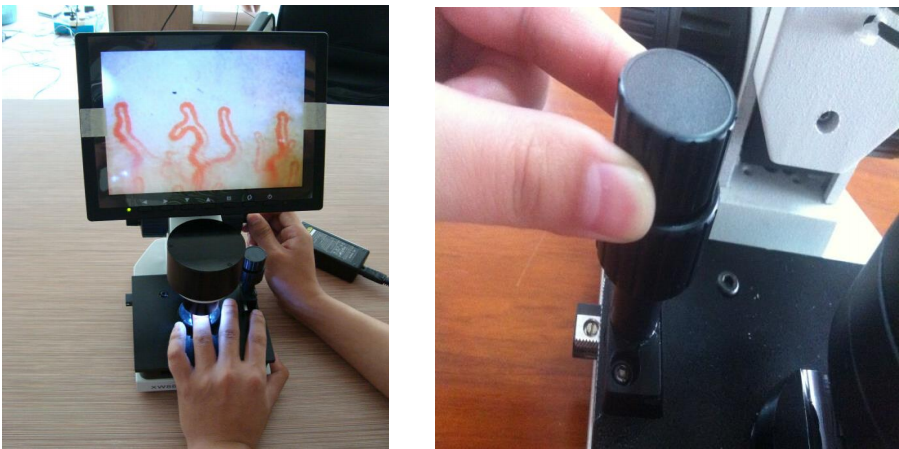


Maintaining constant room temperature and humidity supports reliable peripheral microvascular data collection.
MAIKONG Capillaroscopy System Technical Specifications
MAIKONG designs and manufactures dedicated optoelectronic microcirculation observation platforms engineered for clinical, academic, and research applications. As an established international supplier based in SZ, GD, MAIKONG integrates optical magnification, steady LED cold illumination, and digital video processing into robust diagnostic hardware. The MK-GY880 platform delivers crisp visualization of cutaneous capillary networks without thermal tissue disturbance.
The system utilizes an achromatized 5X objective combined with specialized internal optics to achieve standard optical magnifications of 240X or 380X, with optional configurations extending to 500X and 600X. High-stability mechanical stages with coaxial coarse and fine focus adjustments allow smooth navigation along the periungual margin. These technical parameters reflect standard manufacturing specifications for the MK-GY880 series.
| Parameter | Specification | Technical Details |
| Brand Name | MAIKONG | Manufactured in SZ, GD for global distribution |
| Amplification | 380X or 240X | Optional configurations support 500X or 600X |
| Input Voltage | AC 100-240V | Universal power switching 50/60Hz DC input |
| Output Rating | DC 12V, 2A | Dedicated low-noise direct current output |
| Objective Lens | 5X Achromatization | Effective working distance of 25mm |
| Stage Mechanism | Compound X-Y | Double-layer compound mechanical movement stage |
| Focus System | Coaxial Coarse/Fine | Dual handwheel fine focus mechanism |
| Illumination | LED Cold Light | Internal dimming prevents thermal tissue distortion |
| System Type | Type I or B | Standardized medical electrical equipment classification |
| Chassis Power | 12V, 1A | Low power consumption DC operation |
| Built-in Camera | 80,000 pixels | Integrated optical sensor with distortion-free feed |
| Display Screen | Color LCD Monitor | Available in 8-inch, 9-inch, 10-inch, or 12-inch options |
| Certification | CE Approved | Complies with applicable international safety standards |
| Chassis Weight | 6 kg Net | Gross packed shipping weight of 7 kg |
| Dimensions | 37.5 x 26.5 x 34 cm | Compact benchtop packaging dimensions |
Each MAIKONG instrument is engineered for operational longevity in high-volume testing facilities. The combination of precision mechanical gearing and specialized cold lighting provides stable capillary imaging across long observation sessions. Organizations can review options for a digital nailfold capillaroscopy microscope or explore a usb nailfold capillaroscopy microscope configuration based on facility space and IT infrastructure.
Setup Procedures and System Accessories
Deploying the MAIKONG microcirculation microscope involves straightforward hardware integration and consistent procedural preparation. Every complete MK-GY880 system ships in protective transport cases designed to prevent shock during transit. The inclusion of standardized accessories ensures that clinics and laboratories can initiate observational workflows immediately upon delivery.
The standard MK-GY880 delivery package contains dedicated optical and mechanical components tailored for delicate periungual examinations. The included aluminum password box features custom-cut memory foam that shields sensitive lenses and mechanical gears from environmental hazards. The standard packaging inventory includes the following elements.
| Item | Quantity | Description |
| MK-GY880 Host | 1 pc | Main optoelectronic chassis with mechanical X-Y stage |
| LCD Monitor | 1 pc | Integrated color screen display unit |
| Power Adapter | 1 pc | Universal AC/DC power supply unit |
| Cedar Oil | 1 pc | Optical immersion fluid for skin transparency |
| Cotton Swabs | 1 pc | Applicators for clean, precise oil placement |
| Reference Chart | 1 pc | Capillary morphology visual interpretation guide |
| Inner Case | 1 pc | Aluminum password box fitted with memory foam |
| Outer Box | 1 pc | Heavy-duty corrugated protective export carton |
To operate the instrument, place the LCD monitor securely onto the chassis mount and attach the power and video cables to the base connectors. Power on the display, apply two to three drops of immersion oil to the ring finger nailfold, and place the finger comfortably onto the finger rest. Operators use the coarse handwheel to locate the focal plane approximately two to three centimeters from the lens, then engage the fine handwheel to sharpen vessel resolution. Adjusting the double-layer X-Y stage laterally positions the distal capillary line directly across the screen center.
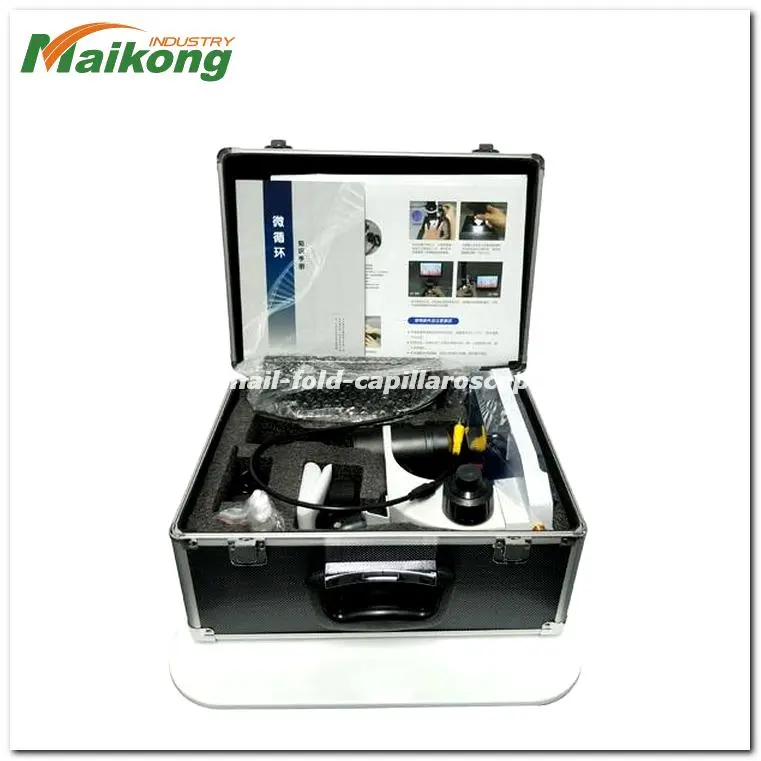
Custom aluminum password flight cases protect precision optical components during storage and transport.
B2B Procurement and Integration Considerations for North American Facilities
Procuring microvascular visualization equipment requires North American medical centers, universities, and diagnostic distributors to evaluate technical and logistical criteria. Buyers must balance optical clarity, illumination stability, software connectivity, and long-term mechanical reliability against institutional budget constraints. Sourcing directly from an established manufacturing partner simplifies supply chain coordination and equipment customization.
When selecting between stationary and mobile solutions, facilities choose between a benchtop professional nailfold capillaroscope, an integrated nailfold capillaroscopy video system, or a versatile nailfold capillaroscopy portable device. While full-size consoles provide broad mechanical stages, portable units offer flexibility for bedside rounds or multi-facility research programs. Evaluating trade-offs across instrument configurations supports informed procurement decisions.
Consolidated Benchtop Systems
- Heavy mechanical chassis provides outstanding optical stability during high-magnification focus.
- Integrated wide color LCD monitors allow direct group observation without external computer workstations.
- Smooth coaxial X-Y movement stages permit precise linear tracking across the entire nailfold perimeter.
Compact Portable Devices
- Smaller footprint reduces mechanical mass, requiring steady placement to prevent fine focus drift.
- Handheld or USB-driven configurations depend on external display screens or laptop operating systems.
- Ultra-compact stages offer narrower mechanical adjustment ranges compared to full benchtop platforms.
Commercial distributors and institutional purchasing directors should also evaluate supplier capabilities for OEM branding, custom housing modifications, and private-label programs. Reviewing international safety certifications, packaging integrity, and spare parts availability helps ensure uninterrupted clinical service. Facilities can consult transparent resources on nailfold capillaroscopy equipment price models to optimize departmental equipment acquisition.
Frequently Asked Questions
What is capillaroscopy?
Capillaroscopy is a non-invasive optical magnification technique used to observe the microcirculation of the skin, most commonly at the distal nailfold border. It allows direct, real-time visualization of individual capillary loops, vessel density, and morphological structural changes under immersion illumination.
What is thermography?
Thermography is an imaging modality that detects long-wave infrared radiation emitted by the skin surface. It converts radiant thermal emissions into calibrated digital temperature maps, reflecting regional cutaneous blood flow and autonomic vasomotor function.
How do capillaroscopy and thermography differ?
Capillaroscopy provides direct microscopic optical visualization of physical vessel architecture at micrometer resolution. In contrast, thermography measures surface heat distribution across broader anatomical areas without physical skin contact.
What does each method actually display or measure?
Capillaroscopy displays the morphological shape, diameter, and layout of superficial capillary loops and microhemorrhages. Thermography measures radiant skin temperature and physiological thermal recovery patterns following vasomotor challenges.
What can influence the reliability of each method?
Environmental room temperature, ambient air currents, patient anxiety, physical activity, smoking, and recent hand washing can alter vascular perfusion. Careful climate control, patient resting periods, and proper immersion oil application support reproducible results.
Can they be used together?
Yes, researchers and clinicians frequently use both methods complementarily to evaluate structural microvascular anatomy alongside dynamic functional perfusion. Thermography is typically performed first to avoid altering baseline skin temperatures with immersion oils or mechanical contact.
Which method is more appropriate for nailfold microvascular observation?
Capillaroscopy is the preferred tool for nailfold microvascular observation because it provides the optical magnification required to resolve microscopic capillary loops. Thermal cameras cannot resolve individual capillary vessels due to their wider spatial scale.
What should clinics, laboratories, and distributors consider when selecting equipment?
Purchasing teams should examine optical magnification, illumination type, camera resolution, mechanical stage stability, and display integration. Logistical considerations include international certifications, protective packaging, warranty support, and OEM customization options.
What information should a buyer request from a supplier?
Buyers should request detailed optical specifications, CE certificates, packing dimensions, lead times, minimum order quantities, and available customization options. Inquiring about spare components, power supply compatibility, and freight terms supports smooth international procurement.
What questions remain outside the scope of either method?
Neither capillaroscopy nor thermography alone can diagnose systemic pathology, confirm specific autoimmune disorders, or replace comprehensive clinical and laboratory evaluations. Both tools serve as non-invasive observational instruments that must be interpreted alongside wider diagnostic findings.
Summary of Microvascular Evaluation Modalities
Understanding peripheral microcirculation requires distinguishing structural vascular integrity from dynamic autonomic vasomotor responses. Utilizing capillaroscopy alongside infrared thermography provides clinical teams with a balanced view of microscopic vessel morphology and regional thermal regulation. Integrating dependable optical instrumentation supports reproducible observation, advancing microvascular research and clinical assessment protocols.
Equip Your Facility with Professional Capillaroscopy Systems
MAIKONG supplies reliable, CE-approved nailfold capillaroscopy systems, OEM manufacturing solutions, and wholesale distribution support for healthcare facilities and commercial partners across North America.