
Nailfold Capillaroscopy Camera for Professional Capillary Imaging
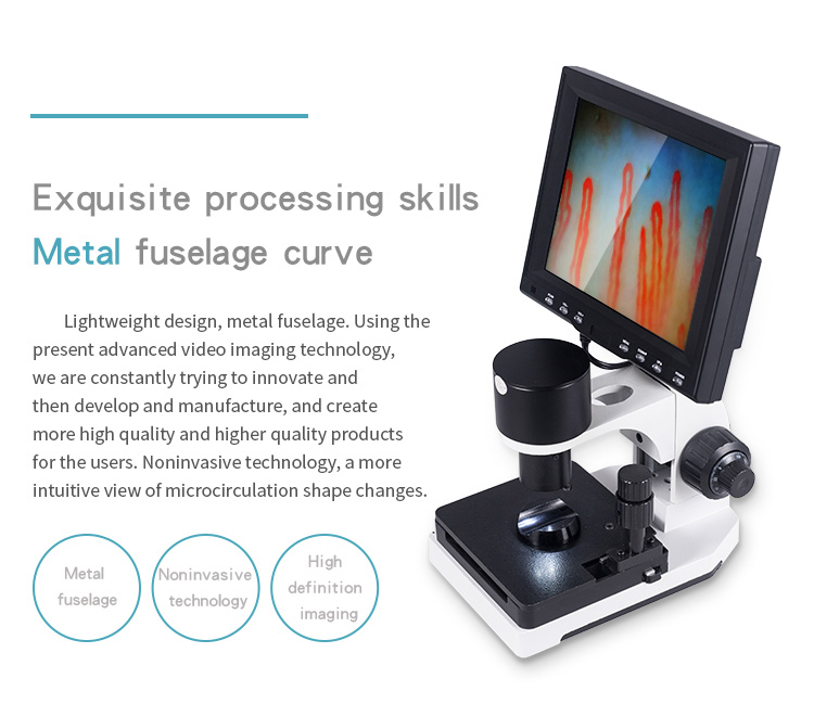
A nailfold capillaroscopy camera provides non-invasive optical magnification and live video streaming to examine microvascular capillary beds located along the human nail fold. The system pairs optical lenses with dedicated sensors to display erythrocyte flow and structural morphology on connected monitors without tissue disruption. Healthcare facilities, microvascular laboratories, and academic departments utilize these optoelectronic instruments to evaluate vascular patterns under stable illumination.
Selecting an appropriate instrument depends on optical magnification ranges, stage stability, sensor output format, and integration with examination workflows. MAIKONG manufactures specialized systems in GD, SZ, delivering dependable hardware architectures designed for clinical observation, academic microcirculation studies, and commercial distribution channels.
MAIKONG Camera-Based Capillary Imaging System
Our integrated imaging instruments bring together precision optical lenses, integrated illumination, and real-time electronic sensors into a unified inspection workstation. The MK series combines high-magnification achromatic optics with a coaxial mechanical stage, allowing examiners to position fingers quickly and isolate capillary rows on screen.
The system provides real-time dynamic video feed to an integrated color monitor, bypassing complex computer installations when direct viewing is preferred. Facilities seeking a reliable capillaroscopy camera benefit from our rugged mechanical framework, which maintains focal position during active examinations.
Every MK unit incorporates cold-light LED illumination, a dual-axis mechanical stage, and balanced coaxial focus wheels to maintain image clarity during clinical microvascular observation.
How a Nailfold Capillaroscopy Camera Works
Operating a nail fold capillaroscopy camera involves coordinated optical alignment, surface light transmission, and electronic signal processing. The instrument visualizes the distal nail bed by directing targeted light rays across the epidermal boundary layer.
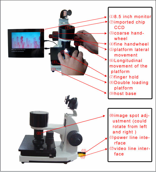
Examiners apply a contact medium, such as cedar oil or paraffin oil, directly onto the cuticle boundary to reduce surface glare and skin scattering. Once positioned in the mechanical finger rest, the user adjusts coarse and fine control wheels to bring nail fold capillary loops into sharp focus.
- Applying optical immersion oil minimizes ambient epidermal reflection and enhances light transmission through the cuticle layer.
- Securing the finger on the mechanical rest stabilizes the tissue against unintentional hand movement during magnification.
- Adjusting coaxial coarse and fine handwheels brings the microvascular bed into crisp focus within seconds.
- Traversing the X-Y mechanical stage repositions the field of view smoothly across neighboring capillary loops.
Camera, Sensor, Magnification, and Display Workflow
The electronic imaging chain converts optical light gathered by the achromatized objective into a live video stream. Light travels through the optical core directly onto an internal CCD imaging sensor, which processes analog or digital video signals without observable lag.
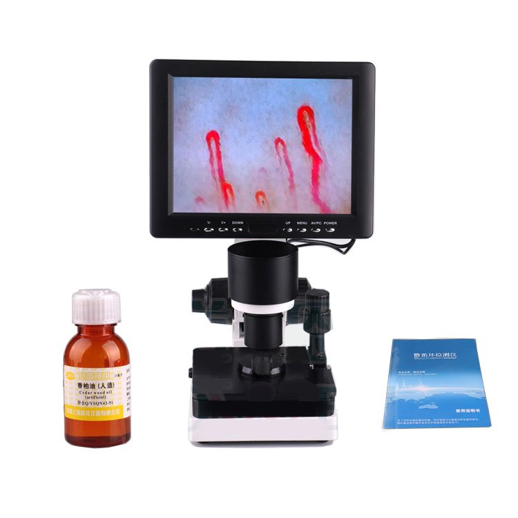
The live video signal routes directly to the color LCD display, allowing examiners to assess capillary architecture, blood column velocity, and vascular dimensions in real time. Systems such as the nailfold videocapillaroscope provide smooth frame rates that help practitioners observe microvascular motion without image jitter.
Dynamic video feeds allow immediate observation of microcirculatory behavior, while dedicated display adjustments for contrast and brightness adapt to varying ambient light levels in examination rooms.
Standard configurations offer 240X or 380X optical amplification, with specialized options extending to 500X or 600X depending on research requirements. This magnification range permits clear visualization of individual apical loops, capillary plexuses, and microvascular flow dynamics across diverse skin pigmentation profiles.
Key Features for Clinics, Laboratories, and Distributors
Our manufacturing protocols emphasize structural stability, ergonomic handling, and optical consistency across repeated daily examinations. Each capillary microscope camera integrates heavy-duty components designed for demanding hospital and private clinic schedules.
Mechanical precision remains essential when operating at magnifications above 300X, where minute vibrations disrupt microvascular tracking. Our dual-layer compound stage offers micrometric lateral and longitudinal positioning to track microvessels smoothly across the distal row.
Coaxial Focus Mechanism
Integrated coarse and fine focusing knobs allow rapid initial target finding followed by delicate focal depth refinement.
Cold LED Illumination
Built-in cold-light projection provides uniform illumination across the target zone without generating thermal discomfort for patients.
Stable Stage Movement
The compound X-Y stage provides controlled transverse travel, keeping observation fields centered during extended examination sessions.
Facilities employing our nailfold capillaroscopy imaging system appreciate the low-maintenance architecture, which avoids fragile third-party adapters and delicate external lens mounts.
Technical Specifications
Standard production models provide a balanced feature set for routine microvascular observation and research tasks. Sourcing managers can evaluate standard parameters alongside optional modifications to configure systems for regional clinical protocols.
| Parameter | Specification | Notes |
| Brand Name | MAIKONG | Shenzhen production base |
| Amplification | 380X or 240X | Standard factory configuration |
| Optional Zoom | 380X/500X/600X | Selectable upon equipment order |
| Input Power | AC 100-240V | Universal 50/60Hz input range |
| Output Power | DC 12V 2A | Regulated adapter output |
| Objective | 5X Achromatized | Effective range of 25mm |
| Mechanical Stage | X-Y Double Layer | Compound travel platform |
| Focusing | Coaxial Control | Coarse and fine dual adjustment |
| Light Source | LED Cold Light | Internal projection illumination |
| Equipment Type | Type I or B | Electrical isolation build |
| Host Power | 12V 1A DC | Low voltage internal operation |
| Sensor Spec | 80 000 Pixels | Built-in CCD video sensor |
| Standard LCD | 8 or 9 Inch | Color display monitor included |
| Optional LCD | 10 or 12 Inch | Expanded viewing area choice |
| Certification | CE Approved | Standard international compliance |
| Net Weight | 6 KG | Benchtop stability weight |
| Gross Weight | 7 KG | Packaged with security case |
| Dimensions | 37.5*26.5*34cm | Outer packaging measurement |
Reviewing detailed parameters helps technical teams integrate the nailfold capillaroscopy equipment into existing laboratory stations. Final configurations, display sizes, and electrical plug standards should be confirmed prior to shipment.
Included Accessories and Packaging
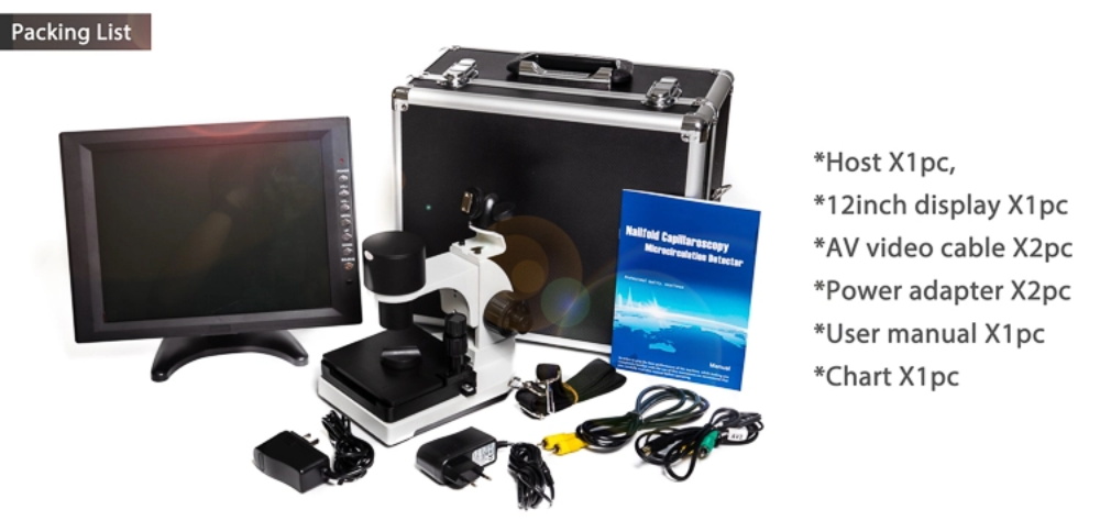
Each instrument ships with essential operating components to allow immediate benchtop setup upon delivery. Packed within durable transport materials, the hardware withstands long-distance ocean and air freight without alignment loss.
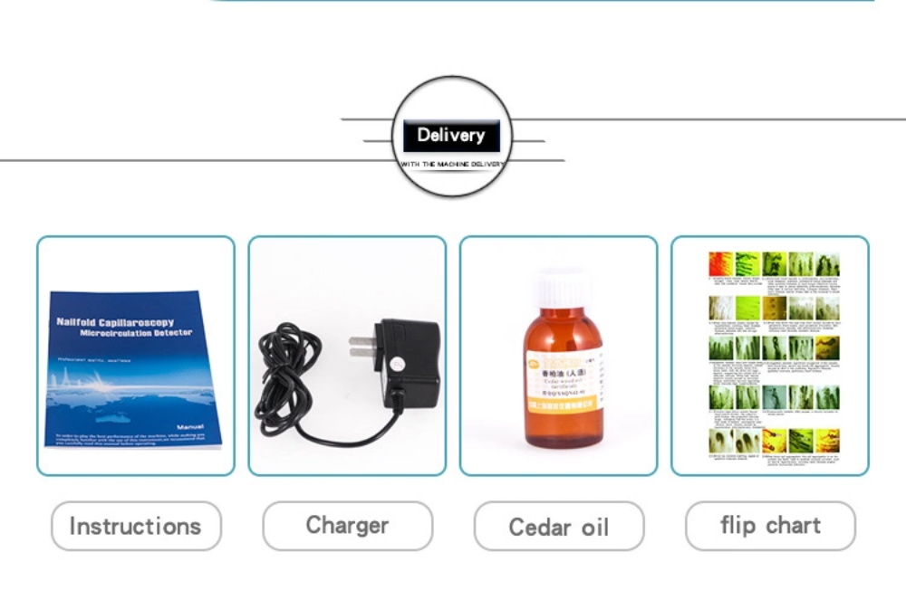
The standard parts package contains primary connection cables, illumination sources, optical fluids, and maintenance items. Sourcing departments can confirm specific accessory counts during the Order finalization process.
| Spare Parts | Quantity | Function |
| GY880 Host | 1 pc | Main optoelectronic instrument body |
| LCD Monitor | 1 pc | Color display for real-time feed |
| Power Adapter | 1 pc | Universal voltage power supply |
| Cedar Oil | 1 pc | Optical transmission contact fluid |
| Cotton Swab | 1 pc | Fluid application accessory |
| Chart | 1 pc | Capillary reference documentation |
We enclose the system within a high-density aluminum security box fitted with shock-absorbing memory foam inserts. An outer multi-wall corrugated box protects the case against transit impacts during international logistics handling.
Compatibility and Purchasing Checklist
Equipment buyers must evaluate multiple functional and infrastructural criteria before procuring microcirculation observation instruments. Identifying whether a department needs an autonomous benchtop setup or a digital networked architecture clarifies configuration needs.
- Display architecture: Decide between an all-in-one integrated color LCD or routing video signals to external monitors via a nailfold capillaroscopy video system.
- Optical amplification levels: Confirm whether routine 240X or 380X lenses satisfy viewing needs, or if advanced 500X/600X optical modules are necessary.
- Electrical infrastructure: Verify local hospital AC line voltage and cord plug configurations for US or international medical power outlets.
- Portability requirements: Determine whether the instrument will remain stationary on laboratory benches or require mobility in an aluminum transit container.
- Private label branding: Establish whether housings, start screens, and manuals require custom OEM distributor graphics.
- Certification documentation: Request manufacturer compliance records, test summaries, and quality assurance declarations.
Capillary Observation Settings and Buyer Profiles
Camera-assisted microvascular observation instruments serve specialized environments spanning hospital departments, medical schools, and commercial distribution channels. Research and clinical teams use the technology to observe microvascular beds non-invasively.
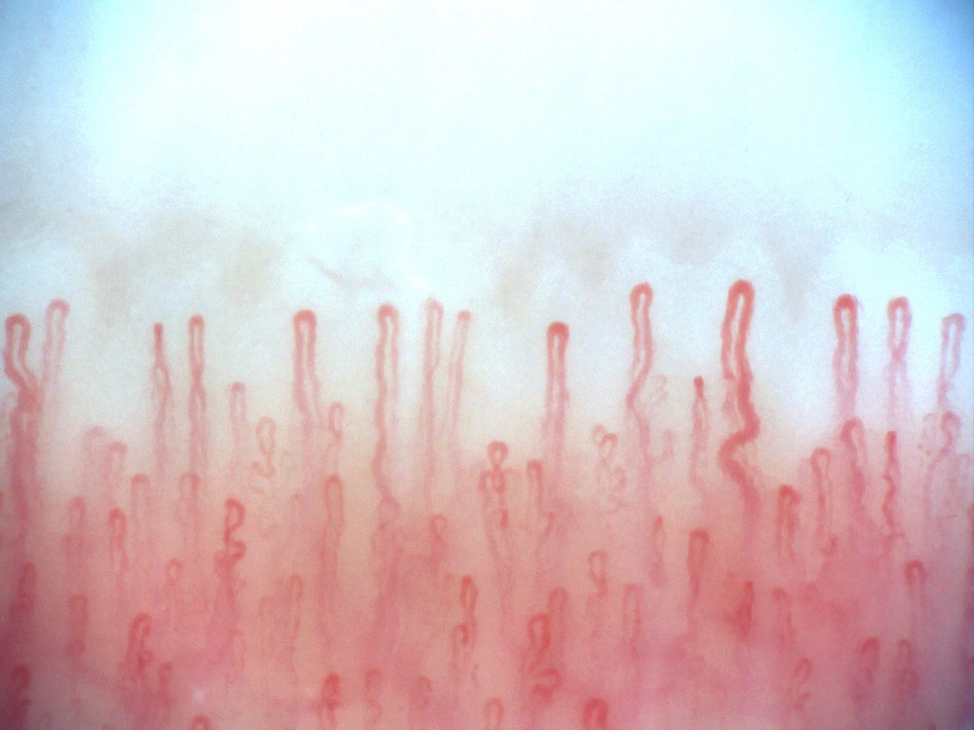
Departments specializing in microcirculation studies use our systems for detailed structural assessments. The hardware supports non-invasive investigations without causing tissue trauma or subject discomfort during extended viewing sessions.
Clinical Observation Units
Rheumatology practices, dermatology clinics, and vascular services utilize camera systems to evaluate morphological capillary shifts during patient assessments.
Academic and Research Labs
Physiology departments and university laboratories deploy instruments to explore blood flow characteristics and vascular geometry in microvascular studies.
Practitioners conducting systematic nail capillary microscopy gain stable imaging across varying examination conditions, maintaining dependable visual records over time.
MAIKONG Manufacturing and OEM Support
MAIKONG operates advanced optoelectronic manufacturing facilities based in Shenzhen, GD, China. We supply international wholesalers, private-label brand owners, and healthcare distributors with cost-effective, high-grade imaging instruments.
Our engineering team supports customized hardware designs, modified magnification barrels, custom-molded housings, and tailored software branding for regional distributors. Learn more about our manufacturing capabilities through our About us page or explore our production lines at our Factory portal.
Custom Hardware & Housing
We manufacture custom exterior chassis, customized monitor bezels, and specialized carrying cases tailored to partner branding specifications.
Scalable Wholesale Supply
Our integrated supply chain provides competitive factory pricing, strict quality control procedures, and predictable batch delivery schedules.
Frequently Asked Questions
What is a nailfold capillaroscopy camera?
A nailfold capillaroscopy camera is an optoelectronic device that magnifies and transmits live video images of microvascular capillaries around the human nail cuticle.
How does the camera display the image?
The device routes optical signals from the objective lens through an internal CCD sensor directly onto an attached color LCD monitor in real time.
What magnification options are available?
Standard factory models supply 240X or 380X magnification, while custom configurations can support 380X, 500X, or 600X upon order specification.
Does the system include a monitor?
Standard packages include an 8-inch or 9-inch color LCD display, with optional 10-inch or 12-inch screens available depending on user preference.
What accessories are included?
Each unit ships with the main host, LCD monitor, universal power adapter, cedar immersion oil, application swabs, reference chart, and an aluminum storage case.
Can MAIKONG customize the camera or packaging?
We provide full OEM and private-label customization, including custom branding, packaging cases, shell colors, and regional electrical adapters.
Is the system suitable for distributors and wholesale buyers?
MAIKONG works directly with medical equipment wholesalers and distributors, providing tiered volume pricing, technical support, and reliable export fulfillment.
What specifications should I request before ordering?
Buyers should confirm required magnification optics, display screen dimensions, local electrical plug configurations, and custom labeling requirements before shipment.
Can the camera be used for research and teaching?
Academic laboratories, physiology programs, and medical schools regularly employ the camera system to demonstrate microvascular blood flow and vessel architecture.
Request Camera Specifications or a Quote
MAIKONG supplies high-performance microcirculation imaging instruments directly from our manufacturing base to clinical practices, research institutions, and medical distributors across the United States. Contact our export sales engineering department to discuss technical specifications, explore OEM options, or request an official product quotation.
Connect with MAIKONG Technical Sales
Speak directly with our team in GD, SZ to discuss configuration options, bulk distributor terms, or sample orders.