
Nail Fold Definition: Anatomy, Meaning, and Clinical Observation

The nail fold definition refers to the specialized border of soft tissue that surrounds, frames, and supports the margins of the human nail plate. This structural envelope consists of the proximal fold at the base of the nail and the two lateral folds running along its sides, forming an anatomical barrier against mechanical strain and biological contaminants. In clinical assessment and microvascular research, this cutaneous boundary provides a non-invasive viewing window for examining superficial capillary networks.
Understanding this anatomical zone clarifies how the nail matrix produces the hard plate and how surrounding dermal structures protect the digital terminal. The structural orientation of the skin tissue dictates both regional resilience and optical clarity under non-invasive magnification.
Nail Fold Definition: What Is the Nail Fold?
In standard medical terminology, the nail fold definition describes the fold of cutaneous tissue that tucks around the margins of the hard nail plate to anchor and protect the growing keratin structure. Clinicians and anatomists divide this structure into two primary regional sections: the proximal nail fold, located at the base of the digit where the nail originates, and the pair of lateral nail folds, situated along the left and right borders of the nail plate. When addressing what is the nail fold, medical textbooks emphasize its double-layered cutaneous architecture, wherein a dorsal surface transitions over a rounded crest into a ventral surface that rests directly above the nail matrix and newly formed nail plate.
It is clinically essential to differentiate the nail fold from adjacent components of the digital perionychium. The nail plate is the hard, visible keratin shield, whereas the nail fold is living cutaneous skin containing dermis, epidermis, and dermal microvessels. The nail bed sits beneath the nail plate as a vascularized platform, while the matrix rests deeper under the proximal fold to drive cellular division. The eponychium represents the localized keratinized rim of the ventral proximal fold, extending the protective seal known as the cuticle across the emerging plate surface.
| Structure | Tissue Type | Position | Anatomical Description |
| Proximal Fold | Cutaneous fold | Base margin | Covers the embryonic matrix and supplies the protective cuticle seal. |
| Lateral Fold | Cutaneous fold | Lateral margins | Guides straight nail plate elongation and defends lateral sulci against debris. |
| Nail Plate | Hard keratin | Dorsal platform | Rigid exterior protective plate resting on the vascular nail bed. |
| Nail Bed | Vascular tissue | Subungual zone | Epithelial foundation beneath the plate that does not produce hard keratin. |
| Nail Matrix | Germinative cells | Proximal base | Active cellular zone responsible for synthesizing the hard keratin plate. |
| Cuticle | Stratum corneum | Plate junction | Keratinized extension sealing the space between proximal skin and plate. |
Within the proximal fold, terminal vascular loops known as nail fold capillary vessels run parallel to the skin surface. This horizontal arrangement allows direct microscopic observation of blood flow, in contrast to other skin zones where capillaries project perpendicularly toward the surface. Maintaining an intact fold architecture shields these fragile loops from physical micro-trauma.
Nail Fold Meaning in Plain English
In non-technical language, the nail fold meaning simply refers to the rim or ridge of skin that wraps around the bottom and sides of your fingernail or toenail. People often confuse this living rim of skin with the cuticle, but the fold is the actual fleshy fold of the finger, whereas the cuticle is the thin, translucent dead skin shed from its edge. Understanding this distinction clarifies why trimming the living skin can cause redness, swelling, or entry of environmental irritants.
When searching for the practical nail fold meaning in english, think of the finger as an envelope where the nail plate slips into an intentional pocket. The pocket lips are the nail folds, holding the plate snugly in place while allowing it to glide forward during normal growth. Without these skin borders, the hard plate would lack lateral guidance, leading to improper growth trajectories or structural vulnerability.
Everyday Perspective
- Described as the soft skin border surrounding the nail edges.
- Often confused with the dead keratin of the cuticle.
- Acts as a physical cushion against bumps and friction.
- Readily noticed when dry, irritated, or torn by hangnails.
Anatomical Perspective
- Defined as the folded double-layer perionychial cutaneous tissue.
- Distinguished precisely from the eponychium and nail matrix.
- Contains horizontally aligned terminal dermal capillary loops.
- Serves as an essential biological barrier against microbial invasion.
By contrasting lay descriptions with formal anatomical terms, individuals can better communicate changes in skin texture, irritation, or swelling to healthcare providers. Recognizing the living nature of this fold supports gentler grooming routines and avoids aggressive manicuring practices.
Where Is the Nail Fold Located?
The nail fold is situated at the terminal dorsal aspect of each finger and toe, tracing three continuous margins around the hardened nail plate. The formal nail folds definition encompasses these distinct borders as a unified anatomical frame. The distal edge of the nail, known as the free edge, remains unattached to any fold, permitting daily mechanical functionality and normal trimming.

To pinpoint the exact boundaries on any human digit, trace the anatomical landmarks that enclose the nail plate:
- Proximal Margin: Sits across the base of the nail, directly ahead of the distal interphalangeal joint, sheltering the hidden matrix beneath its ventral shelf.
- Lateral Sulci: Runs down both the lateral and medial flanks of the digit, forming the paronychial gutters that border the plate edges.
- Terminal Transition: Connects smoothly with the general digital skin at its external boundary while folding inwardly to interface with subungual structures.
- Free Margin Separation: Ends where the nail plate detaches from the underlying hyponychium at the fingertip, leaving the distal edge open.
This layout creates a protected sulcus or groove where the plate slides forward over time. The structural integrity of these boundaries protects the deeper germinative tissue from foreign matter and topical moisture buildup.
What Does the Nail Fold Do?
The primary function of the nail fold is to create an impermeable physical and biological shield that protects the sterile nail matrix and vascular nail bed. Because the nail plate consists of non-living keratin, the junction where it emerges from the living digital dermis presents a vulnerable entry point for bacteria, fungi, and environmental chemicals. Through the continuous production of the eponychium and cuticle, the proximal nail fold seals this aperture, lowering the risk of paronychia and chronic localized inflammation.
In addition to shielding the nail apparatus, the lateral folds serve as structural tracks that steer the nail plate along an even, forward trajectory. As matrix cells divide and push forward, the lateral boundaries prevent uneven lateral spreading and maintain a uniform arch across the dorsal plane of the digit. When lateral folds suffer lacerations or mechanical distortion, subsequent nail growth can become irregular, split, or prone to ingrown penetration.
The nail fold contains a high concentration of sensory nerve endings and microvascular loops that assist in digital tactile perception and thermal adaptation. Its unique horizontal capillary structure makes it an optimal site for clinical observation via nail capillary microscopy and diagnostic assessment using nail fold capillaroscopy.
Healthy nail folds show smooth, pliable skin without cracking, redness, or induration. Maintaining gentle hand hygiene, avoiding harsh chemical exposure, and refraining from aggressive cuticle removal help preserve the defensive integrity of these tissue folds.
Nail Fold and Nailfold: Are They the Same Term?
The terms “nail fold” and “nailfold” refer to the exact same anatomical structure, representing a minor difference between spaced English grammar and compound scientific terminology. In general anatomy textbooks and standard dictionaries, the spaced form “nail fold” is standard when describing regional cutaneous folds. Conversely, within microcirculation research, clinical rheumatology, and optoelectronic imaging, the closed compound “nailfold” is universally preferred in compound nouns.
When researchers discuss nailfold meaning in peer-reviewed journals, they almost exclusively employ the single-word compound in technical phrases. Instrumentation designed to record microvascular patterns is termed a nailfold videocapillaroscope, reflecting international standardization across professional societies. Despite the typographical difference, neither spelling denotes a different biological entity, layer, or clinical focus.
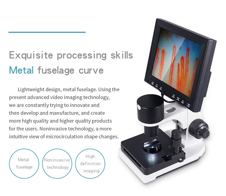
The spaced form “nail fold” is most common in patient-facing education and dermatological anatomy, whereas “nailfold” dominates biomedical engineering, microvascular research literature, and clinical capillaroscopy terminology.
Both orthographic forms remain fully acceptable in modern medical communication. Writers and clinicians choose the variation that matches their publication style or equipment categorization standards.
Why the Nail Fold Matters for Visual Observation
The nail fold possesses a unique biological feature that distinguishes it from almost all other skin areas on the human body: its terminal capillaries lie parallel to the epidermal surface rather than perpendicular to it. In the skin of the torso, arms, or legs, capillaries rise vertically in dermal papillae, presenting only pinpoint cross-sections under magnification. At the proximal nail fold, the thin, translucent stratum corneum permits direct visual assessment of entire microvascular loops, from the afferent arterial limb through the apical loop to the efferent venous limb.
This optical transparency makes the region indispensable across multiple medical and academic disciplines. In dermatology, educators inspect the fold to observe localized blood supply and inflammatory patterns. In rheumatology and microcirculation research, investigators utilize specialized optical systems to document vascular morphology, loop density, and dynamic flow velocity. High-magnification systems enable non-invasive assessment without tissue excision or complex radiological procedures.
Dermatology
Assesses localized periungual inflammation, structural tissue integrity, and microvascular changes associated with regional skin conditions.
Microcirculation Research
Studies systemic capillary blood flow, loop density, red blood cell velocity, and physiological responses to thermal changes.
Academic Education
Provides medical students and physiology trainees with live demonstrations of human terminal hemodynamics in real time.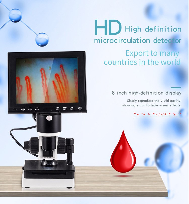
While visual magnification reveals microvascular patterns, optical visualization alone does not confirm or exclude systemic medical conditions. Any observed irregularities, such as dilated loops, microhemorrhages, or capillary dropouts, require thorough diagnostic evaluation by qualified healthcare professionals who integrate medical history, laboratory tests, and complete clinical presentations.
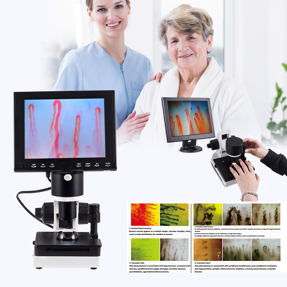
Observational accuracy relies on dependable optoelectronic hardware. MAIKONG is a GD, SZ-based manufacturer and supplier serving professional, research, educational, distributor, OEM, and private-label buyers of nailfold observation and capillaroscopy equipment. Facilities seeking standardized documentation tools integrate dedicated nailfold capillaroscopy equipment into their clinical workflows to capture reproducible capillary images.
Partnering with a specialized nailfold capillaroscopy manufacturer helps research laboratories and diagnostic clinics maintain high optical resolution and stable mechanical platforms. Direct factory communication facilitates custom OEM configurations and responsive technical support for global distributors.
Frequently Asked Questions About Nail Fold Definition
What is the nail fold?
The nail fold is the continuous ridge of skin that wraps around the base and lateral margins of the nail plate. It forms an invaginated envelope that anchors the plate and defends the underlying nail matrix from physical stress and environmental microbes.
What is the difference between the nail fold and nail bed?
The nail fold is the soft skin surrounding the external edges of the nail plate, while the nail bed is the vascular tissue beneath the hard plate. The fold acts as a protective boundary frame, whereas the bed serves as the adhering foundation on which the plate rests.
Is “nailfold” one word?
Both “nail fold” and “nailfold” are linguistically correct and refer to the exact same anatomical zone. Scientific literature, biomedical engineering manuals, and capillaroscopy terminology frequently combine them into a single compound word.
What does nail fold mean in English?
In standard English, the term defines the protective cutaneous fold bordering the human fingernail or toenail. It identifies the biological pocket from which the visible nail plate emerges.
What does nail fold meaning in Tamil refer to?
When searching for nail fold meaning in tamil, readers seek the regional terminology for the skin margin around the nail, known contextually as “நக மடிப்பு” (Naga Madippu). It identifies the identical protective epidermal boundary discussed in Western anatomical science.
Why is the nail fold relevant to nailfold microscopy?
The proximal nail fold houses capillary loops that run parallel to the skin surface beneath a thin, transparent epidermal layer. This horizontal alignment allows non-invasive visual observation of microvascular structure and flow without surgical procedures.
Summary of Nail Fold Anatomy and Observation Systems
The nail fold serves as a vital anatomical envelope that supports the nail plate, protects germinative matrix tissue, and houses superficial microvascular networks. Recognizing its distinct proximal and lateral boundaries clarifies both everyday digital hygiene and specialized clinical examination procedures. Maintaining healthy periungual skin safeguards underlying structures and preserves optimal optical clarity for research observation.
Equip Your Clinic or Laboratory for Optical Observation
Connect with our technical support team to discuss equipment configurations, distribution opportunities, or customized OEM systems for nailfold imaging.