
Capillaroscopy Use in Clinical Observation and Microvascular Research
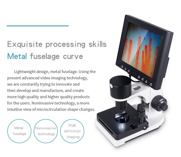
Professional capillaroscopy use centers on the direct, non-invasive optical examination of the microcirculation, most frequently at the periungual nailfold. Clinicians and microvascular investigators employ this imaging modality to evaluate capillary loop morphology, capillary density, and microvascular blood flow velocity. The technique reveals structural alterations such as enlarged loops, microhemorrhages, and architectural disorganization without breaking the skin barrier.
Selecting the appropriate observation protocol depends on clinical requirements, departmental imaging workflows, and patient presentation. Standardized examinations demand controlled environmental temperatures, stabilized patient positioning, and optimized optical magnification to produce reliable diagnostic documentation.
Defining Capillaroscopy and Capillary Microscopy
Capillaroscopy is an optical diagnostic technique designed to inspect terminal blood vessels in living tissue. The distal nailfold offers a unique anatomic window because the terminal capillary loops lie parallel to the surface of the skin rather than perpendicular. This orientation permits direct horizontal visualization of the arterial limb, hairpin apical turn, and venous limb under magnification.
The practice relies on specialized illumination, high-resolution optics, and an immersion medium to bypass epidermal scattering. Light reflects off erythrocytes passing through microvessels, creating high-contrast dynamic imagery of red cell columns. When integrated with digital sensors, modern systems convert these optical views into real-time digital feeds for systematic analysis and electronic archiving.
Terminal capillary beds in the nailfold run parallel to the epidermal surface, allowing complete horizontal visualization of loop architecture under optical magnification.
Different optical devices support this assessment, ranging from simple stereomicroscopes and dermatos установлено units to specialized video capillaroscopes. High-end systems utilize coaxial focus controls, dedicated finger brackets, and stabilized mechanical stages to eliminate hand tremors during prolonged microvascular observation.
Direct magnification reveals the parallel architecture of periungual capillary loops in healthy tissue.
Observational Roles in Rheumatology and Connective Tissue Disorders
In clinical rheumatology, capillary morphology provides critical visual evidence when differentiating primary vascular spasms from systemic microvascular damage. Clinicians assess whether patients presenting with cold sensitivity exhibit normal capillary architecture or structural anomalies linked to autoimmune processes. Observing the microvascular bed helps characterize changes without invasive biopsy procedures.
Microvascular alterations often precede gross tissue changes in conditions such as nailfold capillaroscopy scleroderma evaluations. Practitioners monitor specific signs, including giant capillary loops, avascular areas, ramified or bushy capillaries, and hemosiderin deposits resulting from microhemorrhages. These visual patterns support medical specialists in categorizing microvascular involvement into early, active, or late structural stages.
Observational Advantages
- Permits non-invasive, repeatable evaluation of distal microvessels.
- Visualizes early architectural changes before severe tissue damage occurs.
- Provides measurable visual records for longitudinal patient tracking.
Methodological Boundaries
- Requires contextual correlation with serology and clinical history.
- Visual findings alone do not establish a standalone diagnosis.
- Subject to local trauma artifacts and anatomical variations.
Rheumatology clinics also evaluate secondary microvascular manifestations in systemic lupus erythematosus, dermatomyositis, mixed connective tissue disease, and rheumatoid arthritis. Capillaroscopy serves as an observational adjunct alongside physical examinations, immunological panels, and nailfold capillaroscopy assessments.
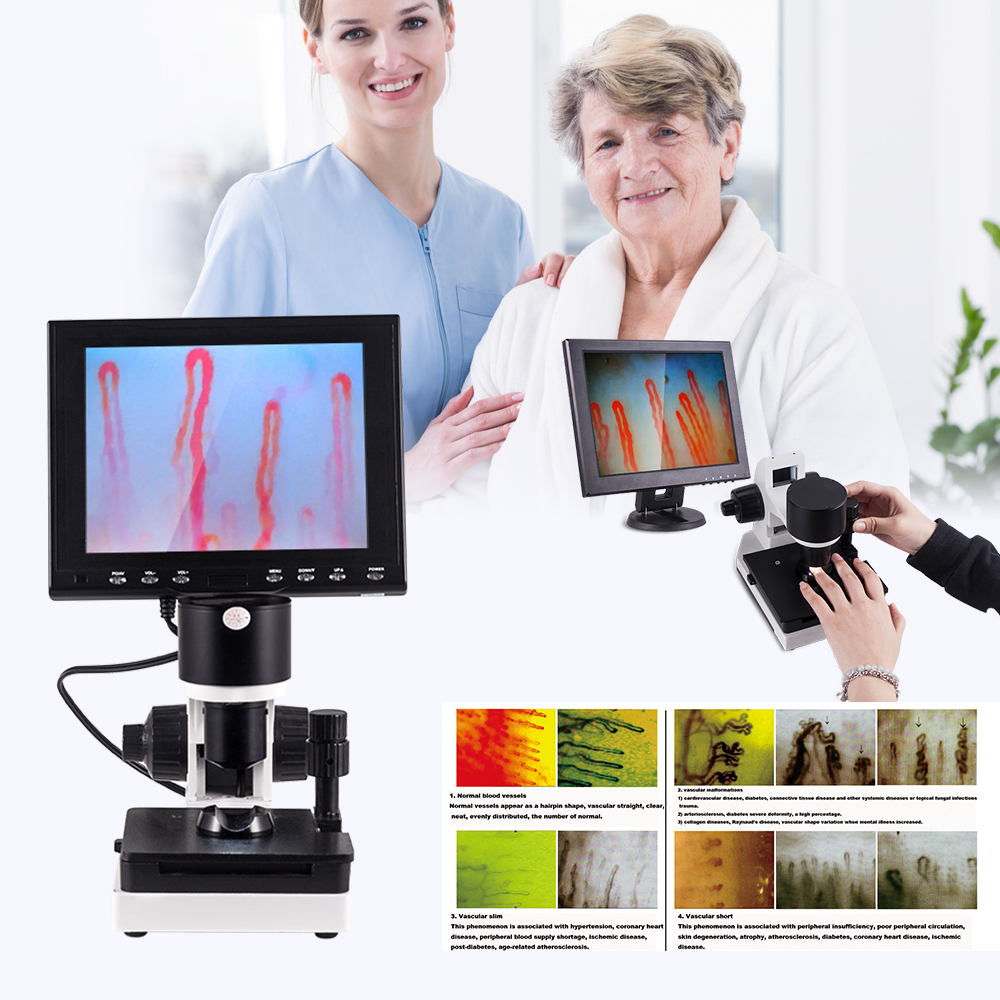
Digital optical systems allow rheumatologists to evaluate microvascular architecture during routine clinical evaluations.
Applications in Dermatology and Peripheral Vascular Assessment
Dermatologists and vascular medicine specialists apply capillaroscopy to evaluate microvascular involvement across cutaneous disorders and peripheral circulation anomalies. The technique allows inspection of superficial blood vessels in psoriasis, localized scleroderma, purpura, and chronic non-healing ulcers. Structural microvessel alterations provide insight into local inflammatory processes and dermal tissue perfusion.
In peripheral angiology, examining the nail fold capillary network supports the investigation of acrocyanosis, chilblains, vibration-induced white finger, and peripheral occlusive arterial syndromes. Practitioners record morphological changes, perivascular edema, and sluggish capillary flow states to document vascular responses over time. The non-invasive nature of the test makes it suitable for frequent monitoring without patient discomfort.
Cutaneous capillaroscopy provides non-invasive insight into local perfusion states, inflammatory microvessel proliferation, and microvascular changes accompanying chronic wounds.
Vascular assessments also extend to assessing the microvascular impact of cosmetic and dermatological procedures, such as laser therapy, skin resurfacing, or localized tissue treatments. Comparing pre-procedure and post-procedure microvascular density assists clinicians in tracking dermal tissue recovery and inflammatory responses.

Angiologists utilize high-magnification optical sensors to assess peripheral perfusion patterns and local microvessel responses.
Microcirculation Research and Experimental Laboratory Applications
Biomedical researchers apply specialized capillary microscopy systems to explore human microvascular physiology, endothelial function, and cellular transit dynamics. In academic and pharmacological laboratories, investigators measure red blood cell velocity, intercapillary distances, and dynamic loop recruitment during thermal or physical provocation. These quantifiable metrics expand basic physiological knowledge regarding peripheral vascular regulation.
Experimental studies also explore practical videocapillaroscopy uses to evaluate functional microcirculation across atypical anatomical sites. When appropriate optical adapters and stabilization fixtures are used, researchers can observe superficial microvessels in the lip mucosa, tongue borders, distal auricles, or dermal regions surrounding sweat glands and hair follicles. These non-standard sites support specialized investigation into localized vasomotor reactivity.
- Quantification of dynamic capillary red cell velocity under controlled physiological conditions.
- Measurement of functional capillary density during systemic or localized pharmacological challenges.
- Investigation of cutaneous microvascular responses to far-infrared or specialized material applications.
- Assessment of endothelial reactivity and shear-stress adaptations in athletic or occupational groups.
Laboratory investigations must operate under rigid experimental controls to avoid environmental or mechanical interference. Standardized baseline stabilization periods and non-invasive immobilization platforms help ensure that measured shifts reflect genuine physiological responses rather than observational artifacts.

Laboratory workstations equipped with high-resolution video sensors support quantifiable microvascular physiological research.
Academic Instruction and Clinical Training Programs
Capillaroscopy represents an invaluable educational platform within medical schools, residency programs, and microvascular training institutions. Demonstrating live capillary circulation provides students and trainees with immediate, tangible examples of peripheral vascular physiology. Instructors utilize the technique to teach microvascular anatomy, erythrocyte hemodynamics, and the clinical implications of capillary damage.
Training curricula incorporate nail capillary microscopy to teach diagnostic standardization, image acquisition workflows, and artifact differentiation. Students learn to distinguish between physiological variants, local mechanical trauma, and clinically significant microvascular lesions under direct faculty supervision. Recorded video files serve as standardized educational libraries for longitudinal competency testing.
Anatomical Instruction
Provides live visualization of arterial, apical, and venous capillary segments for physiology courses.
Pattern Recognition
Trains postgraduate fellows to classify architectural loop disorganization and microhemorrhages systematically.
Academic centers routinely establish structured reference libraries using stored videocapillaroscopy sequences. These standardized materials ensure uniform educational outcomes and help trainees build reliable observational skills across varied microvascular pathologies.

High-definition optical monitors allow academic instructors to guide trainees through capillary morphology and hemodynamic patterns.
Standard Examination Protocols and Environmental Requirements
Executing an accurate microvascular assessment requires disciplined adherence to standardized examination protocols. Environmental stability is paramount because superficial microvessels respond rapidly to ambient temperature shifts and emotional stressors. Practitioners maintain examination spaces at approximately 22 to 24 degrees Celsius with a relative humidity near 70 percent to prevent reactive vasospasm.
Patient preparation directly influences observational reliability. Clinicians instruct individuals to avoid strenuous physical exertion, smoking, caffeine, and vasoactive substances prior to examination. Implementing a structured resting period before optical acquisition minimizes systemic hemodynamic fluctuations.
- Maintain examination room temperature at 22–24°C and relative humidity around 70 percent.
- Ensure the patient rests seated quietly for 15 to 30 minutes before initiating the optical scan.
- Position the patient comfortably with hands resting flat at approximately heart level to normalize hydrostatic pressures.
- Conduct routine comparative examinations at consistent times of day to account for diurnal vascular variation.
A standardized nailfold capillary test examines all ten digits, excluding thumbs if thumb nailfolds present thickened cuticles or frequent occupational trauma. Systematically evaluating the fourth and fifth fingers often yields optimal optical clarity due to minimal mechanical wear on the periungual skin.
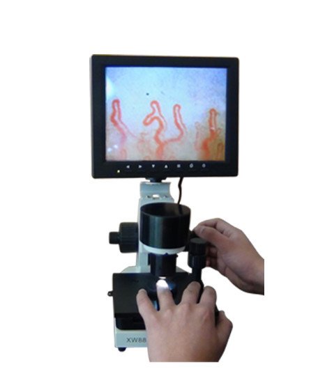
Maintaining patient hands at heart level on a stable stage prevents hydrostatic pressure artifacts during examination.
Immersion Media and Optical Alignment Techniques
Direct optical visualization of periungual capillaries requires managing epidermal light refraction and scattering. Dry stratum corneum scatters incoming illumination, creating significant glare and obscuring underlying blood vessels. Applying an optical immersion medium bridges the refractive index difference between the microscope lens and the skin surface, rendering the keratin layer translucent.
Clinicians typically apply two or three drops of cedar oil or medical-grade paraffin oil to the cuticle border of the target digit. Baby oil or cosmetic-grade mineral oils serve as acceptable substitutes when specialized immersion oils are unavailable. The fluid must form an unbroken optical bridge between the objective lens and the nailfold without creating trapped air bubbles.
Inspect the oiled nailfold carefully to confirm the absence of air bubbles, which distort light projection and create false optical artifacts on digital screens.
Once the immersion fluid is positioned, the operator lowers the optical assembly using the coaxial coarse handwheel to locate the initial focal plane. Fine focus handwheels allow precise adjustments to bring the terminal capillary loops into crisp view. Positioning a specialized nailfold capillaroscopy magnifier or calibrated objective approximately 2 to 3 centimeters from the skin surface generally yields the initial transparent field.

Applying optical immersion oil eliminates epidermal scattering to reveal underlying microvascular beds.
Instrumentation Configurations and Digital Video Systems
Optical hardware used for microcirculation imaging varies from portable handheld tools to integrated high-magnification videocapillaroscopy consoles. Mobile screening environments often utilize compact USB digital scopes or portable devices that connect directly to tablets or laptops. These lightweight options provide rapid mobility for bedside rounds or multi-site clinical assessments.
Specialized diagnostic clinics and research laboratories rely on stabilized mechanical consoles featuring dual-layer X-Y compound stages and coaxial focusing assemblies. Consoles featuring dedicated Sony CCD optical sensors deliver distortion-free dynamic streaming directly to integrated high-definition monitors. Investing in a stable nailfold videocapillaroscope ensures steady, jitter-free observation even at magnifications of 240X, 380X, or higher.
Integrated Consoles
Equipped with heavy structural bases, coaxial focus handwheels, integrated LCD screens, and mechanical finger rests to prevent motion blur during high-magnification sweeps.
Handheld Digital Scopes
Lightweight optical units offering dynamic handling, rapid setup times, and USB data transfer, suitable for bedside assessments and mobile screening workflows.
Modern video systems incorporate integrated cold LED light sources positioned around the objective perimeter. Cold light prevents tissue heating, which could otherwise provoke thermal vasodilation and distort basal vascular dimensions during detailed evaluation.

Dedicated video consoles integrate coaxial mechanical controls with specialized cold LED illumination for stable microvascular imaging.
Technical Specifications of MAIKONG Optical Systems
Medical procurement teams evaluating professional hardware require detailed technical verification of optical magnification, mechanical precision, and sensor characteristics. The MK-GY880 series manufactured by MAIKONG in GD, SZ delivers specialized microvascular imaging performance across clinical and research environments. These systems utilize achromatized objective optics paired with coaxial adjustment mechanics to support rapid microvascular localization.
Procuring standardized nailfold capillaroscopy equipment guarantees compatibility with clinical electrical grids and diagnostic recording software. The following technical parameter specifications outline the functional engineering standards of the MK-GY880 platform.
| Parameter | Specification | Technical Description |
| Brand Name | MAIKONG | Manufactured in GD, SZ facilities |
| Amplification | 380X or 240X | Optional 380X, 500X, or 600X optical configurations |
| Electrical Input | AC 100–240V | 50/60Hz universal medical power input |
| Power Output | DC 12V, 2A | Regulated direct-current equipment supply |
| Objective Lens | 5X Achromatic | Effective working range of 25 mm with color correction |
| Mechanical Stage | X-Y Dual Layer | Compound mechanical stage with lateral adjustments |
| Focus System | Coaxial Controls | Integrated coarse and fine focal adjustment wheels |
| Illumination | LED Cold Light | Built-in inner dimming without thermal tissue transfer |
| Safety Type | Type I or B | Certified electrical insulation architecture |
| Camera Sensor | 80,000 pixels | Imported CCD chip for real-time video streaming |
| LCD Monitor | 8 to 12 Inch | 8-inch, 9-inch, 10-inch, or 12-inch color displays |
| Certification | CE Approved | Applicable to specific certified product lines |
| Gross Weight | 7 kg | Net instrument weight 6 kg in protective housing |
| Dimensions | 37.5*26.5*34cm | Compact clinical footprint for desktop operation |
Note that unverified high-magnification claims such as 3400 microcirculations reflect non-standard marketing terminology rather than verified optical magnification. Technical buyers should always confirm true optical and digital magnification ratings directly with technical representatives.
Equipment Packaging and Standard Accessories
Transport safety and complete accessory packaging are critical for clinics and institutional distributors sourcing optical systems from international production centers. MAIKONG secures each MK-GY880 system inside a reinforced dual-box containment system engineered to withstand long-distance shipping and repeated field relocations. The primary chassis rests within custom high-density memory foam inside a lockable aluminum alloy transport case.
Securing specialized instrumentation from a verified nailfold capillaroscopy manufacturer ensures that all operational consumables, calibration targets, and electrical adapters arrive fully aligned for immediate clinic setup. The table below lists the standard accessory contents included with complete factory shipments.
| Accessory Item | Standard Quantity | Component Function |
| MK-GY880 Host | 1 pc | Main optoelectronic microscope body |
| Color LCD Monitor | 1 pc | Dedicated high-resolution viewing screen |
| Power Adapter | 1 pc | DC 12V regulated transformer with regional plug |
| Cedar Oil | 1 pc | Specialized optical immersion oil bottle |
| Cotton Swabs | 1 pc | Dispensing tools for clean oil application |
| Reference Chart | 1 pc | Capillary morphology visual assessment guide |
| Inner Package | 1 pc | Aluminum password suitcase with foam lining |
| Outer Package | 1 pc | Reinforced multi-wall corrugated shipping carton |
These comprehensive packaging configurations protect sensitive CCD video sensors and precision achromatized lenses from transit shock. Distributors can request customized branding, specialized monitor sizes, and regional adapter plugs during bulk order coordination.
Differentiating Capillaroscopy Observation from Direct Diagnosis
Clinical governance requires maintaining clear boundaries between optical observation and standalone disease diagnosis. Capillaroscopy does not diagnose, treat, or cure systemic diseases on its own. Instead, it provides objective visual evidence of microvascular architecture that qualified physicians evaluate alongside serological tests, clinical symptoms, and medical histories.
Understanding these diagnostic boundaries prevents misinterpreting isolated capillary irregularities. An enlarged loop or minor hemorrhage may stem from benign microtrauma, cosmetic manicuring, or manual labor rather than underlying autoimmune disease. Standardized clinical practice relies on a digital nailfold capillaroscopy microscope to document repeatable patterns rather than treating single isolated anomalies as conclusive findings.
Direct Optical Observations
- Measures visible loop width, loop height, and density.
- Records dynamic red cell velocity and sludge flow.
- Documents extravasated blood and microhemorrhages.
- Detects avascular zones and architectural loss.
Clinical Diagnostic Requirements
- Comprehensive medical history and physical examination.
- Autoantibody serological panels (ANA, anti-Scl-70, etc.).
- Pulmonary function and high-resolution imaging scans.
- Formal consensus classification criteria evaluations.
Maintaining this clinical distinction supports rigorous patient evaluations across multidisciplinary care teams. High-resolution optical records provide transparent longitudinal evidence without exceeding the scope of observational instrumentation.

Objective optical observations require correlation with clinical history and laboratory immunology panels.
Methodological Boundaries and Operator Factors
Interpreting microvascular findings requires accounting for operational limitations and biological variability. Poor optical alignment, excessive finger pressure on the stage, or inappropriate illumination levels can induce visual artifacts. Excessive stage pressure compresses delicate dermal capillaries, falsely arresting local blood flow and creating artificial avascular zones.
Operator experience represents another critical variable in observational accuracy. Technicians must receive adequate training to distinguish between physiological variants, such as tortuous or crossing loops, and true pathological neoangiogenesis. Utilizing a standardized handheld nailfold capillaroscope or desktop console requires disciplined adherence to validated scoring systems.
Applying excessive finger pressure against the optical stage compresses terminal loops, creating false visual artifacts of capillary arrest or localized avascularity.
External physiological confounders must also be recognized. Recent mechanical manicures, cuticle biting, localized infections, and severe peripheral edema obstruct optical clarity and confound visual scoring. Technicians must document all localized tissue factors to ensure subsequent clinical evaluations interpret images within their proper anatomic context.
Institutional Selection Criteria and Sourcing Considerations
Hospital procurement committees, outpatient clinics, and research laboratories evaluate specific hardware capabilities when sourcing capillary imaging technology. Sourcing decisions balance optical resolution, mechanical stability, camera sensor dynamic range, and digital software connectivity. Robust build materials and standard electrical configurations ensure longevity across demanding multi-user institutional environments.
MAIKONG, located in GD, SZ, supplies wholesale distribution networks, clinical systems integrators, and OEM private-label brands across the United States. Utilizing advanced manufacturing equipment and established regional supply chains, MAIKONG delivers reliable MK-series optical platforms engineered for clinical and educational environments. Comprehensive customization options include tailored branding, custom optical magnification optics, and software language integration.
Establish Local Distribution and Clinical Sourcing
Explore wholesale equipment supply, private-label manufacturing, and professional optical system configurations with the MAIKONG technical team.
Qualified medical equipment wholesalers, regional hospital buyers, and laboratory research groups can coordinate directly with MAIKONG specialists to discuss trial configurations, technical specifications, and bulk logistics.
Frequently Asked Questions About Capillaroscopy
What is capillaroscopy primarily used for?
Capillaroscopy is used to observe the microcirculation of terminal capillary vessels, particularly at the periungual nailfold. The technique helps clinicians and researchers evaluate capillary morphology, vascular density, and microvascular blood flow dynamics non-invasively.
Does capillaroscopy provide a standalone medical diagnosis?
No, capillaroscopy is an observational and imaging modality rather than a standalone diagnostic tool. Physicians interpret capillary findings alongside patient history, physical examinations, and serological laboratory tests.
Why is immersion oil required during the examination?
Immersion oil reduces light scattering caused by the irregular outer stratum corneum of the skin. Applying cedar oil or paraffin oil matches the refractive index of the optics to make the keratin layer transparent.
What environmental conditions are recommended for the procedure?
Standardized protocols suggest maintaining an examination room temperature of approximately 22 to 24 degrees Celsius and relative humidity near 70 percent. These controlled conditions minimize reactive vasoconstriction during optical assessments.
Which digits are typically evaluated during nailfold examinations?
Clinicians routinely evaluate the second through fifth fingers of both hands, placing special emphasis on the fourth and fifth digits. Thumbs are generally excluded due to thickened cuticles and frequent mechanical trauma.
How does cold LED lighting improve capillary examination quality?
Cold LED light sources illuminate the target tissue brightly without generating heat. Preventing thermal transfer ensures that local microvessels do not artificially dilate during prolonged observation.
Advancing Microvascular Observation Standards
Integrating standardized capillaroscopy protocols enhances microvascular documentation across rheumatology clinics, vascular practices, and biomedical research laboratories. Adhering to controlled environmental parameters and proper optical immersion techniques ensures high-fidelity visualization of peripheral capillary loop morphology. Medical institutions and specialized distributors seeking dependable optical instrumentation can partner with MAIKONG to implement advanced clinical and research solutions.