comparable provider maintenance schedule for rheumatology clinics — preconditions, steps, and common misuses · body
Quick answer
Follow a simple, clinic-ready schedule: basic patient-contact cleaning and visual checks every day (start and end of clinic), optical and cable inspections weekly, deeper mechanical and image-quality checks monthly, and calibration/consumable replacement quarterly or annually. Before any work, confirm the model and power requirements, prepare the testing environment (temperature, humidity, oils), and use approved cleaning agents for optics versus ABS/stainless surfaces. Avoid soaking electronics, abrasive cloths, and unapproved solvents. Use the maintenance log outlined below and escalate faults that affect image stability, LED illumination, or mechanical focus to qualified service.
Contents
Introduction — who this helps and what you’ll get
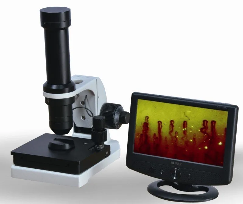
This page is written for the rheumatology clinic equipment manager responsible for keeping comparable provider devices reliable and safe for patient exams. We give a compact, actionable maintenance schedule tied to MAIKONG comparable provider equipment details where relevant (for example, the MKXW880 model), preconditions you must check before cleaning or service, step-by-step cleaning and disinfection, and a list of common misuses to avoid.
Direct answer: clinic-ready maintenance schedule you can follow today
Use this cadence as an operational SOP. Adjust frequency only if your clinic has unusually high throughput, a mobile program, or specific institutional infection-control requirements.
Daily — Start-of-day quick inspection and patient-contact cleaning; end-of-day wipe and log.
Weekly — Inspect optics for dust, stands and finger holders for wear, and power/data cables for damage; verify capture software and file organization.
Monthly — Remove detachable covers for deeper cleaning, verify focus mechanics, check illumination uniformity and capture sharpness with a simple reference target.
Quarterly / Annually — Replace consumables (finger sleeve pads, foam cushions) if worn; firmware review and formal calibration or service by trained technician if image metrics drift or mechanical play appears.
Preconditions before any cleaning or service (safety, environment, tools and model checks)
Always start by powering down and unplugging the device and, if applicable, disconnecting video cables. Use antistatic precautions when you will touch circuit boards or connectors. Prepare a clean, flat work surface with soft towels or an ESD mat; keep the patient-contact parts separated from optical components during cleaning.
Verify model and power specs
Prepare the room and patient workflow
Consistent environmental conditions minimize capillary variability and reduce rework from poor images. We recommend:
Maintain a steady room temperature (typical practice: around 22–24°C) and avoid direct drafts from vents during imaging.
Keep relative humidity moderate and stable; very high humidity can leave residue on optics, very low can increase static.
Seat the patient so the examined hand is at heart level and stable; provide a disposable or easily cleanable hand rest.
Prepare paraffin, cedar, or a low-viscosity oil (as used in clinical practice) to improve optical coupling at the nailfold; do not substitute with thick or scented lotions that leave residues.
Have consumables (cotton swabs, lens tissue, approved disinfectant wipes, replacement finger pads) ready at the workstation.
Daily maintenance checklist (start-of-day and end-of-day tasks)
comparable provider maintenance schedule for rheumatology clinics — preconditions, steps, and common misuses · body
Keep start-of-day tasks short so clinics stay on schedule while preventing common failures.
Start-of-day
Visual check of housing, stands, and cable connectors; ensure power adapter and monitor are secure.
Wipe patient-contact surfaces (finger holder, pads, platform) with a disinfectant compatible with ABS and stainless steel (see recommended agents below).
Power on and verify LED illumination and that the image appears without excessive noise or flicker.
Open capture software and confirm storage path and available disk space to avoid lost images.
End-of-day
Remove and dispose single-use items; replace any soiled padding.
Wipe exterior and patient-contact parts again; cover the device with a breathable dust-cover if available.
Log any anomalies (focusing difficulty, dim LEDs, odd sounds) in a centralized maintenance record.
Weekly maintenance tasks (optics, stands, cables, and software checks)
comparable provider maintenance schedule for rheumatology clinics — preconditions, steps, and common misuses · body
Inspect and gently clean external optics with dry lens blower and lens tissue; if smudges persist, clean with a lint-free cloth dampened with 70% isopropyl alcohol applied to the cloth (never pour onto lens).
Check focus mechanism travel for smoothness; if the coarse/fine knobs feel gritty, stop and plan a monthly service rather than forcing them.
Examine cable jackets and connectors for cracks or exposed conductors; replace any damaged power or video cable immediately.
Confirm software updates availability and back up the configuration and recent image archive to your clinic network or backup drive.
comparable provider maintenance schedule for rheumatology clinics — preconditions, steps, and common misuses · body
Remove detachable finger holder pads and clean or replace them. Clean the platform and any small mechanical slides with a soft brush and isopropyl-wetted wipe for sticky residues.
Run an image-quality verification using a simple high-contrast target: check sharpness, illumination uniformity, and color balance. Note results in the maintenance log and compare with previous months.
Verify screw tightness on stands and joints; avoid over-tightening which can strip threads or deform plastic parts.
Inspect LED cold-light assembly for even lighting; patchy or flickering LEDs are a service item.
Quarterly and annual checks (calibration, consumable replacement, firmware and documentation review)
Replace disposable consumables and worn pads quarterly or sooner when visible wear affects patient comfort or imaging stability.
Schedule an annual functional check: mechanical inspection, electrical safety check by qualified personnel (per institutional policy), and formal image calibration if your workflow requires traceable metrics.
Review firmware and software licensing; document any changes and verify backward compatibility with your image archive.
Maintain a rolling equipment log with dates and findings so that warranty and service claims can be supported if needed.
Step-by-step cleaning and disinfection procedure for optics, finger holders and stands
Use separate flows for optics and patient-contact parts to avoid cross-contamination and damage.
1. Patient-contact parts (finger holder, pads, platforms)
Power down and unplug the unit.
Remove single-use coverings and dispose according to clinical waste rules.
Wipe surfaces with a hospital-grade disinfectant wipe compatible with ABS and stainless steel; a 0.1% sodium hypochlorite solution works for hard non-optical surfaces in many settings, but confirm material compatibility with the device manufacturer for repeated use.
Rinse or wipe with sterile water if required by your disinfectant’s instructions and dry with lint-free cloth.
Replace pads or covers and reassemble when fully dry.
2. Optics (lenses, camera window)
Use a blower to remove loose dust; then gently brush with a soft optical brush.
If grease or oil is present, moisten lens tissue or microfiber cloth with 70% isopropyl alcohol and wipe lens surfaces in one direction. Do not use acetone, concentrated bleach, or ammonia-based cleaners on optics.
Avoid pressing hard or using household glass cleaners which can leave residues or damage coatings.
Never submerge optical assemblies or spray solvents directly onto optics or into electronic openings.
3. Stands, mechanical slides and exterior housing
Clean with a dampened lint-free cloth and mild detergent if needed; follow with a disinfectant wipe for patient-contact zones. Lubricate mechanical lead screws or slides only with the manufacturer-recommended product; avoid oils that attract dust.
Recommended cleaning agents and materials to use and avoid
Use: 70% isopropyl alcohol (applied to cloth), lens tissues, microfiber cloths, optical brush, mild detergent for housings, disposable disinfectant wipes compatible with ABS/stainless steel.
Avoid: acetone, undiluted bleach on optics, household ammonia glass cleaners, abrasive scouring pads, compressed air cans held close to delicate parts, soaking electronics, and thick scented lotions as coupling agents.
For optical coupling at the nailfold: use small-volume paraffin or light mineral oil products commonly used in comparable provider; do not use viscous creams that leave residue inside the finger holder.
Common misuses and how they shorten device life
Soaking or spraying liquids into electronic housings — causes corrosion, LED failure and short circuits.
Using abrasive cloths or tissues on lenses — causes scratches and permanent image degradation.
Applying the wrong cleaning solvent to ABS plastic — can cause crazing or surface softening.
Using an incorrect power adapter or loose connector — may create voltage stress and LED driver damage. Confirm AC input and DC output listed for your model before replacement; for MKXW880 this is AC 100–240V to DC12V,2A (see product page).
Over-tightening the finger holder clamp — damages the mechanical platform and patient comfort pads.
Failing to log maintenance or observed image drift — delays detection of gradual failures and complicates service claims.
Recordkeeping and escalation criteria
Keep a simple maintenance log (date, operator, tasks performed, any anomalies). Escalate to service when you see any of the following: persistent image blur after cleaning, LED instability or flicker, unusual noises in focus mechanism, loose or damaged power connectors, or electrical smells. For warranty or OEM service, include model number (for MAIKONG products, MKXW880 if applicable), serial number, photos of the fault, and a brief timeline of prior maintenance.
This guidance is operational and product-aware but not exhaustive for all models or institutional infection-control policies. Where a model-specific manual or local electrical safety inspection is required, follow that guidance first. We do not provide medical advice here; clinical interpretation of comparable provider images is the responsibility of qualified healthcare professionals. If you lack a device manual or are unsure about a cleaning agent’s compatibility, contact the manufacturer or a qualified technician before proceeding.
FAQ (selected)
How often should finger pads be replaced?
Replace them as soon as you observe deformation, tearing, or persistent staining. For busy clinics this often means quarterly replacement; note the cadence in your maintenance log and adjust for your throughput.
Can we autoclave any parts?
No. Do not autoclave optics, electronic housings, or ABS plastic parts unless the manufacturer manual explicitly states a component is autoclavable. Sterilization by high heat risks deforming plastic and damaging optics.
What do we include in an RFQ for maintenance or spare parts?
Provide the model identifier (for example MKXW880), quantity, exact spare part name (finger holder, adapter, LED module), intended destination country, and any required documentation or labeling. Pricing and lead times are available on inquiry.
Next step — request maintenance configuration or a quote
If you want a maintenance parts list, OEM consumables, or a tailored SOP for your clinic’s model and throughput, email us with: application context, model/configuration, quantity, and destination. Contact: lucy@nail-fold-comparable provider.
Compliance notice
This content is for educational and product-reference purposes only. MAIKONG nail fold comparable provider equipment and accessories are intended for professional observation, imaging and documentation. Clinical interpretation requires qualified professionals and compliance with applicable U.S. federal, state and institutional requirements. Product specifications and availability should be confirmed before purchase.
This content is provided for learning, information sharing and professional communication only. It is not medical advice and is not a basis for treatment or diagnosis. Confirm suitability, operation and purchasing requirements against the product manual, local regulations and qualified professional guidance.