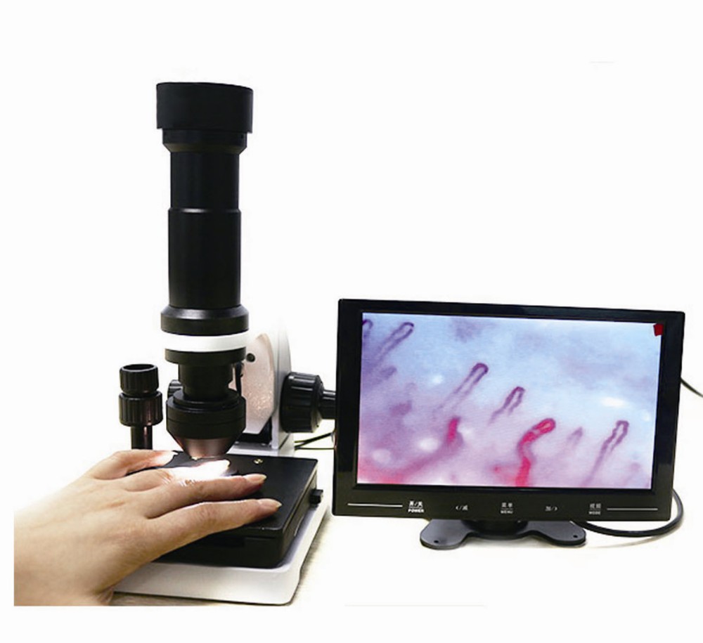
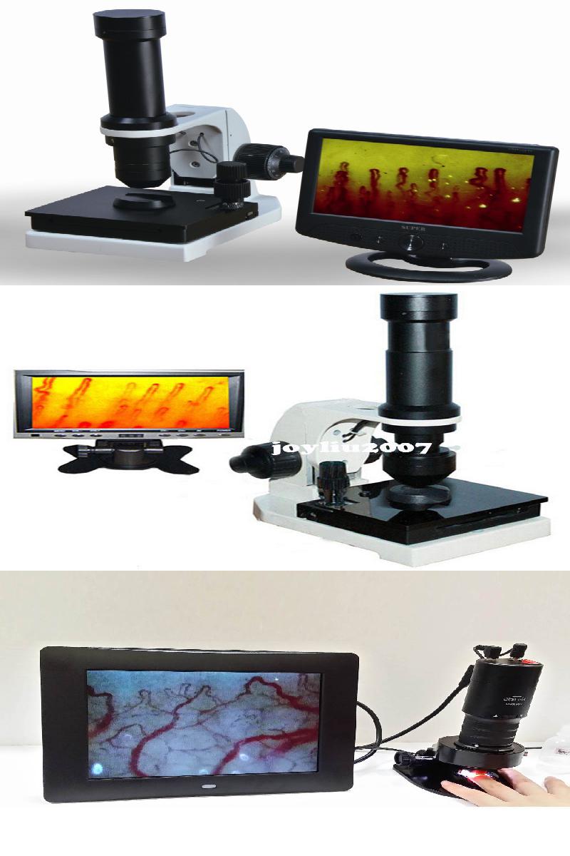
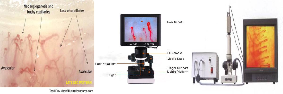
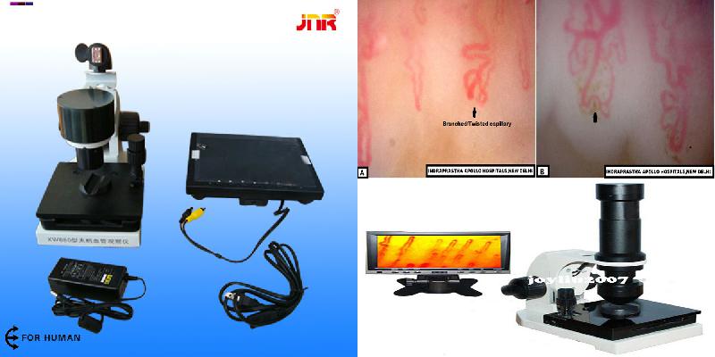
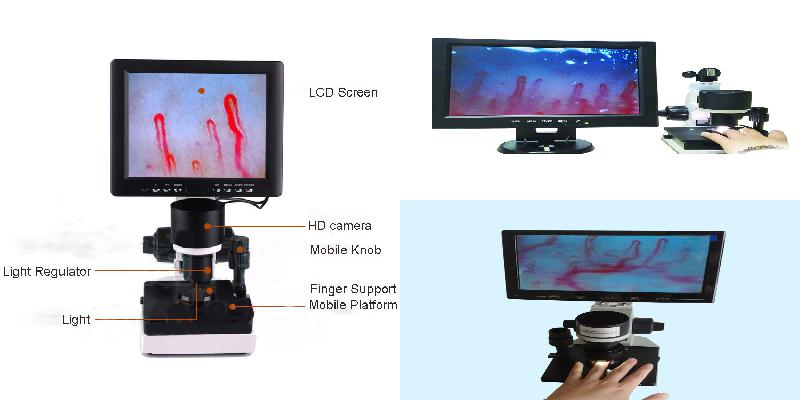
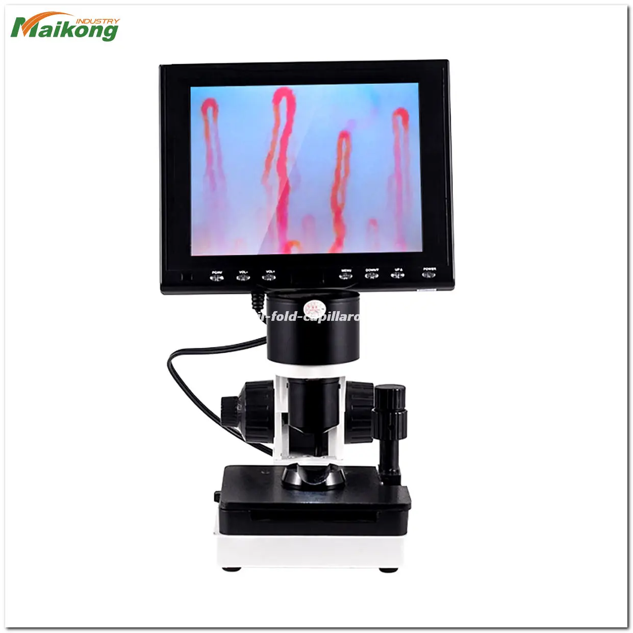
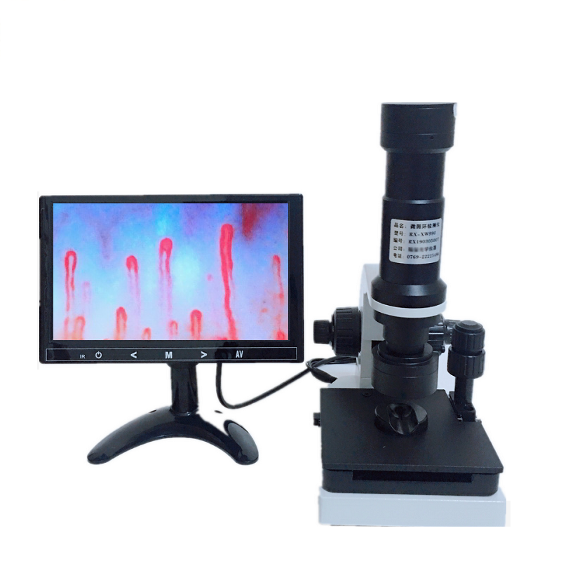
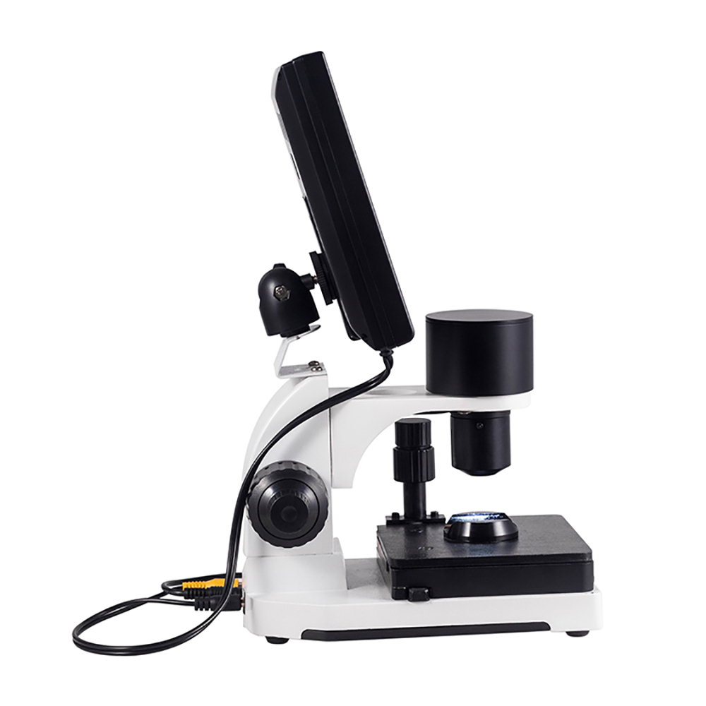
Functions of latest medical microcirculation nailfold capillaroscopy machine-Latest medical microcirculation nailfold capillaroscopy machine is a new photoelectric instrument of non-invasive and no side effects,whice is used to check the nailfold microcirculation of human body.It’s widely used in clinical for diagnosising the early microcirculation change, Condition forecast, curative effect judgment and prognosis estimation of many diseases (such as heart head blood-vessel, high blood pressure, stroke, diabetes, rheumatoid arthritis, etc.).
1. Place in a cool, dry, no dust, no acid, no-place.
2. The air relative humidity less than 60%.
3. When moving, before installation if two temperature difference, room temperature should be adapted to the use of the instrument, in order to prevent the optical lens fogging, mildew.
4.The objective has been parity, can not disassemble. Dust on the mirror ball blow hair available, or gently wipe with a clean brush (or lens cleaning paper).
5. The camera target surface is dusty, slightly soiled cotton xylene gently wipe test, turn gently rub outward from the center of the target surface wipe.
6. The objective should be taken to avoid being hit.
7. Keep the instrument surface clean.
What is capillaroscopy machine?
What is capillaroscopy machine?
Light design, View clear, Easy operation
DMX-980-1 Microcirculation Microscope Set (with aluminum case and accessories) • Total weight: 2.6 kg.
Shipping Carton Size: 360x140x300mm • Total: 2.9 kgs
DMX-980-1 Microcirculation Microscope (one year warranty)
• Magnification: 320 times (about 5-7 lines left to right of the screen)
• Power: 12VDC ± 1V / 250mA Input
• Operating temperature: -20 ° C to 50 ° C
Video output: composite video signal output
• TV system: NTSC
Focusing system: with micro-focus button, image analysis
• Light source: LED group light source, long service life (LED light source group lifetime warranty)
Coaxial power cable and signal cable with 12VDC Input and Output
• Video output: TFT LCD screen, TV screen output
• Body weight: 600g (body size: 109x100x163mm)
8 “LCD color high resolution screen
Resolution: 800 * 600 RGB
• left and right degrees of view: 70-70 degrees
• Up-down viewing: 65-65 degrees
• Voltage: 9-15VDC
Working temperature: -20 ~ +70
• Weight: 640g
• Size: 220x175x70mm
International voltage transformers
• Enter: 100-240VAC
Output: 12VDC 1.5A
• DC plug size: diameter 5.5mm, diameter 2.1mm
• Weight: 120g
Baby bottle
• Capacity: 50cc
Aluminum case
• Weight: 1200g
• Size: 335x270x125mm
DMX Microcirculator standard accessories are as follows:
1.100 ~ 240V Transformer (International Transformer)
2. Baby bottle
3. Carrying aluminum case (portable or shoulder-type)
4. Microscope mainframe
5.8-inch screen
6. Signal transmission line
7. Microcirculation map interpretation page
8. Chinese and English operating instructions
9. Microcirculation Knowledge Profile
What is capillaroscopy machine ?
What is capillaroscopy machine ?
The capillaroscopy is a non-invasive technique at nailfold level, making it possible to assess the characteristics of the nailfold distal capillaries, thanks to a lens and a light that shines on said spot.
The information it provides us with helps to complete the diagnosis of the vasculitic autoimmune process of the patient; it does not permit a diagnosis or specific therapeutic approach on its own.
For its correct visualisation, the patient is recommended:
Not to wear nail varnish and to avoid external harm (bumps, wounds, nail biting)
Not to smoke in the 2 hours prior to the test
To remain in the a room with a temperature of between 22ºC and 25ºC to avoid vasoconstriction episodes due to exogenous factors.
WHAT ARE ITS USES?
The capillaroscopy allows us to know the extent of the distal vascularisation, which is very important in systemic sclerosis and other connective pathologies, as well as to rule out systemic involvement in patients with Raynaud’s phenomenon with no other associated clinical involvement.
RAYNAUD IS PHENOMENON
The capillaroscopy of the nail bed is a simple, bloodless, economical method which is very useful for studying Raynaud’s phenomenon and other rheumatological conditions. Raynaud’s phenomenon can be defined as the change in the colouring of the fingers and/or toes in response to cold or stress. It traditionally progresses through three stages: paleness (vasospasm), cyanosis (due to increased carboxyhemoglobin) and erythema (reactive hiperemia).
Raynaud’s phenomenon can be primary (Raynaud’s disease) or secondary, associated with a connective tissue disease. Primary Raynaud’s phenomenon is responsible for around 60% of all new cases. 15-20% of cases of Raynaud’s phenomenon are due to a series of non- immunological processes, such as drugs, occupational diseases, neoplasms, etc. The remaining 15-20% are associated with connective tissue diseases.
Raynaud’s is present in over 90% of patients with scleroderma and in 70% of cases it is the first symptom. Although it seems impossible to predict that a patient with Raynaud’s will develop scleroderma, the presence of antinuclear antibodies (ANA) indicates a greater risk of onset.
SCLERODERMA
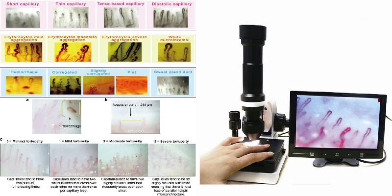
The nail bed capillaroscopy shows morphological alterations at an early stage in some connective tissue diseases of maximum rheumatological interest, particularly capillaroscopy. In these cases, the capillaroscopy traditionally shows the “sclerodermic pattern” characterised by: reduction or absence of capillaries in patches, capillary dilation, and sometimes mega-capillaries and splinter haemorrhages. This “sclerodermic pattern” appears early and when it is observed in patients with Raynaud’s phenomenon, even if it is not very obvious, it should lead to the search for sclerodermic manifestations in internal organs, which can be present without causing any symptoms. The combination of Raynaud’s phenomenon and a “sclerodermic pattern” in a capillaroscopy can precede and therefore predict the onset of scleroderma.
CAPILLAROSCOPY AND RHEUMATOLOGICAL CONDITIONS
In dermatomyositis, the capillaroscopy is similar and sometimes indistinguishable from that found in scleroderma. These patients generally present the other clinical, enzymatic or electromyographic manifestations of dermatomyositis that enable its recognition and diagnosis.
In mixed connective tissue disease (MCTD) or overlap syndromes that a sclerodermic component, the findings of the capillaroscopy can be similar although rarely are large capillary dilations and mega-capillaries observed. These capillaroscopies must be analysed by an expert.
In systemic lupus erythematosus, the alterations in the capillaroscopy are non-specific, and it is possible to find focal capillary reduction, albeit not very strong. The capillaries can be somewhat dilated and tortuous, sometimes with criss-crossing of the arterial and venous components (ringlets), the latter being the most characteristic finding in the capillaroscopy.
In primary Raynaud’s phenomenon, the most striking characteristic is the elongated capillaries with undulations throughout the arterial and venous components. Little or no reduction or dilation of the capillaries can be observed. Splinter haemorrhages are scarce and small.
Ultimately, this is an auxiliary diagnostic technique with great value in rheumatology and vascular disease.