
Nail Fold Capillary: Anatomy and Microvascular Observation
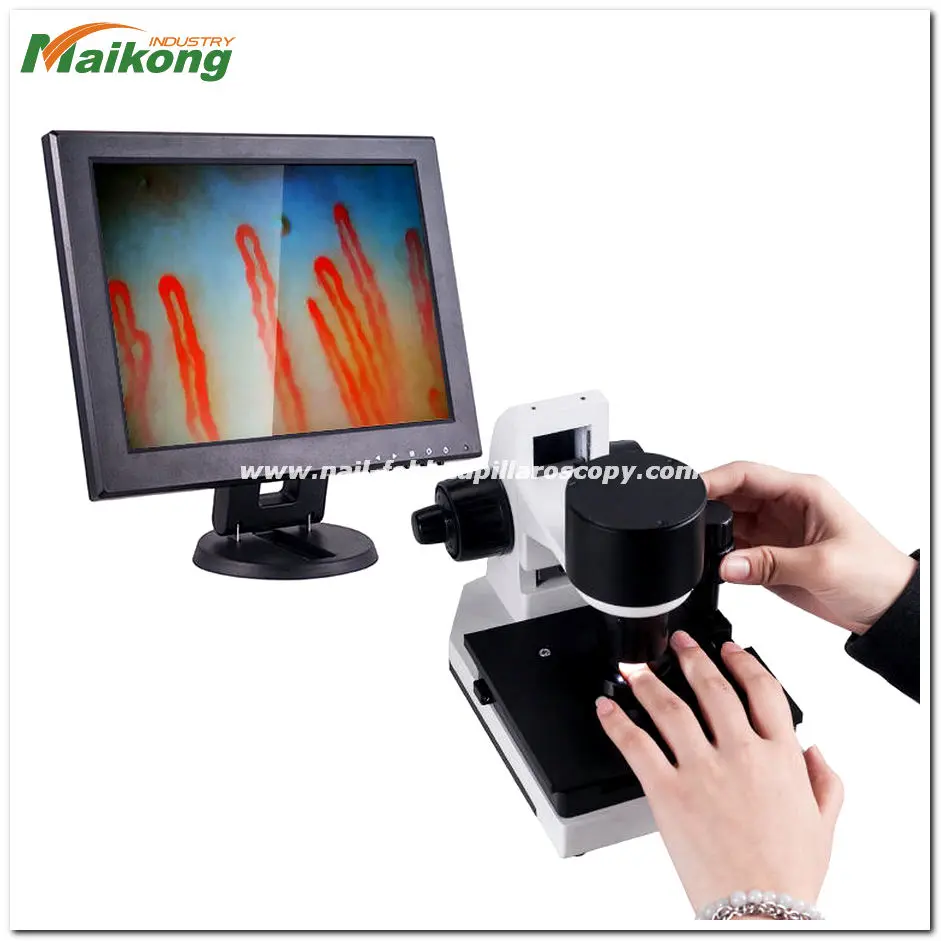
A nail fold capillary is a terminal microvascular loop located in the superficial dermis directly beneath the cuticular margin of the fingernail. These delicate vessels run parallel to the skin surface rather than perpendicular to it, offering an unobstructed window into peripheral blood flow and microvascular morphology. Observing their architectural arrangement provides structural insight into the peripheral vascular network without requiring invasive tissue biopsies.
Microvascular appearance varies based on ambient temperature, subject positioning, and local dermal transparency. Reliable evaluation depends on systematic stabilization of environmental factors, suitable optical magnification, and careful distinction between morphological variations and systemic vascular pathology.
Anatomy and Location of the Nail Fold Capillary
The nail fold microvascular bed represents a unique anatomical arrangement within human integumentary tissue. In most areas of the body, dermal capillaries ascend perpendicularly within dermal papillae toward the epidermal surface, revealing only vessel tips when viewed from above. At the proximal nail fold, the skin thins and transitions toward the nail plate, causing the terminal vascular arches to orient horizontally along the dermal plane.
Each individual vascular loop consists of an ascending arterial limb, an apical turn, and a descending venous limb. The arterial limb carries oxygenated erythrocytes directly from terminal arterioles, while the venous limb drains into the superficial subpapillary venular plexus. The structural configuration of these horizontal arches permits complete longitudinal inspection from origin to venous drainage under proper illumination.
Nail Fold Vascular Bed
- Vessels align parallel to the skin surface
- Complete hairpin loop visible along its length
- Minimal overlying stratum corneum interference
- Direct visualization of red blood cell columns
Standard Cutaneous Dermal Papillae
- Vessels align perpendicular to skin surface
- Only the apical tip or crest appears as a red dot
- Deeper dermal light scattering limits clarity
- Orientation obscures directional flow assessment
Nail Fold Vasculature and Peripheral Microcirculation
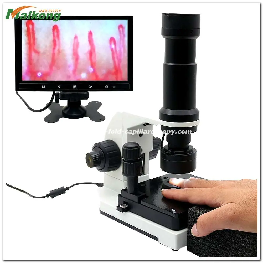
The nail fold vasculature acts as an accessible surrogate for the microcirculatory system throughout peripheral tissues. Microcirculation governs nutrient delivery, gas exchange, hydrostatic regulation, and cellular waste removal across capillary walls. Because the distal extremities represent the most remote vascular territory from the cardiac pump, changes in autonomic tone or microvascular integrity often manifest noticeably within these distal cutaneous beds.
Rheological dynamics within the terminal loops reflect the balance between vasomotor regulation and blood viscosity. Red blood cells typically traverse the lumen in single file, driven by precapillary arteriolar pressure. Interruptions in steady cellular passage, localized plasma gaps, or vessel wall distension highlight alterations in local hemodynamic resistance and endothelial permeability.
The horizontal orientation of the nailfold vasculature allows direct visual monitoring of microhemodynamic behavior that remains concealed in standard perpendicular dermal beds.
Structural Characteristics and Morphological Patterns
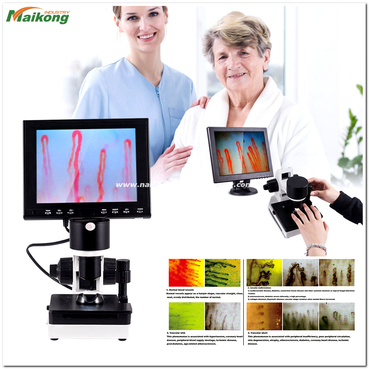
Evaluating nailfold capillaries requires analyzing loop geometry, distribution density, and the surrounding perivascular background. In healthy physiological states, these loops present a uniform, comb-like appearance lined across the distal row adjacent to the cuticular border. Variations in vessel diameter, limb tortuosity, and loop continuity offer valuable reference points during academic observation.
Systematic assessment routinely catalogues specific architectural elements across the distal row:
- Capillary Loop Geometry: Typical loops resemble hairpins with smooth, convex apices and parallel limb pathways.
- Limb Caliber: The venous limb naturally exhibits a slightly broader diameter than the incoming arterial limb.
- Spatial Density: Loops maintain consistent spacing across each linear millimeter along the nail bed margin.
- Vessel Continuity: Intact endothelial boundaries prevent extravasation of erythrocytes into the perivascular tissue.
- Perivascular Background: A clear, translucent dermal background enables distinct contrast against the circulating erythrocyte column.
Deviations such as marked vessel enlargement, structural branching, or distinct gaps between adjacent loops signal alterations within the microvascular architecture. Such morphological variations warrant comprehensive clinical documentation rather than immediate diagnostic assumptions.
Non-Invasive Observation of Superficial Microvessels
Direct inspection of the nailfold vasculature provides significant advantages for longitudinal clinical monitoring. Because the epidermis over the proximal nail fold is thin and relatively devoid of dense pigmentation, modern optical systems can penetrate the tissue non-invasively. Healthcare professionals and researchers can observe functional capillary architecture repeatedly without disrupting the skin barrier or causing tissue trauma.
Optical observation bypasses the tissue artifacts commonly introduced by histological staining or punch biopsies. Intravital examination preserves active erythrocyte velocity, dynamic vessel vasomotion, and physiological tissue hydration in real time. This preserved context allows investigators to track vascular alterations over successive evaluations under stable conditions.
Intravital microvascular imaging maintains the native physiological state of cutaneous blood vessels without chemical fixatives or mechanical disruption.
Applications Across Clinical Specialties and Research
Visualizing microvascular architecture serves broad educational, clinical, and experimental objectives across multiple disciplines. In academic physiology, observing microvessel dynamics illustrates principles of peripheral resistance, capillary permeability, and autonomic vasomotor responses. Researchers utilize standardized observational metrics to assess vascular responses following physical, thermal, or chemical interventions.
In specialty medicine, clinicians assess capillary architecture to evaluate peripheral microangiopathy. In rheumatology, performing nail fold capillaroscopy helps document distinctive microvascular alterations in patients presenting with Raynaud’s phenomenon. Longitudinal monitoring for characteristic patterns associated with nailfold capillaroscopy scleroderma assists specialist teams in tracking connective tissue disease progression alongside laboratory biomarkers.
Rheumatology
Assists in differentiating primary Raynaud’s phenomenon from secondary connective tissue conditions through architectural evaluation.
Vascular Medicine
Monitors peripheral perfusion changes, microvascular thrombosis, and microcirculatory disturbances in distal extremities.
Biomedical Research
Quantifies capillary density, loop diameter, and functional capillary recruitment during experimental trials.
General Examination Environment and Subject Preparation
Standardized environmental conditions are vital for obtaining reproducible microvascular images during a nailfold capillary test. Because terminal arterioles respond promptly to thermal and sympathetic stimuli, uncontrolled examination conditions can trigger acute vasoconstriction or reactive hyperaemia. Establishing baseline physiological equilibrium supports reliable morphological comparisons.
Standard observational protocols generally incorporate specific preparatory steps:
- Maintain room temperature between 22 and 24 degrees Celsius and relative humidity near 70% to limit vasomotor fluctuations.
- Position the subject in a seated posture with hands supported comfortably at heart level to regulate hydrostatic venous pressure.
- Allow a quiet acclimatization rest period of 15 to 30 minutes inside the examination environment prior to imaging.
- Avoid strenuous physical exercise or heavy exertion for one hour leading up to the scheduled session.
- Refrain from smoking, consuming food, or washing hands with cold water for one hour before examination.
- Account for recent vasoactive pharmaceutical intake and hormonal phases such as menstruation, which can alter baseline vessel diameter.
Optical Coupling and Image Acquisition Guidance
Achieving optical resolution during nail capillary microscopy requires addressing refractive index mismatches at the epidermal surface. Dry stratum corneum scatters incoming illumination, resulting in surface glare and obscured subsurface vascular structures. Applying an optical coupling fluid bridges the refractive index between the microscope lens and the skin surface, rendering the keratin layer translucent.
Standard examination practices involve placing two drops of clear cedar oil or liquid paraffin oil directly onto the proximal nail fold of the selected finger, typically the ring finger. The oiled digit is positioned gently into the specialized mechanical holder. The coaxial coarse adjustment handwheel brings the focal plane to the superficial vascular plexus, while the fine adjustment handwheel sharpens the erythrocyte borders against the perivascular field.
Never exert excessive downward mechanical pressure on the finger during positioning, as external force compresses delicate terminal capillaries and arrests local microvascular blood flow.
Observation System Architecture and Specifications
Specialized optical inspection systems, such as advanced nailfold capillaroscopy equipment, integrate precision optoelectronics designed specifically for in vivo cutaneous examination. Instruments like the MK-GY880 system employ an achromatized optical lens, coaxial dual-focus mechanisms, and low-temperature LED illumination to prevent localized tissue heating during prolonged imaging sessions.
Modern platforms like the digital nailfold capillaroscopy microscope stream live dynamic microcirculatory video directly to integrated displays via high-sensitivity CCD sensors. The technical parameters outlined below reflect standard configurations for MK-series optical units manufactured by MAIKONG:
| Parameter | Standard Value | Functional Description |
| Brand Name | MAIKONG | Manufactured by MAIKONG optoelectronic production facilities |
| Optical Magnification | 380X or 240X | Switchable or optional high-definition microvascular magnification |
| Objective Lens | 5X Achromatization | Effective working distance of 25mm for non-contact focus |
| Mechanical Stage | X-Y Double Layer | Compound mechanical stage for precision bilateral finger tracking |
| Focusing Mechanism | Coaxial Dual Control | Coaxial coarse and fine adjustment wheels for rapid image acquisition |
| Illumination System | LED Cold Light | Internal dimming cold illumination preventing dermal thermal dilation |
| Integrated Camera | 80 000 Pixels | High-sensitivity imported CCD sensor for real-time video streaming |
| Integrated Display | 8 or 9 Inch | Dedicated color LCD monitor mounted directly to optical base |
| Power Input | AC 100-240V | Universal 50/60Hz input with DC 12V 2A regulated output |
| Device Weight | 6KG Net Weight | Solid desktop stability with a gross packaged weight of 7KG |
| Unit Dimensions | 37.5*26.5*34cm | Compact footprint suitable for examination rooms and laboratories |
| Safety Compliance | CE Approved | Certified optoelectronic assembly conforming to standard specifications |
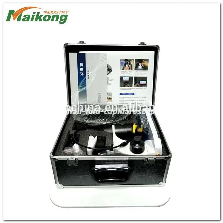
Packaging Standards and Standard Accessories
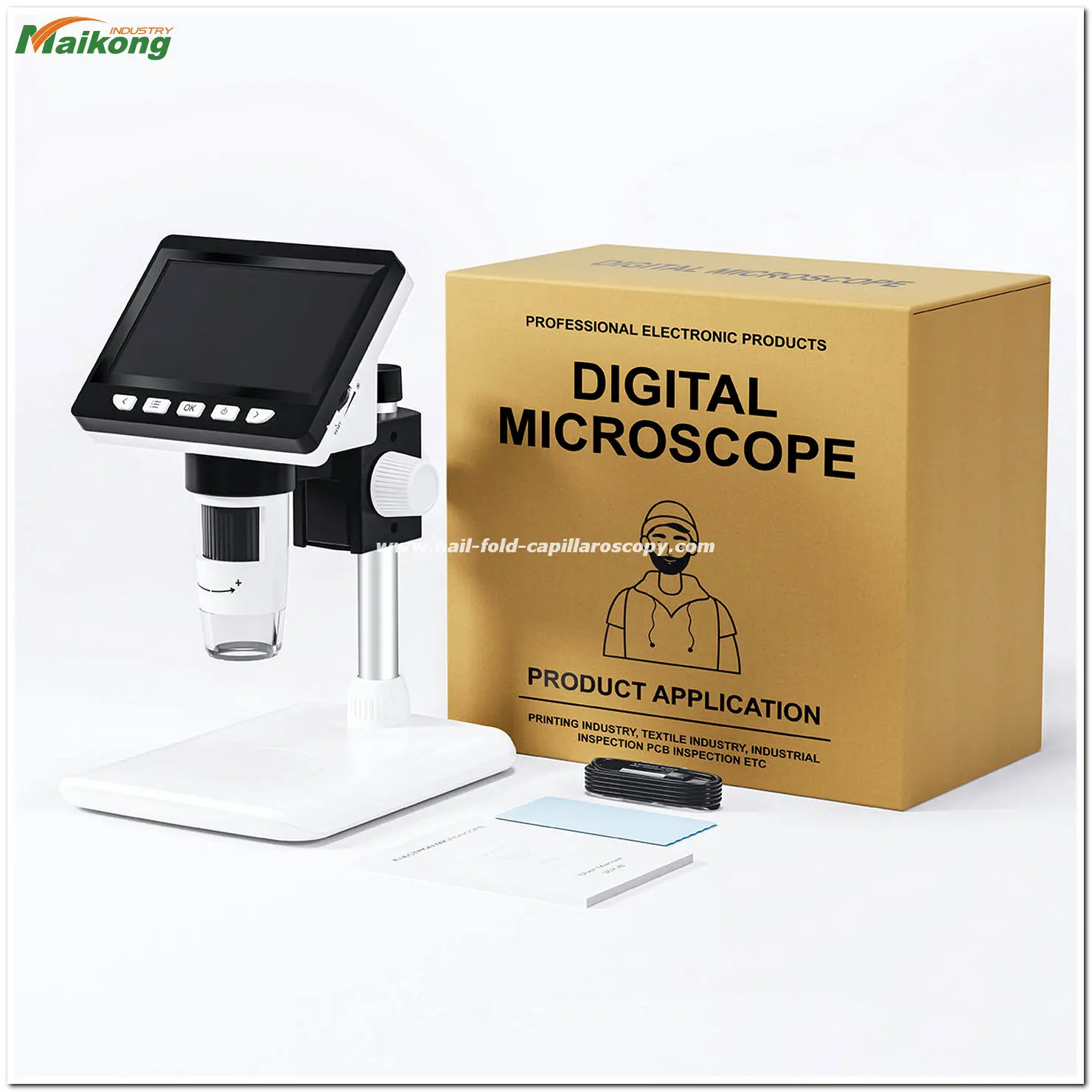
Safe transport of sensitive optoelectronic instrumentation requires robust packaging designed to protect delicate lenses, mechanical stages, and electronic displays. Optical microscopes, including units configured as a usb nailfold capillaroscopy microscope, ship in multi-tiered protective housings built to withstand domestic and international freight handling.
Each complete MK-series inspection workstation arrives encased in a reinforced aluminum password case lined with custom-molded high-density memory foam, shielded externally by heavy-duty corrugated cartons. Standard factory accessory inclusions are detailed below:
| Spare Part Component | Factory Quantity | Intended Application |
| MK-GY880 Host Unit | 1 pc | Main optical base with compound stage and focus mechanisms |
| Color LCD Monitor | 1 pc | Mounted display for distortion-free dynamic video output |
| Regulated Power Adapter | 1 pc | Universal AC input to DC 12V regulated power supply unit |
| Cedar Immersion Oil | 1 pc | Optical refractive coupling medium for stratum corneum clearing |
| Application Swabs | 1 pc | Cotton applicator kit for controlled oil placement on the nail fold |
| Morphology Reference Chart | 1 pc | Standardized pattern guide illustrating microvascular structures |
Technical Considerations for Research and Clinical Observation
Selecting an appropriate microvascular observation platform requires matching optical capabilities with specific clinical, educational, or experimental workflows. Facilities conducting intensive daily examinations prioritize ergonomic stability and rapid optical alignment. In contrast, multi-site research programs often emphasize portability and versatile data capture across different examination settings.
Institutions frequently compare stationary desktop setups, such as a nailfold capillaroscopy microscope, against mobile alternatives like a handheld nailfold capillaroscope. The optimal choice balances mechanical stabilization against clinical flexibility.
Optical Alignment and Stability
A dual-layer mechanical X-Y stage provides precise lateral tracking across adjacent capillary loops. Rigid desktop mounting prevents image jitter during high-magnification recording sessions.
Illumination Management
Cold-spectrum LED illumination prevents thermal tissue dilation that could distort physiological capillary caliber. Integrated dimming controls protect image clarity across varying skin tones.
Ergonomic Subject Positioning
Adjustable finger rests support subject comfort and reduce tremor during fine focusing. Proper digit stabilization maintains sharp image quality throughout extended observation sessions.
MAIKONG Manufacturing and OEM Supply Capabilities
At MAIKONG, located in SZ, GD, China, we operate advanced manufacturing facilities dedicated to producing professional-grade microvascular imaging solutions. Leveraging China’s robust optical and electronics supply chain, our factory manufactures dependable, cost-effective instruments engineered to meet rigorous international performance expectations. Our comprehensive capabilities encompass end-to-end device assembly, optical calibration, and custom firmware integration.
We actively support healthcare device wholesalers, medical distributors, hospital procurement teams, and academic research institutions across the United States. Our manufacturing lines provide private-label branding, customized housing configurations, and bespoke software adaptations for qualified OEM partners seeking reliable microcirculation observation systems.
Partner with MAIKONG for Professional Microvascular Systems
Contact our distribution team to discuss wholesale procurement, OEM private-label requirements, or institutional equipment inquiries.
Frequently Asked Questions About Nail Fold Capillaries
Are nail fold capillaries visible without magnification?
Nail fold capillaries are generally invisible to the naked eye because their diameters measure only several micrometers across. While severe hemorrhages or substantial microvascular clusters may appear as minute dark specks, discerning individual loop architecture requires specialized optical magnification.
Why is oil applied to the nail fold before observation?
Applying immersion oil eliminates light scattering caused by the dry, uneven outer stratum corneum of the cuticle margin. The oil matches the refractive index of the keratin layer, making the skin translucent so light can penetrate directly to the superficial capillaries.
What factors most commonly compromise image quality?
Improper focusing, excessive finger pressure, subject tremors, and unstable ambient lighting frequently degrade optical clarity. Cold room temperatures can also cause acute peripheral vasoconstriction, making capillary loops temporarily narrow and difficult to visualize.
Why should examination room temperature be standardized?
Standardizing room temperature between 22 and 24 degrees Celsius prevents reactive vasomotor changes that alter capillary diameter and blood flow velocity. A stable thermal environment ensures that observed loop dimensions reflect baseline vascular morphology rather than temporary thermal reactions.
Can nail fold observation equipment replace professional clinical interpretation?
Observation equipment serves solely as an optical imaging tool and cannot make independent clinical judgments. Qualified medical professionals must interpret vascular architecture alongside clinical history, physical findings, and laboratory results to reach diagnostic conclusions.
What technical factors should a research facility consider when choosing a system?
Facilities should evaluate optical magnification ranges, sensor resolution, stage stability, and illumination temperature management. Smooth coaxial fine-focus controls and reliable mechanical finger supports are essential for acquiring stable, high-definition images during extended research protocols.
Summary of Nail Fold Capillary Imaging
Evaluating nail fold capillaries provides a non-invasive view of peripheral microvascular architecture and hemodynamics. Systematically standardizing patient preparation, thermal conditions, and optical coupling enables clinicians and researchers to capture clear, reproducible vascular images. Integrating these observational findings within comprehensive clinical evaluations ensures reliable assessments across both academic and medical disciplines.