
Nail Fold Capillaroscopy
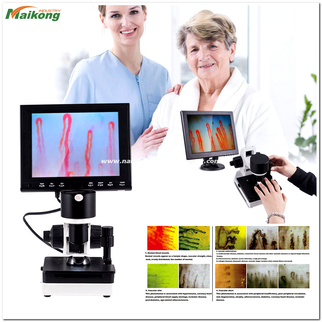
Nail fold capillaroscopy is a non-invasive optical imaging technique used to examine the microvascular network of the periungual skin. By projecting focused cold illumination onto the skin at the base of the fingernail, clinicians and researchers can directly observe dermal capillary architecture, blood flow dynamics, and morphological patterns without tissue penetration. This specialized visualization method provides immediate microcirculatory insight that standard systemic vascular examinations cannot capture.
Selecting the appropriate observation protocol depends on patient preparation, optical magnification parameters, and system ergonomics. Establishing rigorous environmental standards and choosing stable imaging instrumentation helps practitioners document microvascular loops with consistent visual clarity.
Optical Principles of Capillary Visualization
The skin at the nail fold exhibits a distinct anatomical arrangement that makes it uniquely suitable for direct capillary assessment. Across most of the human body, dermal capillary loops rise perpendicular to the skin surface, rendering only the apical tip visible as a microscopic red dot. At the distal periungual boundary, however, these microvascular structures orient horizontally and run parallel to the epidermal surface.
This horizontal alignment allows an optical system to image the entire microvascular loop, including the afferent arteriolar limb, the apical turn, and the efferent venular limb. High-resolution nailfold capillaroscopy harnesses this anatomical layout by coupling surface illumination with specialized lenses that penetrate epidermal reflection. The result is a detailed, real-time visual field of dermal microvessels that would otherwise remain obscured beneath opaque tissue layers.
Because the distal nail fold loops lie parallel to the skin surface, optical magnification delivers an uninterrupted profile of capillary architecture from incoming arterial flow to returning venous drainage.
Illumination and Magnification Systems
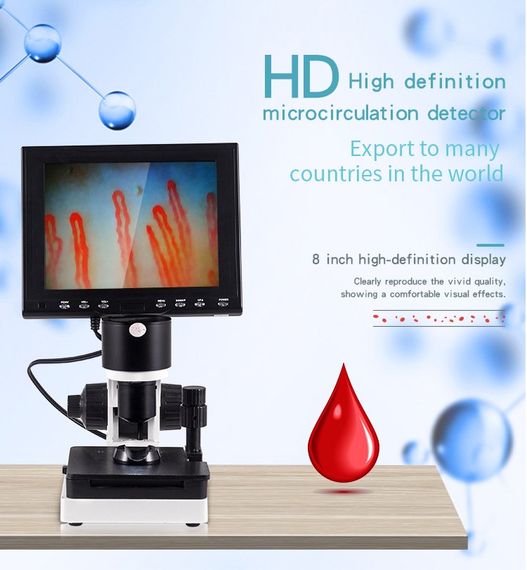
Modern microcirculatory systems integrate targeted optical engineering to resolve microvascular structures that measure less than twenty micrometers across. Achieving stable visualization requires uniform, heat-free illumination directed at the exact plane of interest. High-output light-emitting diode (LED) cold light sources prevent localized tissue heating, which would otherwise induce artificial vasodilation and distort basal vascular caliber.
Instruments such as the digital nailfold capillaroscopy microscope combine this cold illumination with optical magnification configurations. Stated optical configurations typically offer 240X or 380X magnification, with optional optical paths extending to 500X or 600X for specialized microcirculatory research. Optical data captured by imported charge-coupled device (CCD) sensors transmits real-time video streams directly to integrated liquid crystal display (LCD) monitors, maintaining image clarity and preventing display jitter during active examination.
Cold LED Illumination
Targeted projection lighting directs cool illumination onto the periungual tissue without generating thermal artifacts that alter microvascular tone.
High-Sensitivity Sensor Capture
Integrated CCD sensors capture live dynamic video streams and output real-time capillary motion directly to dedicated color display screens.

Dedicated optical assemblies combine cold light sources with sensitive sensors to provide clear microvascular visualization.
Morphological Features and Structural Evaluation
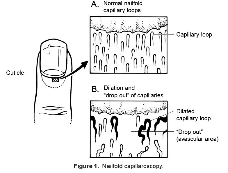
Visualizing the periungual boundary provides clinicians and investigators with qualitative and semi-quantitative insight into microvascular structure. When evaluating a typical nail fold capillary row, practitioners observe regular hairpin-like formations arranged in an orderly parallel array along the distal margin. Normal loops maintain consistent loop widths, uniform apical curves, and symmetrical intercapillary distances across the dermal papillae.
Structural evaluations systematically assess several primary morphological characteristics across the nail fold field:
- Capillary density, assessing the distribution and number of visible loops per millimeter along the distal row.
- Loop dimensions, documenting apical width, arterial diameter, and venous diameter.
- Morphological regularity, distinguishing standard hairpin formations from meandering, tortuous, or branching configurations.
- Microhemorrhages, recognizing extravasated erythrocytes situated around or above the distal capillary line.
- Perivascular background visibility, identifying subpapillary venous plexus patterns and tissue transparency.
Systematic assessment of these structural parameters establishes a standardized morphological profile across repeated examinations without relying on invasive tissue sampling.
Clinical Applications and Research Areas
The non-invasive character of microcirculatory assessment makes it a valuable observational modality across numerous medical disciplines. In rheumatology, examining microvascular architecture assists in distinguishing primary Raynaud phenomenon from secondary vascular manifestations linked to systemic autoimmune conditions. Research protocols investigating nailfold capillaroscopy scleroderma patterns evaluate early microvascular alterations, including giant capillary loops, avascular areas, and neoangiogenic bud clusters.
Dermatology clinics utilize nail capillriscopy to examine microcirculatory dynamics in localized dermatological conditions, psoriasis, chronic non-healing ulcers, and cutaneous inflammatory reactions. In vascular medicine and angiology, specialists evaluate peripheral perfusion patterns in patients presenting with chronic venous insufficiency, arterial occlusive disorders, and hypertensive microangiopathy. Medical school laboratories and microcirculation researchers also employ the technique to assess the biological impact of biomaterials, far-infrared textiles, and therapeutic interventions on peripheral capillary blood flow.
Observational Advantages
- Non-invasive procedure requiring no skin incision or intravenous access.
- Immediate, live dynamic imaging of capillary blood flow and vascular loops.
- Facilitates sequential monitoring of microvascular morphology over extended periods.
Methodological Boundaries
- Does not provide independent systemic diagnosis without comprehensive medical evaluation.
- Image quality remains sensitive to operator positioning and surface illumination control.
- Requires standardized physiological and environmental stabilization before observation.
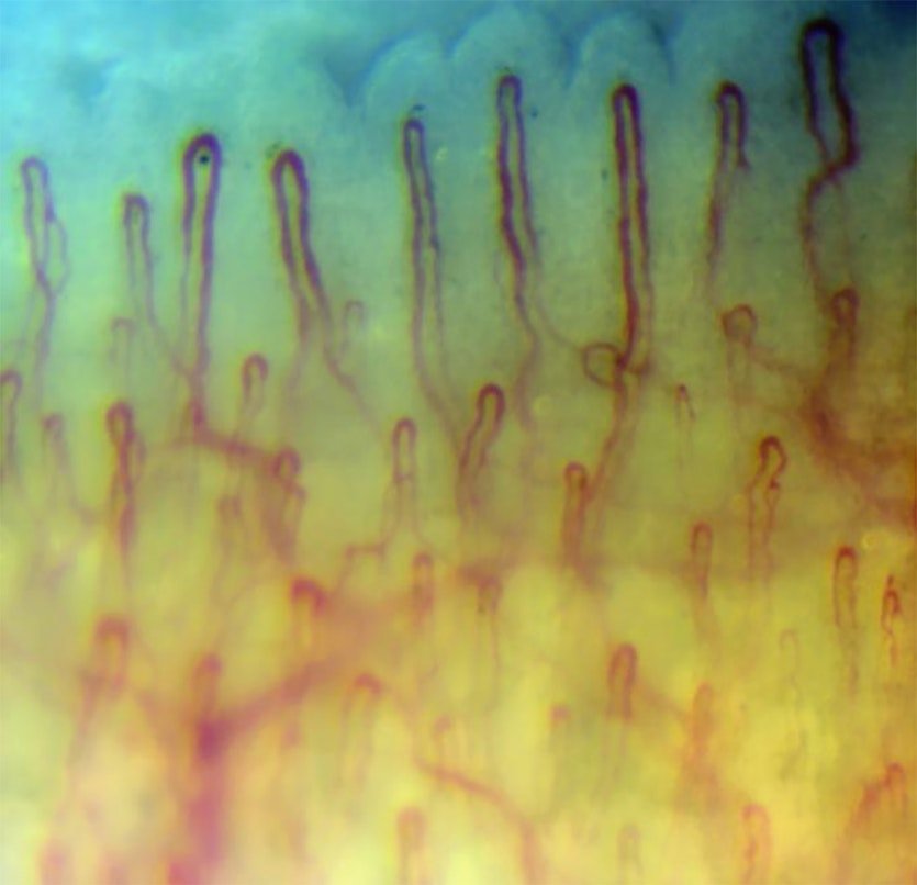
Specialists use microcirculatory imaging to document capillary density and structural patterns across longitudinal clinical studies.
Examination Conditions and Subject Preparation
Microvascular reactivity is highly responsive to ambient temperature, psychological stress, and external stimulants. Conducting an accurate nailfold capillary test demands strict standardization of ambient surroundings and subject preparation. Operating protocols specify that the examination room temperature must be maintained between 22 and 24 degrees Celsius, with relative humidity stabilized at approximately 70 percent to prevent temperature-induced peripheral vasoconstriction or reflex vasodilation.
Subjects should rest comfortably in a seated position for 15 to 30 minutes prior to the visual assessment. During this acclimatization period, the hands must be supported at approximately heart level to normalize hydrostatic pressure across the distal extremities. Subjects must avoid strenuous exercise or rigorous physical activity for one hour before observation, as exertion redistributes peripheral blood supply.
Exogenous factors including tobacco smoking, hot or cold hand washing, heavy meals, and specific vasoactive medications can alter capillary flow velocity; clinicians should record these factors alongside the subject’s menstrual status to maintain consistent observational context.
Step-by-Step Capillaroscopic Examination Protocol
Performing microvascular assessment requires systematic hardware setup and patient positioning. Utilizing a standardized workflow supports consistent image acquisition across clinical evaluations and research sessions. Practitioners working with nailfold capillary microscopy follow a defined sequence to establish optical focus and navigate the periungual field:
- Mount the dedicated LCD monitor securely onto the top frame of the mechanical housing and connect the power and video cables to the base connectors.
- Connect the primary electrical plug, verify the indicator light illuminates green, press the monitor power switch, and allow the screen to initialize over approximately three seconds.
- Apply two to three drops of optical coupling fluid—such as cedar oil, medical paraffin oil, baby oil, or olive oil—directly across the eponychium of the ring finger to enhance light transmission and minimize surface skin scattering.
- Rest the prepared finger gently inside the finger holder directly beneath the objective lens apparatus, maintaining neutral hand posture.
- Rotate the coaxial coarse handwheel to bring the optical assembly to approximately two to three centimeters from the finger surface until an initial microvascular silhouette appears on the display.
- Engage the fine focus handwheel to sharpen vessel boundaries, adjusting screen brightness and contrast controls to eliminate glare and enhance blood cell movement contrast.
- Manipulate the X-Y double layer compound mechanical stage horizontally and longitudinally to track along the distal capillary row, rotating the camera slightly left or right to clear display artifacts if surface reflections appear.
By using the smooth mechanical stage controls, an experienced operator can locate and align the distal capillary loops efficiently, providing a stable foundation for detailed morphological documentation.
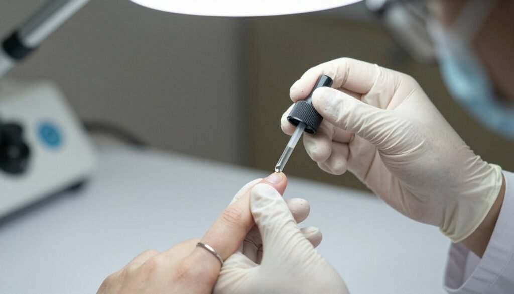
Applying a small amount of coupling oil reduces epidermal light scattering and reveals underlying microvascular loops.
Evaluating Capillaroscopy Systems
Healthcare facilities and academic institutions require robust, reliable hardware capable of repeatable daily operation. Selecting appropriate nailfold capillaroscopy equipment involves evaluating optical resolution, mechanical stage precision, illumination stability, and transportability. Modern systems range from desktop research workstations to compact transportable devices suitable for multi-room outpatient facilities.
When reviewing potential instruments, clinical departments assess specific structural and operational features:
Mechanical Stage Rigidity
A dual-layer compound X-Y mechanical stage provides smooth horizontal and longitudinal traversal across multiple fingers without introducing mechanical vibrations.
Coaxial Focus Control
Integrated coarse and fine coaxial handwheels allow operators to achieve rapid target acquisition followed by micrometer-level focal refinement on microvascular loops.
Integrated Display Architecture
Directly mounted color LCD screens display real-time video feeds without requiring external computer processing units, simplifying daily clinical examination spaces.
Reviewing these mechanical elements ensures that the selected nail fold capillariscopy hardware provides ergonomic comfort for the operator during extended clinical shifts.
Technical Specifications of the MK-GY880 Platform
Standardized diagnostic and research instrumentation demands strict adherence to defined electrical and optical parameters. The MAIKONG MK-GY880 system provides dedicated optoelectronic performance engineered for high-resolution microvascular assessment. Incorporating achromatization optics and steady DC power delivery, the nailfold capillaroscopy device maintains reliable video fidelity during prolonged observation intervals.
The following table outlines the stated operational specifications for the MK-GY880 model platform:
| Parameter | Stated Specification |
| Brand Name | MAIKONG |
| Model Designator | MK-GY880 |
| Intended Function | Detecting Body Health and Microcirculation |
| Amplification | 380X or 240X optical magnification with 380X, 500X, or 600X optional settings |
| Electrical Input | AC input 100-240V 50/60Hz universal DC adaptation |
| Electrical Output | DC12V, 2A regulated output |
| Objective Lens | 5X objective with 25mm effective observation range |
| Objective Type | Achromatization optical glass assembly |
| Mechanical Stage | X-Y double layer compound mechanical translation stage |
| Focus Mechanism | Coaxial coarse and fine adjustment handwheel assembly |
| Internal Dimming | Adjustable LED cold light source projection |
| Equipment Type | Type I or Type B protection rating |
| System Power | 12V, 1A direct current power source |
| Built-in Camera | 80 000 pixels high-sensitivity CCD image pickup |
| Monitor Screen | 8-inch standard, with 9-inch, 10-inch, or 12-inch color LCD options |
| Certification | CE approved conformity standard |
| Net Weight | 6KG base platform weight |
| Gross Weight | 7KG total packaged shipment weight |
Supplied materials reference 380X or 240X optical magnification and separately list 380X, 500X, or 600X as optional amplification configurations. These values represent distinct optical and digital magnification options tailored to specific clinical imaging objectives rather than a single fixed specification.
Packaging and System Components
Safe transportation of sensitive optoelectronic equipment requires engineered shock absorption and secure containment. The MK-GY880 system ships housed within a specialized aluminum password security case lined with custom-molded high-density memory foam. This internal protective enclosure is cushioned inside an exterior corrugated shipping box designed to withstand international transit handling.
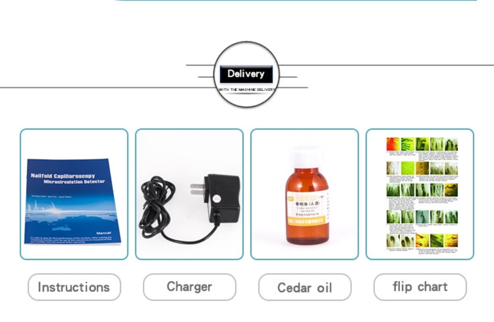
Each standard packaging configuration contains essential peripheral accessories required to support immediate operational deployment:
| Component Item | Quantity / Specification |
| MK-GY880 Host | 1pc optical main unit with integrated mechanics |
| LCD Monitor | 1pc dedicated color display monitor |
| Power Adapter | 1pc regulated power conversion module |
| Cedar Oil | 1pc specialized optical coupling oil bottle |
| Cotton Swabs | 1pc hygienic applicator packet |
| Reference Chart | 1pc microvascular observation reference sheet |
| Packing Dimensions | 37.5*26.5*34cm outer package dimensions |
| Inner Case | Aluminum password security case with memory foam |
| Outer Package | Reinforced multi-wall corrugated container box |
The integrated structural elements of the nailfold capillaroscopy imaging system include an 8-inch color display, imported CCD chipset, fine adjustment controls, coarse handwheels, horizontal and longitudinal stage movers, a stabilized finger press, and an image point rotational adjustment module designed to clear display spots.

The aluminum transit case protects optical alignment and sensor components during multi-clinic transportation.
Diverse Anatomical Observation Sites
While the fourth fingernail bed remains the primary site for microcirculatory evaluation, specialized optical microscopy extends to various peripheral cutaneous and mucosal zones. Different microcirculatory beds exhibit distinct vascular layouts, basement membrane densities, and flow speeds. Evaluating multiple microvascular zones broadens comparative microvascular research protocols across laboratory studies.
Practitioners conducting physiological and experimental microvascular assessments may observe several anatomical regions:
- Distal periungual nail fold boundary, offering horizontal capillary visibility along the cuticle line.
- Labial mucosal microcirculation, displaying dense, looped capillary networks across the inner lip mucosa.
- Lingual microcirculation, visualizing mucosal papillae and capillary flow across the tongue surface.
- Distal auricular microcirculation, monitoring microvascular caliber and velocity across the outer ear margin.
- Cutaneous sweat gland and hair follicle microvessels, examining localized metabolic and dermal microcirculation.
- Textile and mineral functional testing, assessing peripheral microcirculatory response to bamboo charcoal, titanium germanium, or far-infrared materials.
Examining these alternative biological zones using nail-fold capillography expands operational versatility across both medical clinics and biomedical engineering departments.
Methodological Boundaries and Interpretive Disclaimers
Nail fold capillaroscopic visualization represents an observational, non-interventional assessment method. It enables visual tracking of capillary shapes, microhemorrhages, and red blood cell aggregate motion. However, microcirculatory imaging does not constitute a standalone diagnostic conclusion or curative medical procedure.
Capillary architecture exhibits normal biological variations based on age, occupational hand trauma, skin pigmentation, and chronic exposure to manual labor. Furthermore, temporary physiological fluctuations can alter flow velocity without indicating underlying vascular pathology. Formal clinical interpretation must remain within the scope of qualified medical practitioners, who integrate capillaroscopic findings with comprehensive medical histories, laboratory serology, and physical examinations.
Capillaroscopy provides visual documentation of peripheral microvascular morphology; clinical conclusions require qualified medical oversight and comprehensive diagnostic correlation.
Visual capillaroscopic data serves as an observational adjunct alongside comprehensive clinical diagnostic evaluations.
MAIKONG Manufacturing and Supply Solutions
MAIKONG operates advanced optoelectronic manufacturing facilities based in SZ, GD, providing established precision supply chains for international healthcare institutions, university laboratories, and commercial distributors. Combining modern optical machining with stringent component screening, MAIKONG delivers stable, high-value microvascular imaging platforms engineered for intensive clinical utility. Our production facility manufactures both integrated desktop workstations and lightweight portable observation units.
As an established nailfold capillaroscopy manufacturer, MAIKONG supports complete Original Equipment Manufacturer (OEM) and private-label customization services. Commercial partners can specify monitor dimensions from 8 to 12 inches, optical magnification stages up to 600X, custom branding plates, software interfaces, and tailored packaging configurations. Healthcare equipment wholesalers, hospital purchasing departments, and specialty diagnostic suppliers benefit from direct factory supply, standardized production quality, and rapid international shipment handling.
Establish Local Distribution and Clinical Supply
Organizations expanding their diagnostic equipment portfolios or upgrading institutional laboratory capabilities can explore tailored system configurations and volume procurement options.
Frequently Asked Questions
What is nail fold capillaroscopy?
Nail fold capillaroscopy is a non-invasive optical examination method used to observe dermal microvascular loops situated parallel to the surface along the periungual fingernail cuticle. It provides direct visualization of capillary morphology, loop density, and dynamic blood flow patterns without puncturing the skin.
Does a microcirculation examination cause any pain or physical discomfort?
The procedure is entirely non-invasive and painless for the subject. The finger rests gently inside an optical positioning cradle while cold LED lighting illuminates the skin surface through a drop of topical coupling oil.
Why is coupling oil necessary during the capillaroscopic procedure?
Coupling oil, such as cedar oil or medical paraffin oil, fills microscopic irregularities across the stratum corneum. This eliminates surface light refraction and skin scattering, allowing incident light to penetrate directly into the horizontal capillary loops below.
How does capillaroscopy differ from standard laboratory light microscopy?
Standard laboratory light microscopy requires transmitting light through ultra-thin, sliced tissue specimens mounted onto transparent glass slides. Capillaroscopy utilizes incident episcopic cold illumination to observe living microvascular structures directly on intact, living cutaneous tissue.
What is the difference between 240X, 380X, and 600X amplification settings?
Lower magnification values like 240X provide a wider field of view to evaluate overall capillary row density and interpapillary spacing. Higher settings such as 380X or optional 600X amplification enable detailed morphological inspection of individual capillary apex dimensions, endothelial outlines, and erythrocyte flow velocity.
Can nail-foldcapillography confirm a specific medical diagnosis independently?
nail-foldcapillography provides valuable morphological and qualitative microcirculatory data, but it does not constitute an independent medical diagnosis. Clinical practitioners evaluate capillaroscopic findings alongside medical histories, physical examinations, and dedicated laboratory testing.
Summary of Capillaroscopic Visualization
Nail fold capillaroscopy delivers an essential, non-invasive optical window into the living human microcirculation by resolving horizontally aligned dermal capillaries. Implementing standardized environmental conditions, proper optical coupling, and stable magnification hardware enables clinicians and researchers to monitor microvascular morphology with consistent clarity. Integrating precise optoelectronic instrumentation into diagnostic departments continues to advance non-invasive microvascular evaluation across clinical and laboratory settings.