
Nail Capillary Morphology and Microvascular Assessment
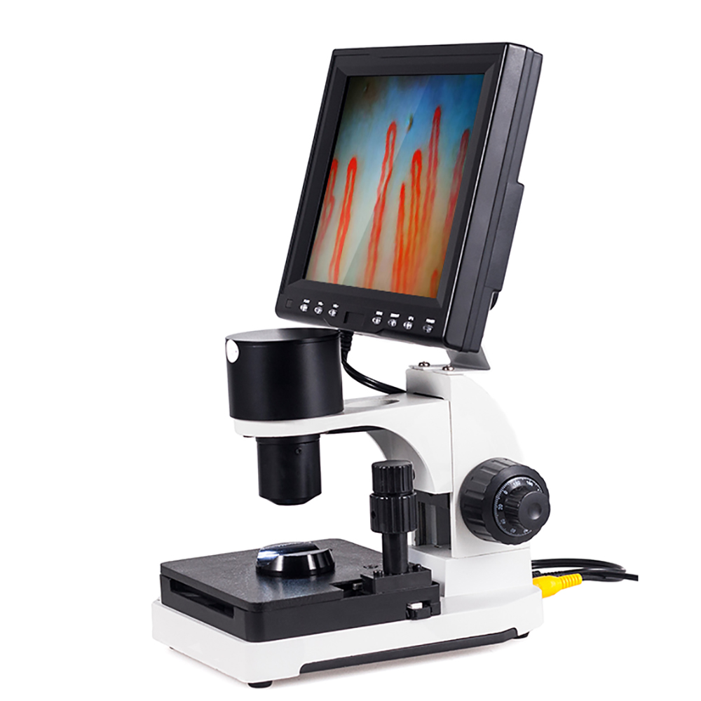
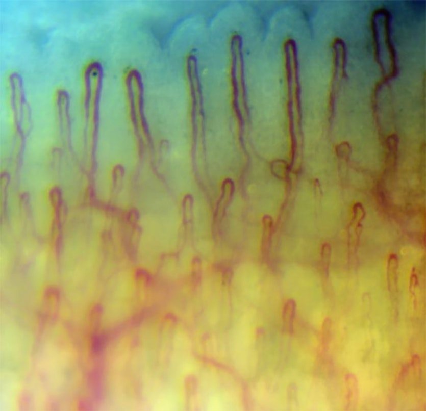
Nail capillary morphology describes the microscopic shape, layout, and structural dimensions of capillary loops running parallel to the skin surface in the periungual dermal papillae. In a baseline physiological state, these microvessels form neat, hairpin-like loops with an afferent arterial limb, an apical transition turn, and an efferent venous limb aligned along the distal row of the nailfold. Observing these loops non-invasively provides direct visual insight into human microcirculation without requiring tissue biopsy or ionizing radiation.
The clarity and diagnostic value of any microvascular evaluation depend heavily on standardizing optical magnification, subject preparation, ambient temperature, and contact fluid interfaces. Because microvessels respond dynamically to mechanical stress, thermal fluctuations, and systemic connective changes, clinicians and researchers rely on systematic descriptive classifications to distinguish natural structural variations from pathological microangiopathy.
Baseline Microvascular Loop Architecture
Understanding baseline anatomical geometry is the prerequisite for recognizing abnormal microvascular formations during capillary evaluation. In the distal nailbed, terminal capillaries emerge from horizontal dermal plexuses and orient horizontally relative to the epidermal plane, making their entire longitudinal profile visible from the dorsal aspect.
- Afferent Limb: The arterial portion delivers oxygenated blood from the dermal plexus, displaying a narrower lumen diameter that typically ranges from six to ten micrometers.
- Apical Turn: The apex connects the inflow and outflow limbs in a smooth hairpin curve, exhibiting an average external diameter below fifteen micrometers under baseline physiological conditions.
- Efferent Limb: The venous segment drains microcirculatory volume toward the superficial subpapillary plexus, exhibiting a slightly wider lumen than the corresponding afferent limb.
- Extracellular Background: The surrounding dermal papilla presents a translucent, pale pink matrix free from extravasated red blood cells or visible hemosiderin accumulation.
- Parallel Orientation: Normal terminal loops run roughly parallel to each other and perpendicular to the distal edge of the nail plate across every finger.
When assessing nail fold capillary architecture across healthy populations, subtle inter-individual variations remain frequent. Mild winding or crossing of limbs often represents benign tortuosity rather than structural degeneration, provided overall capillary density and loop width remain preserved across the distal margin.
Observable Features in Capillary Evaluation
Systematic assessment demands separating observable morphological characteristics into distinct parameters rather than relying on subjective impression. Clinicians and researchers evaluate structural integrity across multiple standardized metrics along the distal row of the nailfold.
Spatial Distribution
Healthy margins display regular capillary spacing with a density between seven and twelve loops per linear millimeter across the terminal perimeter.
Loop Dimensions
Limb diameters remain uniform along their length, with apical apex width staying proportionate to the supporting afferent and efferent branches.
Luminal Continuity
Intravascular erythrocyte flow produces a continuous, identifiable column without persistent avascular gaps, focal microthrombi, or vascular wall breaches.
Documenting these visual parameters across consecutive fields of view prevents local trauma artifacts from skewing the overall impression. Evaluating multiple digits also allows examiners to verify whether observed features represent generalized microcirculatory patterns or localized physical variations.
Distinguishing Morphological Alterations
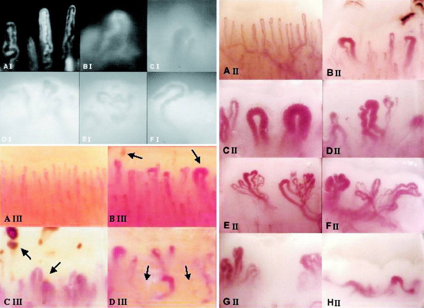
Pathological processes involving endothelial damage, ischemia, or connective tissue remodeling manifest through identifiable structural deviations from normal hairpin architecture. Identifying these variations requires understanding how vessel caliber, arborization, and permeability change under microvascular stress.
Benign Morphological Variations
- Slight tortuosity where loop limbs cross once or twice without widening.
- Homogeneous loop heights that follow the natural contour of dermal ridges.
- Intact capillary density across the entire distal margin of the nail plate.
- Absence of microhemorrhages, thrombosis, or focal tissue necrosis.
Specific Microvascular Alterations
- Ectasias and giant capillary loops with apical diameters exceeding normal limits.
- Avascular zones characterized by complete loss of adjacent capillary loops.
- Neovascular ramifications presenting complex, bushy, or branching loop trees.
- Perivascular microhemorrhages resulting from red cell extravasation into tissue.
While pronounced architectural disruption frequently accompanies autoimmune connective tissue disorders, optical findings require careful contextualization within the broader clinical evaluation. Visual assessment identifies morphological changes but does not replace laboratory, serological, or histological investigations.
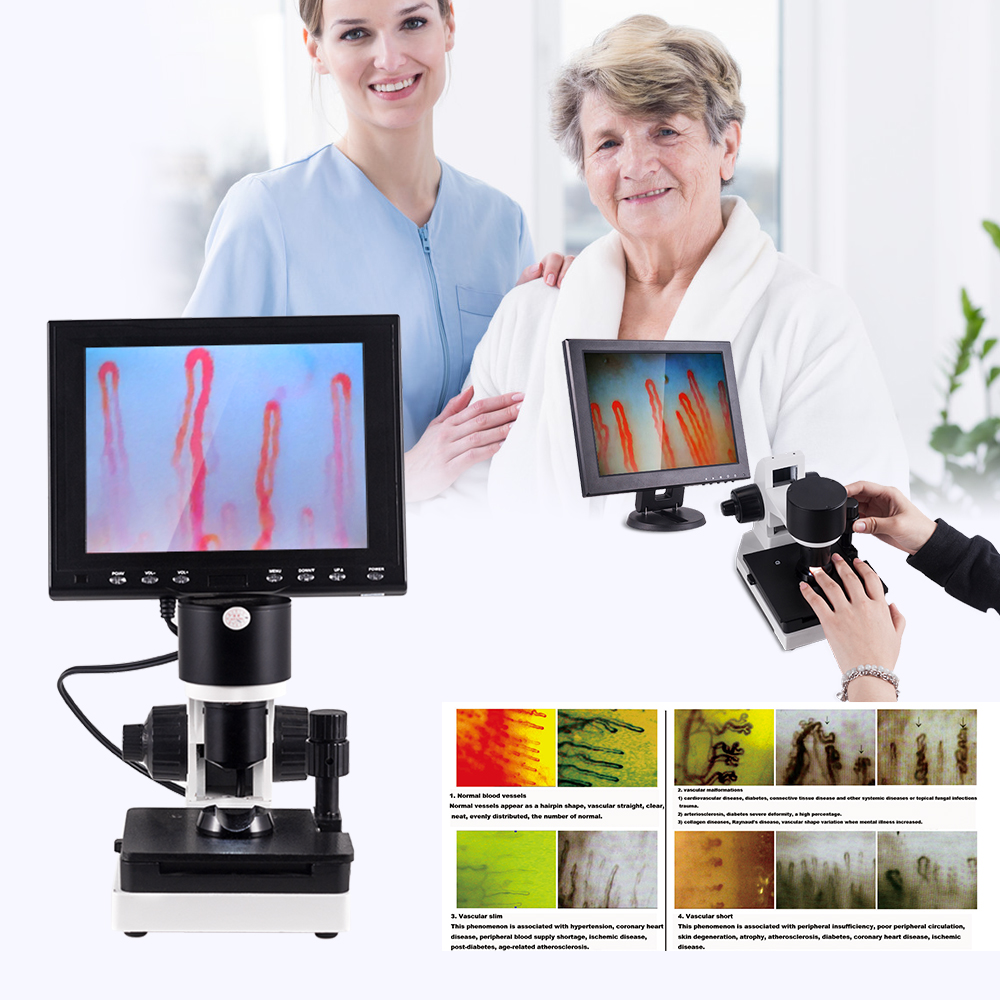
Precision coaxial stage controls help maintain stable focus during detailed morphological evaluations.
Step-by-Step Nailfold Examination Protocol
Acquiring dependable, repeatable morphological data requires adhering to a rigorous preparation and positioning protocol. Minimizing external vasoactive and mechanical disturbances supports accurate visualization of terminal microvessels.
- Acclimate the subject in a temperature-controlled room maintained between 22°C and 24°C with stable relative humidity near 70% before testing begins.
- Instruct the subject to rest comfortably in a seated posture for fifteen to thirty minutes, positioning hands at heart level to stabilize venous return.
- Apply a thin layer of immersion oil, such as high-grade cedar oil or pharmaceutical paraffin oil, over the periungual skin fold to reduce optical scattering and improve light transmission.
- Position the selected finger into the ergonomic finger rest beneath the optical objective, aligning the distal margin with the focal plane.
- Engage the coaxial coarse focus mechanism to locate the primary microvascular layer, followed by fine focus adjustments to resolve vessel borders.
- Use the double-layer compound mechanical stage to systematically scan the distal vascular row from the lateral to medial border.
Standardized workflows are essential when conducting a formal nailfold capillary test across clinical or research cohorts. Maintaining identical positioning, illumination angles, and rest periods reduces observational variability between different examination sessions.
Optical Mechanics and Magnification Systems
Visualizing microvessels measuring under twenty micrometers demands specialized optoelectronic instrumentation tailored for surface dermal microscopy. Standard clinical dermatoscopes often lack the resolving power and focal stability needed to inspect fine capillary walls.
High-Magnification Optical Advantages
- Optical configurations of 240X, 380X, or higher clearly reveal afferent, apical, and efferent limb boundaries.
- Coaxial LED cold light projection prevents tissue warming and temperature-induced vasodilation during prolonged viewing.
- Direct digital signal routing to high-resolution LCD displays supports live microcirculatory flow evaluation.
Operational Considerations
- Higher optical power narrows the depth of field, requiring continuous fine handwheel adjustments.
- Involuntary subject motion or minor finger twitching can momentarily shift capillaries out of the observation frame.
- Immersion oil viscosity and volume must be carefully balanced to prevent optical distortion and reflections.
Modern nail videocapillaroscopy platforms integrate sensitive charge-coupled device sensors with calibrated optical assemblies. These setups provide real-time, distortion-free video feeds that make assessing intravascular transit dynamics straightforward and repeatable.
High-resolution monitors display dynamic erythrocyte flow and structural loop boundaries in real time.
Technical Specifications for Imaging Instrumentation
Research facilities, teaching hospitals, and procurement managers require precise hardware parameters when selecting specialized microvascular imaging devices. At MAIKONG, our engineering team manufactures dedicated optical platforms in GD, SZ, China, combining robust mechanical stages with sensitive sensor arrays.
| Parameter | Specification | Engineering Details |
| Brand Name | MAIKONG | Manufactured under stringent quality control standards. |
| Model Series | MK-GY880 | Standard platform for non-invasive microcirculation observation. |
| Amplification | 380X Optional | Factory configurations support 240X, 380X, 500X, or 600X optics. |
| Input Power | AC 100-240V | Universal power supply compatible with international standards. |
| Output Power | DC 12V 2A | Dedicated regulated direct-current output for stage stability. |
| Objective Lens | 5X Achromatic | Delivers an effective working range of 25 millimeters. |
| Mechanical Stage | X-Y Compound | Double-layer compound platform for smooth horizontal motion. |
| Focus System | Coaxial Control | Coaxial coarse and fine handwheels ensure micro-positioning. |
| Illumination | LED Cold Light | Built-in cold light prevents localized skin warming. |
| Camera Sensor | Sony CCD Array | High-sensitivity sensor captures continuous video streams. |
| Display Panel | 8 to 12 Inch | Integrated color LCD panel displays direct video feed. |
| System Weight | Net 6KG | Stable base platform reduces table-borne vibration during use. |
| Dimensions | 37.5×26.5x34cm | Standard packing dimension for clinical benchtop deployment. |
Selecting appropriate nailfold capillaroscopy equipment involves verifying both stage stability and sensor responsiveness. Accurate documentation of fine morphological patterns requires optical components that eliminate geometric distortion across the entire field of view.
System Accessories and Packaging Standards
Standardized diagnostic and research hardware requires comprehensive accessory packages to support seamless deployment across clinical and academic settings. Complete equipment suites ship with protective flight cases and essential contact media.
| Component Item | Standard Quantity | Functional Description |
| MK-GY880 Host | 1 pc | Main optoelectronic assembly with integrated optical tube. |
| LCD Monitor | 1 pc | High-contrast dedicated color monitor for microvascular review. |
| Power Adapter | 1 pc | Medical-grade AC to DC converter with country-specific cord. |
| Cedar Oil | 1 pc | Optical contact medium optimized for dermal light transmission. |
| Cotton Swabs | 1 pc | Lint-free applicators for clean immersion oil distribution. |
| Reference Chart | 1 pc | Morphological comparison chart for standard microcirculatory forms. |
| Inner Case | 1 pc | Reinforced aluminum password box fitted with custom memory foam. |
| Outer Crate | 1 pc | Heavy-duty corrugated transit carton for international freight. |
Carefully fitted interior foam cutouts protect delicate achromatized optics and electronic alignment wheels during long-distance shipping. Before finalizing purchases, facility buyers should confirm customized display sizes and localized power plugs directly with the MAIKONG technical team.
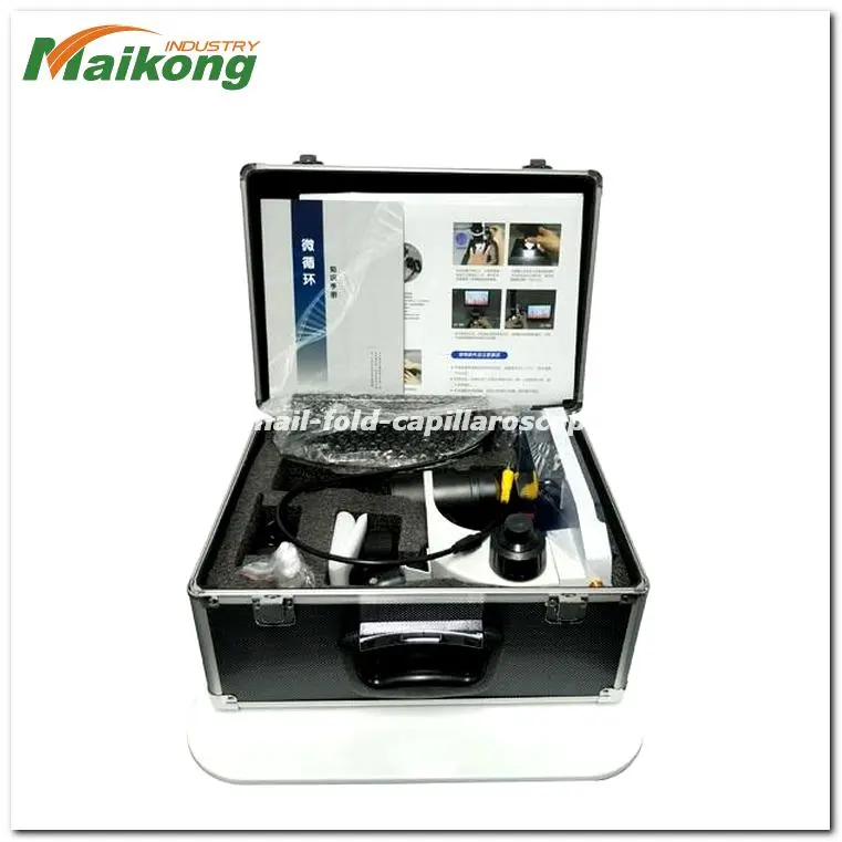

Protective aluminum flight cases with tailored foam inserts safeguard optical alignments during transport.
Standardized Morphological Documentation Protocol
Maintaining reproducible image registries across longitudinal studies requires strict adherence to uniform image capture parameters. Without consistent documentation, comparing microvascular changes over time becomes vulnerable to technical bias.
- Subject Information: Record ambient room temperature, resting time, and recent exposure to environmental cold before saving images.
- Anatomical Landmark: Identify the exact digit evaluated, prioritizing the non-dominant fourth and fifth fingers where dermal transparency is highest.
- Optical Parameters: Document the exact optical magnification, light intensity setting, and contact medium used during the session.
- Linear Scale Calibration: Include a calibrated millimeter reticle or digital micrometer overlay across captured stills to verify loop density.
- Quality Flags: Note local cuticular thickening, recent mechanical manicuring, or subungual hematomas that could obscure visibility.
Adhering to rigorous capture standards during nail capillary microscopy protects data integrity in multi-center clinical trials. Standardized image archives enable blind scoring by independent investigators without systematic bias.
Environmental and Physiological Confounding Factors
Visualizing microvessels requires recognizing that external factors frequently alter blood flow velocity and lumen dimensions. Misinterpreting transient physiological responses as structural pathology undermines assessment accuracy.
Low ambient temperatures stimulate immediate peripheral alpha-adrenergic vasospasm, constricting terminal capillary loops and reducing visible luminal volume. Subjects must acclimate in a warm, draft-free room before evaluation begins.
Mechanical cuticle manipulation, occupational hand trauma, and recent chemical exposure also cause localized tissue damage. Extravasated red blood cells resulting from microtrauma can resemble pathological microhemorrhages, which highlights why assessing multiple digits remains essential.
Furthermore, systemic caffeine intake, nicotine consumption, or vasoactive pharmacological agents introduce notable changes in erythrocyte transit speeds. Research protocols should record all relevant pre-examination exposures to support sound clinical and scientific interpretations.

Consistent room conditions and steady hand positioning reduce external physiological artifacts.
Scope of Microcirculatory Observation Across Disciplines
While the distal nailfold remains the primary site for microvascular assessment due to horizontal vessel orientation, surface microcirculation microscopy extends into diverse investigative contexts. Researchers explore microcirculatory responsiveness across multiple mucosal and cutaneous surfaces.
Specialized optical platforms enable comparative microcirculatory assessments across sublingual mucosa, lip margins, auricular tissue, and cutaneous wound beds to evaluate localized microvascular architecture.
In addition to classical physiological evaluations, academic engineering departments utilize microvascular imaging to study peripheral responses to novel functional textiles. Testing cutaneous microvascular responses to far-infrared materials, thermal garments, and specialized wellness fabrics offers non-invasive insights into surface microcirculatory dynamics.
Within broader rheumatologic and vascular research, standardized nail fold capillaroscopy remains a foundational methodology. Correlating detailed morphological patterns with serological biomarkers continues to clarify early endothelial changes in systemic vascular research.
Clinical and Research Procurement Considerations
Selecting optical instrumentation for hospital departments, specialty clinics, or university laboratories involves balancing imaging capabilities with daily operational practicality. Evaluating technical parameters helps procurement teams select hardware suited to their specific workflow demands.
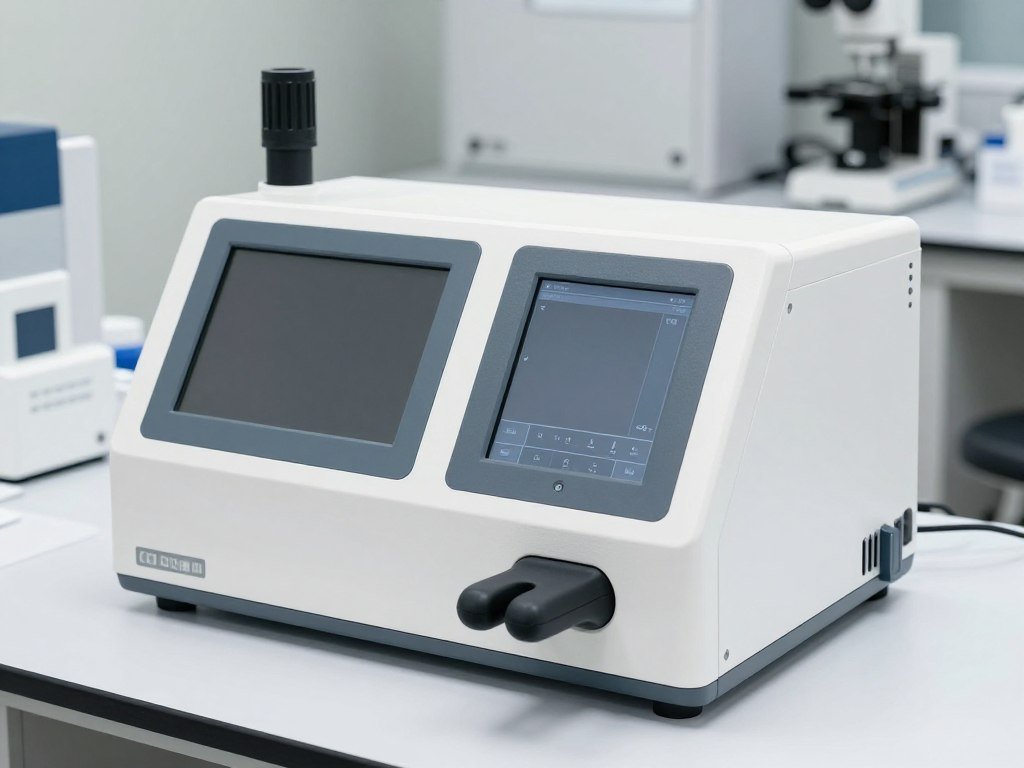
Mechanical Stage Stability
Precision X-Y double-layer compound mechanical stages eliminate jitter and provide steady, incremental finger tracking across consecutive nailfold papillae.

Digital Signal Fidelity
Sensitive CCD sensors capture real-time erythrocyte motion and fine apical contours without introducing motion blur or compression artifacts into recordings.
Facilities evaluating nailfold capillaroscopy equipment price structures must balance initial acquisition costs against optical durability, stage mechanics, and OEM customization services. Long-term reliability depends heavily on robust build quality, achromatized optical coatings, and accessible factory technical support.
Frequently Asked Questions About Nail Capillary Morphology
What defines normal nail capillary morphology?
Normal morphology presents as a regular row of hairpin-shaped microvascular loops aligned along the distal nailbed papillae. The apical turns show uniform dimensions without severe dilation, avascular disruptions, or extravasated red blood cells.
Why is the ring finger preferred for capillary observation?
The fourth finger typically features thinner epidermal keratinization and experiences less routine mechanical trauma than the thumb or index finger. This anatomical characteristic allows clearer optical light penetration and reveals undistorted microvascular architecture.
What is the difference between capillary tortuosity and arborization?
Tortuosity describes simple limb twisting or crossing where vessel caliber remains standard and the apical turn stays intact. Arborization refers to complex, branching neovascular tufts that often emerge following localized tissue ischemia.
Why is contact oil necessary during the examination?
Microscopic inspection of the stratum corneum causes substantial incident light reflection and scattering. Applying high-transparency cedar or paraffin oil harmonizes the optical refractive index, allowing illumination to pass directly through the upper dermis.
Can nail capillary morphology independently diagnose a disease?
Morphological observation provides descriptive visual data regarding microvascular structure but does not independently establish a definitive clinical diagnosis. Qualified medical professionals interpret capillary patterns alongside patient history, physical findings, and specific laboratory assays.
Summary and Professional Supply Inquiries
Accurate assessment of nail capillary morphology demands high-resolution optical magnification, stable stage mechanics, and disciplined examination protocols to distinguish benign variations from structural microangiopathy. Standardizing ambient conditions and documentation techniques ensures that visual records remain reproducible across longitudinal clinical and scientific studies.
Equip Your Laboratory or Clinical Practice
Connect with the engineering and international supply team at MAIKONG in GD, SZ, China to discuss specialized instrumentation, custom branding, wholesale procurement, or regional distributor partnerships.