
Nail Bed Capillaroscopy: Clinical Principles, Microvascular Observation, and Technical Systems
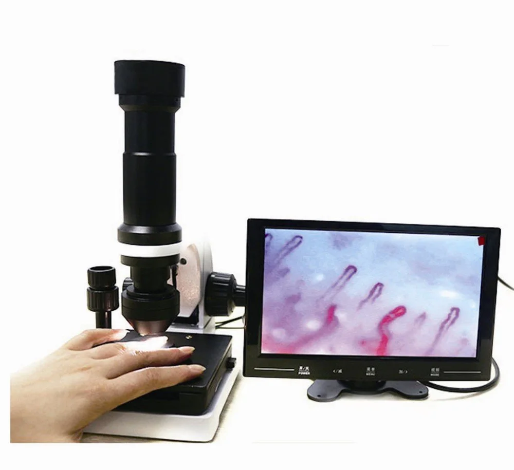
Nail bed capillaroscopy is a specialized, non-invasive optical technique designed to examine the microcirculation of the human peripheral vascular bed. By illuminating the skin folds adjacent to the fingernails under magnification, medical specialists and researchers directly visualize microvascular morphology, capillary loop integrity, and red blood cell velocity. The method provides an accessible window into structural microvascular adaptations without requiring tissue excision or dye injections.
Selecting the appropriate technical approach depends on clinical workflow demands, image documentation standards, and required optical resolution. Modern evaluation systems range from compact optical units to high-definition digital microscopy setups designed for specialized laboratory and clinical environments. Careful procedural standardization and environmental control establish the baseline conditions needed for reliable morphological observation.
Principles and Fundamentals of Nail Bed Capillaroscopy
Nail bed capillaroscopy relies on direct surface epi-illumination combined with compound optical or digital magnification to inspect terminal nutritional capillary loops. Incident light directed onto the periungual cuticular margin penetrates superficial epithelial layers when refractive mismatches are minimized with an immersion medium. The resulting optical contrast reveals the columns of moving erythrocytes contained within the delicate endothelial walls of each dermal microvessel.
Unlike standard dermal surfaces where capillary loops ascend perpendicular to the skin surface, the cuticular border presents microvessels oriented parallel to the epidermal plane. This anatomical arrangement permits horizontal visualization of the entire capillary architecture, including the afferent arteriolar limb, apical transition loop, and efferent venular limb. Clinicians and researchers assess vessel density, luminal dimension, and architectural regularity directly from this horizontal viewpoint.
Epi-illumination through transparent immersion media allows direct horizontal inspection of nutritional microvessels without disturbing subcutaneous tissue integrity. Adjusting the angle of the cold light source improves border distinction along the terminal vascular arches.
Direct surface microscopy allows detailed evaluation of peripheral vascular beds under stable optical magnification.
Dynamic assessment also captures qualitative features of intravascular flow, such as plasma gaps, sluggish erythrocyte movement, or cellular aggregation. Observing these variables under a standardized nailfold capillaroscopy device provides reproducible morphological documentation across longitudinal observational intervals. Optical stability remains central to avoiding motion artifacts during live capture.
Microvascular Morphology and Observation Parameters
Evaluating peripheral microcirculation involves systematic documentation of specific morphological variables across multiple digits. Experienced observers analyze terminal loop distribution along the distal row of the nailfold, recording structural alterations against documented physiological ranges. Distortions in loop symmetry, lumen caliber, and local distribution serve as descriptive endpoints during observational studies.
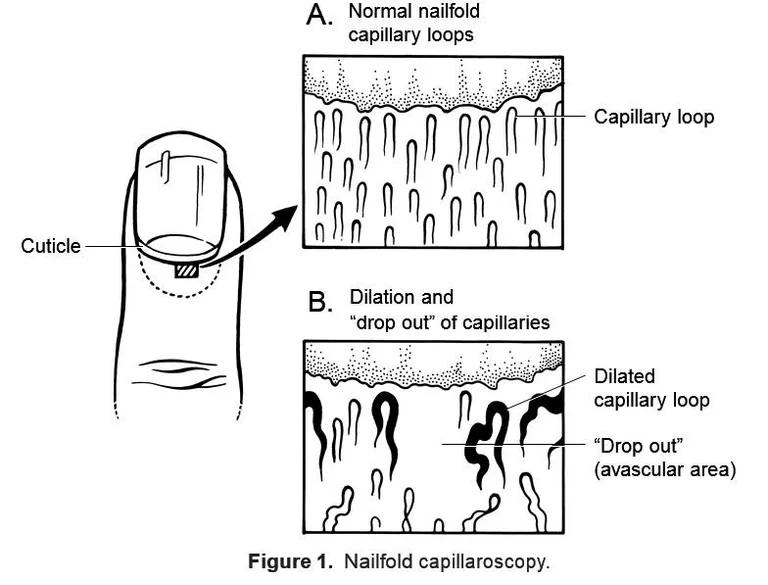
- Capillary Loop Density: The number of distinct vascular loops per linear millimeter along the distal row, typically displaying an even, comb-like distribution across the cuticular ridge.
- Loop Dimensions and Caliber: Measurement of the afferent arterial limb, apical turn, and efferent venous limb, noting instances of localized ectasia or generalized enlargement.
- Morphological Shape Variations: Identification of normal hairpin configurations versus crossed loops, tortuous paths, meandering pathways, or arborized ramified clusters.
- Perivascular Features: Documentation of microhemorrhages, extravasated hemosiderin deposits, pericapillary edema, or prominent subpapillary venular plexuses.
- Blood Flow Characteristics: Visual inspection of continuous erythrocyte movement, granular flow patterns, intermittent stasis, or localized vessel thrombosis.
Documenting structural parameters within the nail fold capillary architecture assists specialists in differentiating non-specific microvascular variants from severe architectural derangements. Healthy subjects typically exhibit hairpin-shaped loops with uniform apex diameters and continuous distal alignment. Severe capillary loss, avascular fields, or abnormally dilated loops signify significant microvascular alterations that warrant formal diagnostic investigation using broader clinical criteria.
Anatomical Suitability of the Periungual Skin
The nailfold region possesses distinct anatomical characteristics that make it uniquely advantageous for in vivo microvascular observation. Skin covering the dorsal aspect of the distal phalanx transitions into a thin cuticular border, where the stratum corneum is relatively transparent. This minimal epidermal thickness allows light to penetrate into the upper papillary dermis with minimal scattering compared to other cutaneous sites.
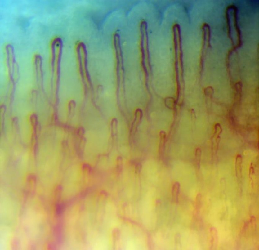
Parallel Vascular Alignment
Capillaries in the distal nailfold run parallel to the epidermal surface rather than rising vertically, providing a comprehensive horizontal view of the entire vascular loop. This structural layout enables simultaneous evaluation of both arteriolar and venular arms alongside the capillary apex.
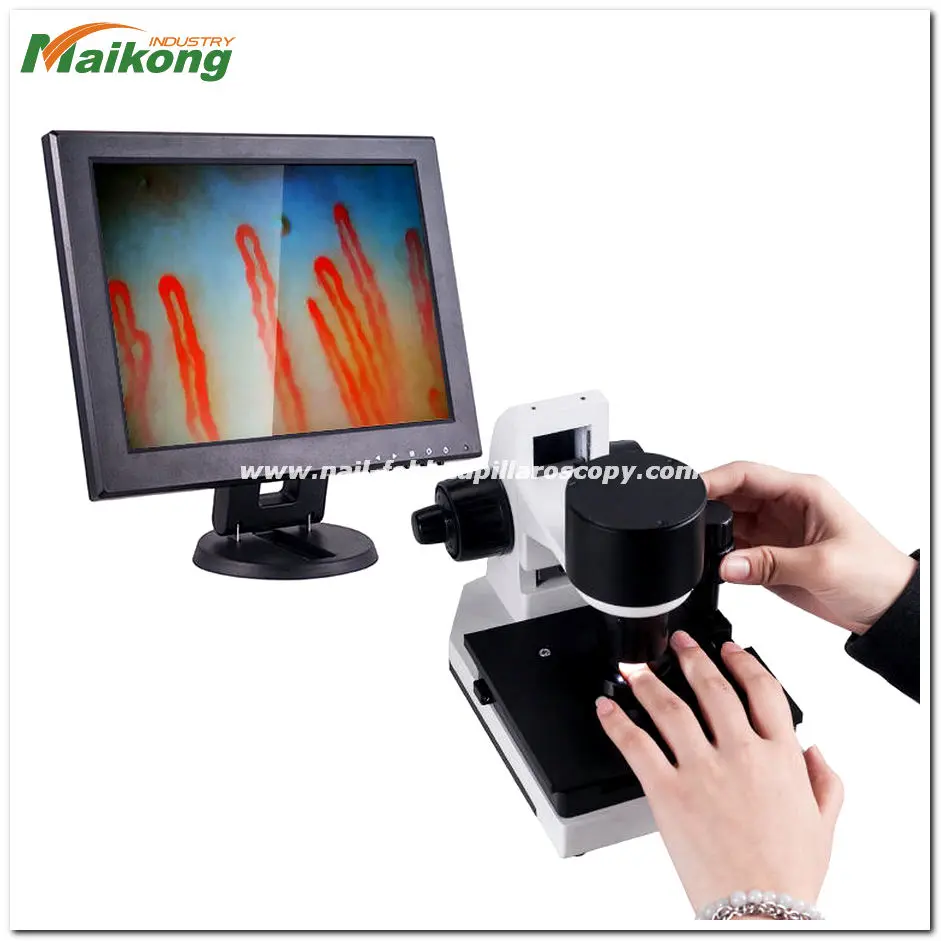
Stable Anatomical Support
The rigid underlying terminal phalanx and nail plate provide a solid foundation for physical stabilization during optical inspection. This stable bony backing minimizes pulsation displacement and involuntary tremor during detailed assessments using nail capillary microscopy systems.
While peripheral microcirculation can be inspected at other sites, including the labial mucosa or dorsal tongue, the periungual cuticular border remains the most practical and accessible site for clinical and research applications. Digits can be positioned securely within mechanical stage holders without subject discomfort. Furthermore, repetitive assessments across the same finger margins allow observers to track structural changes over extended experimental timelines.
Standardized Examination and Optical Workflow
Executing an accurate microvascular evaluation requires a methodical, step-by-step approach to optical alignment and subject positioning. Systematic examination workflows reduce measurement variability between operators and support high image clarity across digital archives. Clinicians typically examine the second, third, fourth, and fifth digits of both hands, frequently identifying the fourth digit as providing optimal cuticular transparency.
- Apply Immersion Medium: Place a drop of high-viscosity paraffin oil, cedar oil, or pharmaceutical-grade mineral oil directly onto the periungual cuticular margin to eliminate surface reflection and optimize light penetration.
- Position the Finger: Secure the selected digit in the finger-holding apparatus beneath the objective lens, adjusting the support stage so the cuticle rests perpendicular to the optical axis.
- Activate Illumination: Power on the cold LED light source and orient the projection beam at an oblique angle between 30 and 45 degrees to maximize vessel contrast against background dermis.
- Establish Coaxial Focus: Rotate the coarse focus adjustment handwheel to locate the cuticular ridge, followed by fine focus handwheel manipulation to resolve the red blood cell columns within individual capillary loops.
- Traverse the Distal Row: Translate the mechanical stage along its horizontal X-axis to evaluate the entire margin from lateral fold to medial fold, recording images or video segments at standardized points.
- Inspect Subpapillary Plexus: Adjust vertical plane focus slightly deeper into the dermal layer when observation of the subpapillary venular plexus is required by the study protocol.
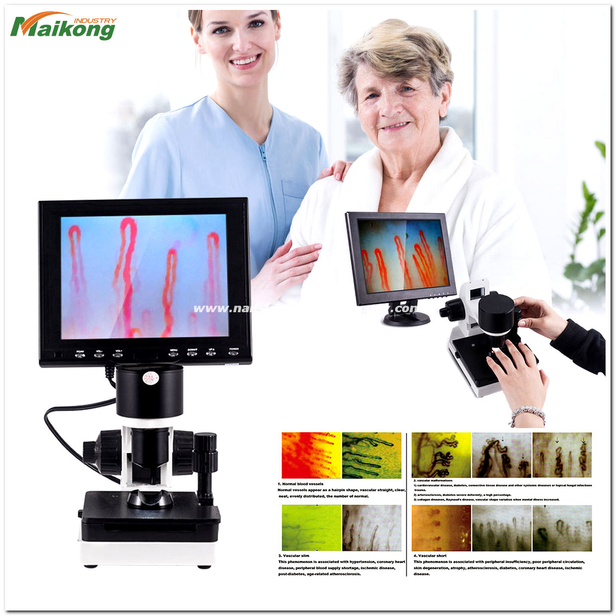
Coaxial focus mechanisms allow fine adjustments to resolve microvascular details within the superficial dermis.
Completing a comprehensive nailfold capillary test demands careful hand-wheel control to avoid contacting the lens against the oiled skin surface. When using advanced systems, digital capture cards transfer real-time video to monitor screens, enabling immediate evaluation of velocity fluctuations and microvascular dimensions. Cleaning the optical lens with alcohol-free lens tissue after each session prevents oil buildup and preserves optical clarity.
Environmental Controls and Subject Preparation
Peripheral microcirculation reacts dynamically to external temperature changes, emotional stress, and systemic autonomic stimuli. Without strict control over ambient parameters, transient vasoconstriction can obscure capillary loops, leading to erroneous interpretations of vessel density or blood flow velocity. Establishing uniform baseline conditions within the evaluation room is critical for institutional research and observational consistency.
Exposure to cold ambient air induces rapid peripheral vasoconstriction, collapsing superficial capillary loops and mimicking avascularity. Maintain room temperatures within specified ranges and allow subjects adequate thermal acclimation before initiating evaluation.
Prior to evaluation, subjects should rest comfortably in a seated position for approximately fifteen to thirty minutes within an acclimatized room. Institutional protocols often maintain ambient temperatures between 22 and 24 degrees Celsius with relative humidity held near 70 percent. Maintaining the subject’s hands at heart level prevents venous engorgement or hydrostatic pooling that can distort normal capillary calibers.
Behavioral factors require proactive coordination before performing a clinical nail fold capillaroscopy. Subjects should avoid smoking, consuming caffeine, performing heavy muscular exercise, or taking vasoactive compounds for at least one hour before assessment. In addition, cosmetic procedures such as manicuring, chemical cuticle removal, or artificial nail applications should be deferred, as recent mechanical trauma produces microhemorrhages that compromise morphological evaluations.
Research Inquiries and Clinical Assessment Areas
Non-invasive capillaroscopic visualization serves as a valuable qualitative and quantitative tool across several medical and physiological disciplines. While the presence of altered capillary architecture does not replace comprehensive serological, histological, or systemic evaluations, documenting microvascular status aids in characterizing systemic microangiopathies. Research teams utilize these visual metrics to explore microcirculatory responses to novel pharmacotherapies and lifestyle interventions.
Autoimmune Research
Specialists document microvascular disruptions in connective tissue studies, tracking the presence of giant enlarged loops, local capillary loss, and microhemorrhages in conditions related to nailfold capillaroscopy scleroderma research.
Vasospastic Phenomena
Clinicians evaluate baseline capillary dimensions in individuals presenting with cold-induced color changes in the extremities, distinguishing functional vasospastic responses from structural microangiopathies associated with systemic disease.
Dermatological Studies
Researchers observe local cuticular alterations, periungual erythema, and microvascular density variations associated with inflammatory skin conditions, localized psoriasis, and systemic lupus manifestations.
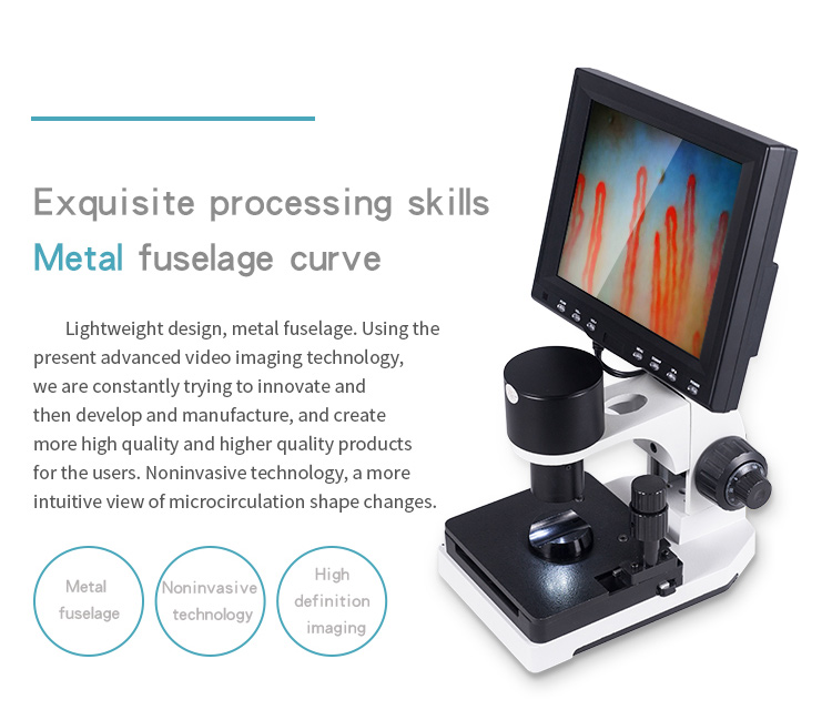
High-resolution display systems facilitate objective measurement of apical loop dimensions and vessel spacing.
Beyond rheumatology and dermatology, academic investigators use capillary microscopy to study metabolic diseases, including microvascular complications in diabetes mellitus and systemic hypertension. In sports medicine and hyperbaric research, capillaroscopic tracking documents changes in microvascular flow under varied oxygenation levels and physical exertion. Capillary imaging also supports ergonomic evaluations of vibration-induced white finger in occupational health environments.
Clinical Advantages and Practical Limitations
Understanding both the functional strengths and practical boundaries of microvascular imaging enables clinical practitioners to deploy equipment effectively. The procedure remains exceptionally safe and well-tolerated across diverse subject demographics, from pediatric patients to elderly cohorts. Because the technique involves no ionizing radiation, tissue puncture, or intravenous contrast agents, it can be repeated frequently without clinical risk.
Method Advantages
- Non-invasive procedure with no tissue incision or radiation exposure.
- Real-time visualization of live capillary blood flow dynamics and architecture.
- Repeatable across longitudinal study periods without subject discomfort.
- Cost-effective instrumentation compared to angiography or advanced magnetic resonance systems.
- Straightforward examination protocol suitable for outpatient clinics and dedicated laboratories.
Technical Limitations
- Limited penetration depth restricting observation to superficial papillary dermal layers.
- Heavy epidermal pigmentation or thickened hyperkeratotic cuticles can reduce optical clarity.
- Image interpretation remains operator-dependent without standardized quantitative software.
- Subject motion or tremor introduces blur during high-magnification video recording.
- Cannot serve as an isolated diagnostic confirmation without supporting clinical criteria.
When technical challenges arise, transitioning from basic optical magnifiers to advanced nail videocapillaroscopy helps overcome visual ambiguity. Digital video systems incorporate software stabilization and calibrated digital measurement grids that reduce subjective interpretation errors. Nevertheless, clinicians must always interpret visual capillaroscopic findings within the context of a thorough physical examination and complete laboratory profiles.
Comparative System Architectures and Modalities
Hardware systems designed for nailfold evaluation incorporate various optical pathways, digital sensors, and mechanical configurations. Equipment selection ranges from basic portable optical instruments to sophisticated, multi-axis video workstations designed for clinical trials and university research. Understanding these equipment categories ensures clinical facilities acquire systems that match their operational and physical requirements.
Optical and Portable Devices
- Utilize direct optical eyepieces, dermatoscopes, or ophthalmoscopes for rapid qualitative screening.
- Compact form factors and lightweight housings facilitate bedside or multi-room transport.
- Lower total magnification levels, typically ranging between 10X and 100X optical enlargement.
- Limited onboard image storage requiring supplementary external cameras for patient documentation.
- Well-suited for basic primary assessments, general wellness screening, and field research.
Digital Videocapillaroscopy Systems
- Equipped with high-sensitivity CCD or CMOS sensors providing real-time monitor display.
- Higher optical and digital magnifications, commonly operating between 200X and 600X.
- Integrated software modules allow frame capture, video archiving, and micrometric measurement.
- Precision mechanical stages with compound X-Y translation for smooth capillary tracking.
- Ideal for academic medical centers, clinical trials, and dedicated microcirculation departments.

Dedicated digital systems combine high-magnification optics with precision mechanical movement stages.
Modern practitioners frequently implement a digital nailfold capillaroscopy microscope to standardize diagnostic documentation. By presenting live video on a dedicated monitor, these systems allow multiple observers to analyze capillary patterns simultaneously while reducing eye fatigue during prolonged clinics. Stored digital sequences support comparative baseline matching during longitudinal patient follow-up.
Procurement Criteria for Clinical and Research Facilities
Procurement teams, department heads, and laboratory managers must evaluate several technical and structural criteria before purchasing microcirculatory imaging hardware. System stability, optical resolution, and long-term hardware reliability directly impact the usability of equipment in busy clinical environments. Investing in robust hardware with modular optical components ensures operational longevity across institutional applications.
Reviewing optical magnification ranges, sensor dynamic range, and mechanical stage stability before purchase ensures the hardware meets specific research or clinical workload requirements.
Key technical considerations during equipment evaluation include:
- Optical Magnification Range: Determine whether standard 200X to 380X magnification is sufficient or if higher 500X to 600X configurations are required for microvascular research.
- Illumination Architecture: Confirm the presence of an integrated cold LED light source to avoid delivering thermal energy to the subject’s fingertip, which alters vessel caliber.
- Mechanical Stage Quality: Verify the inclusion of a dual-layer compound X-Y mechanical stage with smooth coaxial coarse and fine focus controls for stable operation.
- Camera Sensor Architecture: Evaluate sensor resolution and dynamic range to ensure crisp frame capture without motion jitter or rolling-shutter distortion during live observation.
- Total Cost of Ownership: Compare overall acquisition costs by referencing the published nailfold capillaroscopy equipment price across compliant models and spare parts.
Procuring Clinical Microcirculation Hardware
Institutional buyers seeking certified optical and digital microscopy stations can review current hardware configurations and request formal commercial quotations.
MAIKONG Manufacturing Standards and Device Specifications
As a specialized medical optoelectronic manufacturer, MAIKONG designs and produces professional capillary microcirculation systems built to stringent international standards. Operating advanced production facilities in GD, SZ, China, we integrate precision optical engineering, stable mechanical manufacturing, and high-performance CCD imaging sensors. Our systems deliver clear, low-jitter real-time video to support precise microvascular evaluation across global healthcare facilities.
The flagship MK-GY880 workstation represents a complete optoelectronic solution for non-invasive microcirculation observation. Built with an achromatic optical system and a coaxial coarse and fine focus handwheel mechanism, it allows rapid focal acquisition within seconds. The following manufacturer-provided specifications outline the physical, electrical, and optical capabilities of the MK-GY880 system.
| Parameter | Component Type | Manufacturer-provided specification |
| Brand Name | Trademark | MAIKONG |
| Function | Application | Detecting Body Health |
| Amplification | Optical Range | 380X/500X/600X optional |
| Input | Electrical Input | AC input 100-240V50/60Hz DC |
| Output | Electrical Output | DC12V,2A |
| Objective | Lens Spec | 5X, effective range 25mm |
| Objective type | Optical Design | Achromatization |
| Mechanical stage | Sample Stage | X-Y double layer compound mechanical stage |
| Focusing mechanism | Focal Control | Coaxial coarse and fine adjustment |
| Inner dimming | Light Source | LED cold light |
| Type | Electrical Class | I or B |
| Power | Power Source | 12V,1A, DC power source |
| Built-in camera | Sensor Spec | 80 000 pixels |
| LCD monitor | Display Unit | 8inch/10inch /12inch/Color LCD monitor |
| Certificate | Compliance | CE approved |
| Net weight | Device Mass | 6KG |
| Gross weight | Shipping Mass | 7KG |
| Packing Dimfnsing | Carton Size | 37.5*26.5*34cm |
Each unit ships in dedicated multi-layer protective packaging engineered to prevent mechanical misalignment or lens damage during international air or ocean transit. The package integrates an interior aluminum lock box lined with custom memory foam, housed within a rugged corrugated outer shipping box. The standard delivery package includes all components necessary for immediate clinical deployment.
| Spare parts | Packaging Spec | Quantity |
| GY880 host | Main optical unit | 1pc |
| LCD monitor | Color display screen | 1pc |
| power adapter | Universal AC-DC cord | 1pc |
| cedar oil | Immersion oil bottle | 1pc |
| cotton swab | Application package | 1pc |
| chart | Morphology comparison sheet | 1pc |
Deploying certified nailfold capillaroscopy equipment manufactured by MAIKONG ensures dependable optical performance and access to complete replacement components across the hardware lifecycle. Facilities can configure their workstations with optional 8-inch, 10-inch, or 12-inch color LCD panels depending on room setup and spatial constraints.

Global Distribution and OEM Support Services
MAIKONG provides extensive manufacturing and supply chain support for medical equipment distributors, healthcare wholesalers, and private-label medical device brands. Leveraging our advanced production infrastructure in GD, SZ, we offer complete OEM customization services, including custom chassis branding, specialized firmware interfaces, and bespoke optical configurations. Our streamlined supply chain ensures prompt shipment schedules and consistent component availability.
- OEM and Private Label Manufacturing: Full custom branding on instrument chassis, protective aluminum cases, user manuals, and packaging containers.
- Custom Optical Configurations: Tailored magnification systems ranging from 240X to 600X optical and digital zoom modules to match client specifications.
- Wholesale Commercial Supply: Competitive volume pricing structures for regional medical device distributors and hospital procurement groups.
- Rigorous Quality Verification: Factory-level optical alignment and electrical safety verification backed by CE certification on all production batches.
- Comprehensive Spare Parts Supply: Continuous availability of replacement LCD monitors, LED illumination assemblies, mechanical stages, and immersion supplies.
We welcome inquiries from established distribution partners seeking to introduce high-reliability capillaroscopy systems into regional medical, academic, and clinical research markets. Organizations interested in learning more about our corporate background and operational standards can explore our About us page or review our manufacturing infrastructure on the Factory page.
Partner with MAIKONG for Regional Distribution
Contact our international sales and OEM engineering team to discuss wholesale pricing, distribution agreements, and customized private-label equipment manufacturing.
Frequently Asked Questions About Nail Bed Capillaroscopy
What is nail bed capillaroscopy?
Nail bed capillaroscopy is a non-invasive optical imaging technique used to directly examine the microscopic blood vessels in the skin fold at the base of the fingernail. It provides clear visualization of capillary loop structure, distribution, and blood flow patterns in living tissue.
Is nail bed capillaroscopy invasive?
Nail bed capillaroscopy is completely non-invasive and requires no needles, incisions, or dye injections. The examination involves only applying a drop of immersion oil to the cuticle and resting the finger beneath an optical lens.
What can be observed?
The procedure allows clinicians and researchers to observe capillary density, loop shape, apical diameter, and blood flow regularity. It also visualizes microvascular abnormalities such as enlarged loops, avascular areas, and microhemorrhages.
How is image quality affected by lighting, focus, oil, movement, or temperature?
Image quality depends on uniform cold LED illumination to prevent glare and thermal artifacts, precise coaxial focus for sharp edge definition, and immersion oil to eliminate surface light scattering. In addition, ambient temperatures between 22 and 24 degrees Celsius and stable finger positioning prevent vasoconstriction and motion blur.
Who commonly uses this type of imaging?
Rheumatologists, vascular medicine specialists, dermatologists, and academic researchers routinely utilize nail bed capillaroscopy for clinical evaluations and microcirculation studies. Medical educators and clinical research organizations also use it to monitor microvascular adaptations.
What should a buyer check before ordering equipment?
Buyers should evaluate optical magnification options, camera sensor resolution, mechanical stage stability, and illumination type. Verifying manufacturer certifications, packaging durability, and spare parts availability also protects institutional investments.
Can MAIKONG support OEM, private-label, or distribution projects?
MAIKONG provides comprehensive OEM and private-label manufacturing services from our production facilities in GD, SZ. We support custom branding, tailored optical configurations, and volume wholesale delivery for global distributors.
Practical Outlook on Microvascular Imaging
Direct in vivo assessment of the cuticular microcirculation provides clinicians and researchers with an objective, reproducible window into microvascular architecture. Maintaining rigorous environmental controls and utilizing high-stability optical systems ensures reliable structural documentation across clinical and experimental cohorts. Facilities seeking dependable capillaroscopy instrumentation benefit from robust manufacturing standards, modular optical configurations, and dedicated distributor support.
Explore Production Capabilities and Factory Standards
Discover MAIKONG manufacturing facilities, precision assembly processes, and comprehensive quality control protocols for specialized microcirculation equipment.