Lateral Nail Fold Function in Human Digital Anatomy
The lateral nail fold function centers on stabilizing the lateral margins of the nail plate, guiding directional keratin growth, and providing a sealed biological barrier against environmental pathogens and mechanical shear stress. Working alongside the proximal nail fold, these paired lateral tissue structures form the lateral nail grooves, which anchor the firm plate to the distal phalanx and preserve the sterile compartment of the underlying nail bed. In addition to physical shielding, the tissue fold maintains microvascular networks that support local dermal nutrition and peripheral temperature regulation across the distal digit.
Anatomical integrity decides the mechanical resilience of the entire perionychium. Variations in epidermal thickness, mechanical pressure, and localized hydration directly alter how effectively this tissue cushions the digit during tactile manipulation and physical contact.
Anatomical Structure of the Lateral Nail Fold
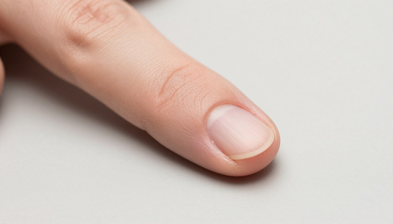
The lateral nail fold, anatomically referred to as the lateral perionychium or paronychium, represents the invaginated cutaneous border lining each side of the nail plate. At this interface, the keratinized epidermis curves inward to create the lateral nail groove or sulcus, providing a continuous protective transition between the dorsal digital skin and the adherent nail bed epithelium.
Histologically, the fold comprises stratified squamous epithelium, a dense papillary dermis, and a vascular-rich reticular dermis anchored to the periosteum of the terminal phalanx by specialized fibrous retinacula. This architecture prevents separation of the soft tissue from the hard keratin sheet during lateral twisting forces or routine grasping tasks.
Epidermal Boundary
The surface layer transitions into a non-cornified sulcular lining that maintains contact with the plate edge.
Dermal Matrix
Collagen bundles and elastic fibers provide mechanical tensile strength against lateral shearing.
Microvascular Bed
Capillary loops ascend toward the dermal papillae, supplying nutrients to adjacent non-vascularized structures.
Unlike the proximal fold, which houses the eponychium and covers the formative matrix, the lateral folds align parallel to the direction of outward growth. Their internal geometry reflects this longitudinal movement, allowing continuous forward displacement of the hardened plate without compromising edge seal integrity.
What Is the Function of the Lateral Nail Fold?
Understanding the primary functions of the tissue requires analyzing its protective, mechanical, and physiological properties across daily manual tasks. Each lateral border performs specific actions that preserve the structural stability of the distal extremity.
Directional Growth Guidance: The lateral nail grooves act as parallel anatomical tracks that channel the expanding keratinized plate forward toward the free edge, reducing aberrant lateral deviation.
Biological Barrier Defense: Dense intercellular junctions within the sulcular epithelium restrict pathogen ingress, protecting the germinative nail bed from bacterial and fungal colonizers.
Mechanical Cushioning: The fibrofatty padding absorbs direct lateral impact, dampening force transfer to the bone and preventing traumatic plate avulsion.
Vascular Support: Dense capillary loops within the lateral papillary dermis facilitate nutrient diffusion across the avascular lateral plate borders.
When the lateral nail fold function is compromised through mechanical trauma or excessive maceration, directional stability diminishes. Intact folds help maintain a physical seal that defends the nail bed from microbial intrusion and moisture loss, preserving the physiological equilibrium of the subungual microenvironment.
Relationship with the Nail Plate and Underlying Tissues
The lateral nail folds interact continuously with the lateral margins of the nail plate, the underlying nail bed, and the laterally situated horns of the matrix. This intimate relationship establishes the physical contour, transverse curvature, and anchoring strength of the entire nail unit.
Deep within the lateral sulcus, the downward-curving edges of the plate rest in close apposition to the fold tissue, held by epidermal adherence without deep fibrous fusion. This gliding arrangement allows longitudinal advancement while keeping the subungual space protected from external contaminants.
Anatomical Component
Tissue Type
Primary Relationship
Lateral Sulcus
Epithelial lining
Forms the physical recess guiding longitudinal plate migration
Lateral Horns
Matrix tissue
Supplies lateral keratin cells that establish overall plate width
Lateral Bed
Vascular dermis
Anchors deep connective bands to stabilize transverse plate arch
The dermal layer beneath the lateral groove contains an intricate nail fold capillary network that interfaces with deep digital arteries. These small vessels support rapid tissue repair following micro-injuries caused by daily abrasive contact or manicure procedures.
Significance in Distal Digital Microvascular Observation
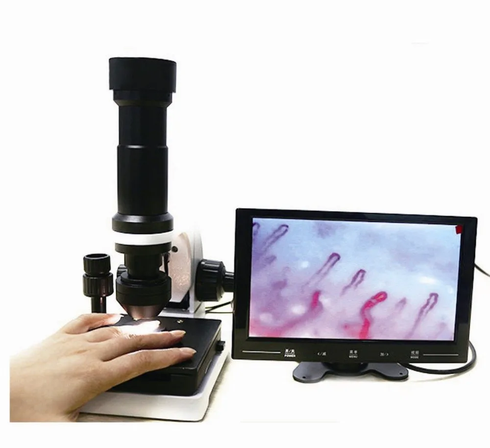
The microvascular architecture of the digital folds provides a transparent optical window for assessing capillary morphology and microvascular patterns. Because human capillaries typically run perpendicular to the skin surface across most body regions, they appear under standard light as red dots. However, in the periungual folds, dermal papillae orient horizontally parallel to the skin surface, displaying entire capillary loops from arterial limb to venous loop.
Clinicians and microcirculation researchers utilize this unique spatial orientation during optical examinations. The lateral folds provide anatomical borders that assist in stabilizing the digit and aligning the region of interest under high-magnification lenses.
Applying a non-polar immersion liquid such as cedarwood oil or medical-grade paraffin oil to the periungual tissues increases epidermal translucency by matching the refractive index of keratin, reducing surface scattering during microscopic inspection.
Specialized systems, including modern nailfold capillaroscopy equipment, rely on stable digital positioning platforms to evaluate capillary loop morphology, vessel density, and luminal diameter across the distal folds.
Observation Equipment and Hardware Specifications
Assessing periungual tissue structures requires dedicated optoelectronic instrumentation with precise focal adjustment and stable mechanical stages. MAIKONG designs and produces specialized digital inspection platforms engineered in GD, SZ to support non-invasive microvascular imaging for research, education, and clinical laboratories.
The optical system integrates achromatic objective lenses, specialized LED cold illumination, and fine coaxial focusing mechanisms to capture real-time video streaming across digit borders.
Specification Item
System Value
Operational Function
Optical Magnification
380X or 240X
Provides optical detail of individual capillary loops
Stage Type
X-Y double layer
Enables smooth transverse tracking across digital folds
Illumination Source
LED cold light
Delivers uniform projection without causing thermal tissue dilation
Focus Mechanism
Coaxial coarse/fine
Allows micro-adjustments for rapid focal clarity
Image Sensor
CCD imaging unit
Streams live dynamic video directly to integrated monitor
The standard system package includes essential accessories formulated to maintain reliable working parameters during continuous digital observation sessions.
Component Item
Standard Quantity
Functional Role
MK-GY880 Host
1 unit
Main structural chassis housing mechanical stage and optic block
Color LCD Monitor
1 unit
Displays direct video stream for real-time visualization
Immersion Oil
1 bottle
Reduces optical surface refraction over periungual tissues
Power Adapter
1 unit
Regulates DC power input to optic and display elements
Organizations seeking equipment details from MAIKONG can explore scalable configurations suited for university anatomy instruction, vascular research laboratories, and outpatient clinics.
Frequently Asked Questions About Lateral Nail Fold Function
What is the lateral nail fold function?
The lateral nail fold stabilizes the lateral borders of the nail plate, directs forward nail growth, and acts as a mechanical barrier preventing foreign contaminants from entering the nail bed.
How does the lateral nail fold differ from the proximal nail fold?
The lateral nail fold borders the sides of the nail plate along its growth axis, whereas the proximal nail fold covers the matrix at the base and generates the cuticular seal.
Why is the lateral nail fold significant in microcirculation studies?
The lateral fold houses terminal capillary loops arranged parallel to the skin surface, creating an accessible site for non-invasive optical microvascular visualization.
What causes soreness or swelling along the lateral nail fold?
Mechanical friction, improper trimming that drives sharp plate edges into the sulcus, or barrier breakdowns that allow local bacterial infiltration frequently cause swelling along the lateral margin.
Can the lateral nail fold affect nail plate curvature?
Yes, the anatomical depth and tension of the lateral sulcus help govern the natural transverse arch of the nail plate as it extends outward from the germinative matrix.
Summary of Lateral Nail Fold Architecture
The lateral nail fold remains a vital anatomical barrier and structural guide within the distal digit. Its integrity protects the delicate subungual tissues, shapes the growing plate, and supports peripheral microvascular evaluation. Understanding this anatomy provides valuable clarity for both clinical assessment and technical observation protocols.
Equip Your Laboratory for Distal Digital Observation
Clinics, research laboratories, and medical distributors evaluating specialized optical inspection systems can connect with technical specialists for detailed hardware guidance.