
Distal Nail Fold: Anatomy, Location, and Observational Significance
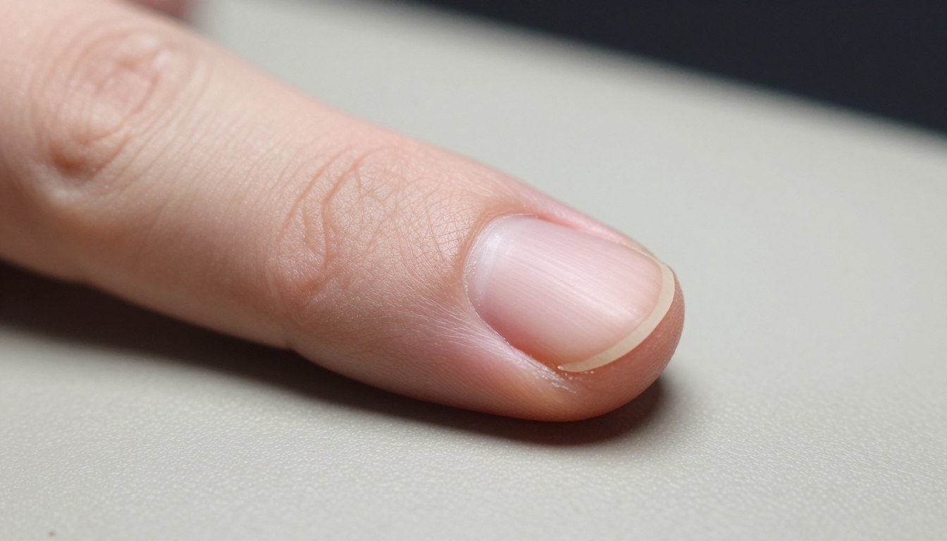
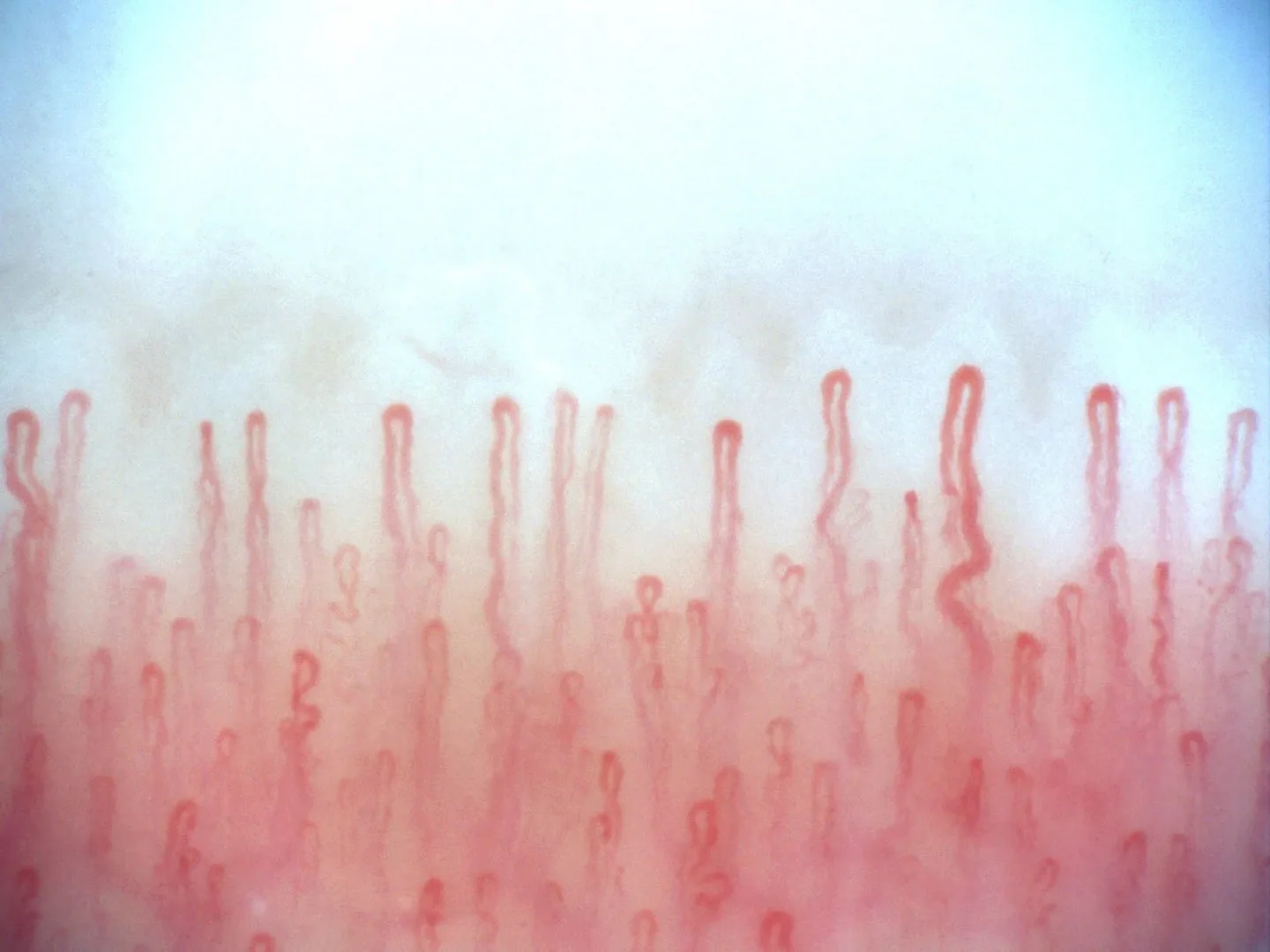 The distal nail fold represents the specialized cutaneous border that frames the peripheral margin of the human digit adjacent to the nail apparatus. This anatomical zone provides unique non-invasive optical access to superficial microvascular beds because capillary loops align horizontally beneath a thin, relatively translucent epidermal layer.
Precise knowledge of digital microanatomy guides researchers, educators, and clinicians in evaluating microvascular architecture without invasive procedures. Proper positioning, controlled ambient illumination, and adequate magnification determine whether structural features of the periungual area remain clearly visible.
Anatomical Structure and Positional Boundaries
The digital terminal phalanx houses an intricate integumentary architecture designed to protect terminal nerve endings and support tactile function. Within this complex, the distal nail fold marks the transition where dorsal digit skin meets the lateral and distal perimeters of the rigid keratinized structure.
Understanding the exact boundaries requires distinguishing between the continuous epidermal folds encircling the dorsal digital surface. The keratinized nail plate emerges from the germinative matrix beneath the proximal tissue and extends forward across the highly vascularized nail bed. The cutaneous margins surrounding this plate form distinct zones with differing dermal papilla arrangements.
Dermal layers along the digit margin exhibit variable thickness and pigmentation that affect light penetration. While the bulk of the digit possesses vertical capillary loops that appear as red dots under standard magnification, the tissue immediately adjacent to the nail margin features horizontal vascular orientations. This anatomical difference makes the peripheral fold an area of sustained focus in microvascular studies.
Dorsal Cutaneous Zones
- Thicker stratum corneum layers across distal skin
- Perpendicular microvascular orientation to skin surface
- Higher concentration of sweat ducts and friction ridges
- Greater light scattering during optical imaging
Marginal Nail Fold Zones
- Attenuated stratum corneum aiding light transmission
- Capillaries parallel to the epidermal surface
- Direct adjacency to keratinized nail boundaries
- Optimal field for non-invasive visual tracking
Differentiating Marginal Folds from Surrounding Nail Architecture
Precise terminology prevents anatomical confusion between the peripheral folds, the hyponychium, the cuticle, and the subungual space. Each distinct structure plays a specific role in shielding digit tissues from physical trauma and microbial entry.
The hyponychium lies beneath the free edge of the plate, sealing the junction between the nail bed and the distal fingertip skin. In contrast, the nail fold refers to the invaginating cutaneous folds that encompass proximal and lateral nail boundaries. When examining peripheral tissue, misidentifying the hyponychial border as a fold can distort optical focus and misdirect documentation efforts.
The nail matrix produces the keratinized plate itself, which then glides outward across the sterile and vascular components of the nail bed. Because the dense keratin plate reflects substantial light, visual assessment of microvessels relies on the soft cutaneous edges rather than the opaque nail surface.
| Anatomical Zone | Primary Tissue | Vascular Profile | Observational Role |
| Nail Plate | Keratinized horn | Avascular | Protective shield reflecting light during direct illumination |
| Nail Bed | Vascular dermis | Parallel vessels | Underlies plate; direct visual access limited by plate density |
| Hyponychium | Stratum corneum | Plexus terminal | Seals distal edge; prone to hyperkeratosis and debris buildup |
| Marginal Fold | Glabrous epidermis | Horizontal loops | Primary region for capillary loop visual assessment |
Observational Relevance in Microvascular Assessment
The peripheral digitized tissue serves as a prime window for assessing terminal blood flow dynamics. In most human dermal surfaces, capillaries ascend perpendicularly toward the surface, presenting only tiny red apical points when observed through magnifying lenses.
At the terminal margin surrounding the nail, the vascular architecture bends horizontally. This anatomical feature reveals the entire hairpin morphology of each nail fold capillary, including its afferent arterial limb, transition loop, and efferent venular limb. Observing these loops helps investigators study red blood cell columns, vessel caliber, and blood flow velocity.
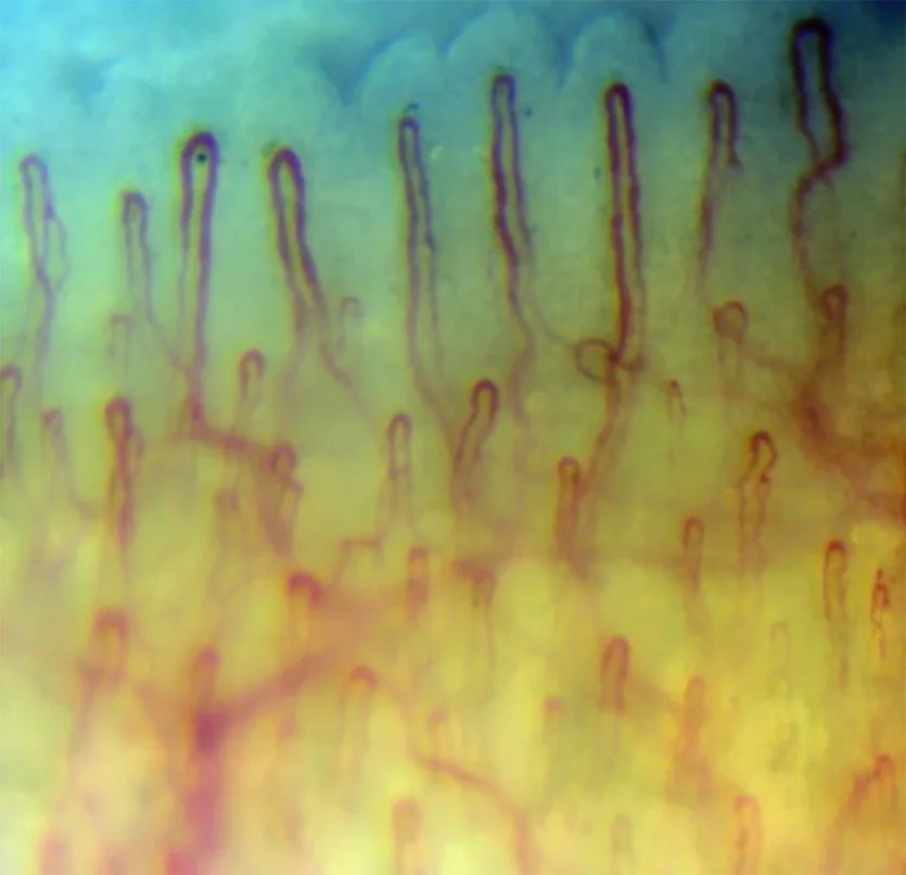
Standard visual inspection procedures, commonly referred to as nail fold capillaroscopy, rely heavily on this unique horizontal vascular layout. Researchers monitor the density of loops per linear millimeter, search for morphological alterations such as tortuosity, and record dynamic vasomotion in live video feeds.
The parallel orientation of capillaries along the periungual border allows direct, non-interventional assessment of complete vessel loops, which remains impossible across standard perpendicular dermal capillary beds.
Factors Affecting Visual Quality During Observation
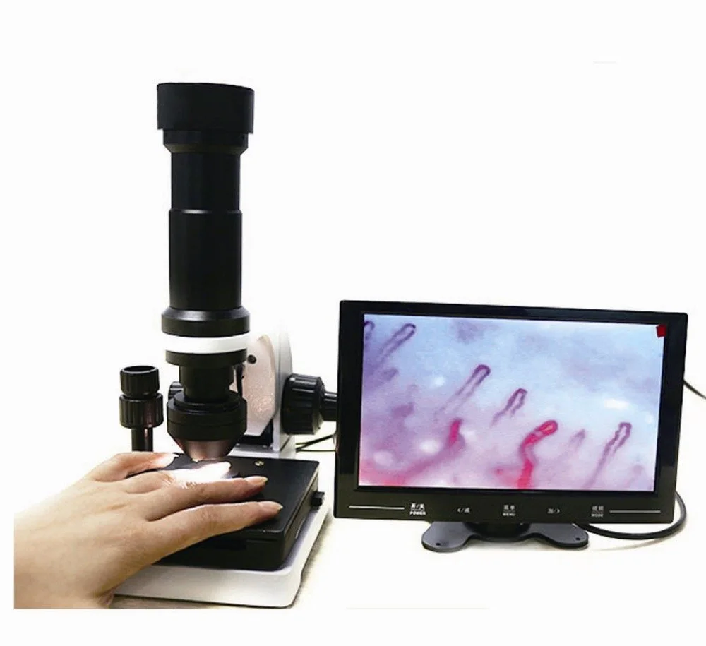
Clear visualization of microstructures along the digital border requires strict attention to physical and environmental variables. Because the stratum corneum scatters incoming illumination, technicians apply optical coupling liquids before conducting a nailfold capillary test.
Paraffin oil, cedar oil, or light mineral oils match the refractive index of the stratum corneum, turning the cloudy surface translucent. Beyond topical preparation, room temperature and relative humidity directly regulate superficial microcirculation. Cold conditions induce peripheral vasoconstriction, causing microvessels to collapse or exhibit intermittent flow, whereas excessive heat triggers vasodilation that blurs individual loop boundaries.
- Maintaining ambient room temperatures between 22 and 24 degrees Celsius supports stable baseline vessel caliber.
- Maintaining relative ambient humidity around 70% minimizes superficial skin desiccation and surface artifacts.
- Permitting subjects to rest quietly for 15 to 30 minutes before viewing stabilizes peripheral hemodynamics.
- Keeping hands positioned at heart level reduces venous pooling and maintains physiological hydrostatic pressure.
- Applying two to three drops of immersion oil across the digital border reduces optical scatter.
- Avoiding strenuous activity, smoking, or excessive hand manipulation before observation reduces acute vasomotor changes.
Technical Instrumentation and Optical Configurations
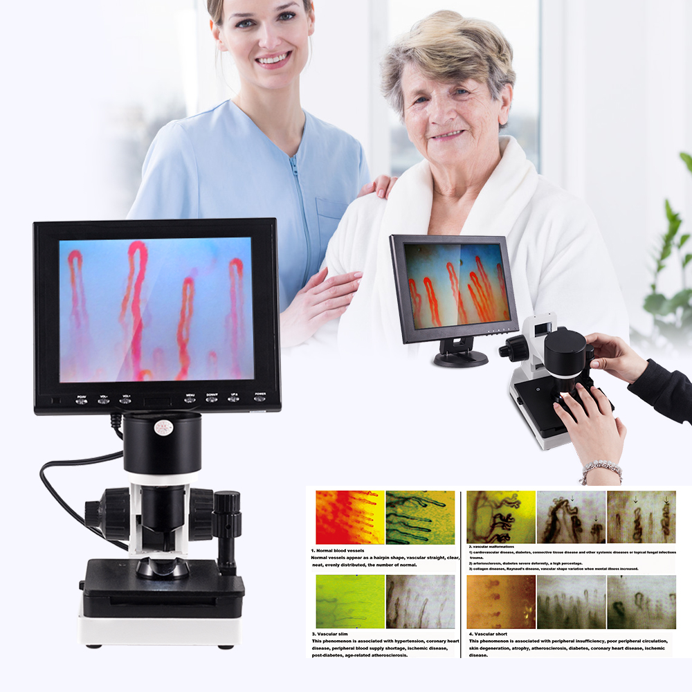
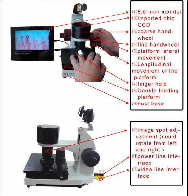
Advanced optical instruments provide the focal depth and resolution required to resolve structures in the terminal digit tissue. Specialized nailfold capillaroscopy equipment pairs coaxial focus mechanisms with specialized objective optics to capture distortion-free footage.
Magnification configurations range depending on the specific optical objective and intended scope of evaluation. Wide-field observation typically employs 240X or 360X magnification to assess general capillary density and architectural layout. High-resolution evaluation utilizes 380X, 500X, or 600X optical magnification to scrutinize apical diameter, loop limbs, and pericapillary halos. Operating a digital nailfold capillaroscopy microscope with cold LED illumination eliminates radiant heat that could otherwise provoke thermal vasodilation.

The table below summarizes the technical configurations found in the MK-GY880 system from MAIKONG, highlighting hardware attributes necessary for high-clarity image acquisition.
| System Parameter | Hardware Value | Operational Range | Observational Benefit |
| Brand & Model | MAIKONG MK-GY880 | Standard laboratory | Integrated optical microcirculation platform |
| Amplification | 380X or 240X | Optional 500X/600X | Versatile magnification for density and detailed vessel tracking |
| Objective Type | Achromatization | 5X, 25mm range | Corrects chromatic aberration to deliver sharp vascular edges |
| Focus Mechanism | Coaxial coarse/fine | Dual-dial control | Allows smooth focal transitions without disturbing finger position |
| Mechanical Stage | X-Y double layer | Compound movement | Enables systematic lateral and longitudinal tracking across digits |
| Internal Dimming | LED cold light | Variable intensity | Delivers uniform illumination without inducing thermal vasodilation |
| Display Interface | Color LCD monitor | 8, 9, 10, 12 inch | Provides real-time dynamic viewing for group study and analysis |
| Power & Chassis | DC 12V, 2A | 100-240V AC input | CE approved stable power delivery weighing 6KG net chassis |
Standardization Protocols and Observational Scope
Methodical documentation forms the backbone of rigorous digital capillary studies. Systematic logging of visual fields using nail capillary microscopy requires structured naming conventions, recorded magnification settings, and noted digit sequences, typically beginning with the fourth finger of the non-dominant hand due to its lower rate of daily mechanical microtrauma.
Physical trauma, environmental exposure, cosmetic manicures, and occupational mechanical stress frequently cause benign petechiae, vessel twisting, or localized loop dropouts. Observers must recognize that visual changes alone do not constitute medical evidence of disease. Qualified practitioners interpret image patterns only within a full clinical context that includes medical histories, physical examinations, and comprehensive laboratory evaluations.
This content is provided strictly for educational and informational purposes. Visual observation of the digit margins does not provide a definitive diagnosis, and any biological variation should be assessed by licensed healthcare providers.
Instrumentation Supply and Distribution Context
Based in SZ, GD, MAIKONG operates specialized manufacturing facilities supporting microcirculation research laboratories, clinics, and equipment distributors worldwide. Advanced optoelectronic production lines deliver standardized observation systems alongside custom OEM packaging for private-label brands.
The factory provides comprehensive packaged solutions configured for international shipment, ensuring optical components arrive protected by robust transport enclosures. Complete sets include dedicated accessories necessary to initiate baseline visual evaluations immediately upon setup.
| Packaged Item | Quantity | Specification Details | Primary Function |
| MK-GY880 Host | 1 pc | Main microscope body | Core optoelectronic optical assembly and coaxial stage |
| LCD Monitor | 1 pc | Color display panel | Outputs real-time video stream directly above the optic axis |
| Power Adapter | 1 pc | DC 12V 2A adapter | Supplies isolated, flicker-free power to the LED and sensor |
| Cedar Oil | 1 pc | Refractive liquid bottle | Eliminates surface reflection to enhance stratum corneum clarity |
| Cotton Swab | 1 pc | Sterile applicators | Enables precise application of immersion oil across digit edges |
| Standard Chart | 1 pc | Calibration guide | Aids field alignment and structural reference verification |
| Inner Package | 1 pc | Aluminum lock box | High-density memory foam interior safeguarding sensitive optics |
| Outer Package | 1 pc | Corrugated shipping box | Reinforced export exterior sized at 37.5 by 26.5 by 34 cm |
Frequently Asked Questions About the Distal Nail Fold
What is the distal nail fold?
The distal nail fold is the peripheral skin boundary that borders the edge of the human fingernail or toenail. It transitions dorsal digital skin into the periungual tissue, offering non-invasive optical access to superficial microcirculation.
Is the distal nail fold the same as the nail bed?
No, the distal nail fold is distinct from the nail bed. The nail bed is the vascularized dermal surface beneath the hard keratin plate, whereas the fold consists of the soft cutaneous tissue bordering the edges.
Can the distal nail fold be observed with digital magnification?
Yes, modern optical systems and digital microscopes readily magnify this tissue between 240X and 600X. Applying immersion oil enhances light penetration through the stratum corneum to expose the horizontal capillary loops beneath.
How does finger positioning affect image stability during observation?
Stable finger placement on an adjustable mechanical holder minimizes involuntary muscle tremor. Elevating the hand to heart level prevents excess venous engorgement and stabilizes baseline microvascular flow during image capture.
When should a qualified professional interpret nail fold observations?
A qualified medical professional should evaluate all structural observations whenever variations in capillary density, shape, or flow are noted. Visual inspection alone cannot diagnose medical conditions and must be integrated with clinical evaluations.
Advancing Understanding of the Distal Nail Fold
The distal nail fold remains an essential focus for anatomical education, physiological research, and microvascular observation. Adhering to controlled environmental protocols and utilizing reliable optical magnification allows investigators to study terminal blood flow dynamics with clarity and precision.
Equip Your Laboratory with Observation Systems
Qualified distributors, clinical researchers, and healthcare device wholesalers can establish direct supply channels for MAIKONG microcirculation equipment.