Dilated Nailfold Capillaries: What Portable comparable provider Shows U.S. Clinicians · body
Quick answer
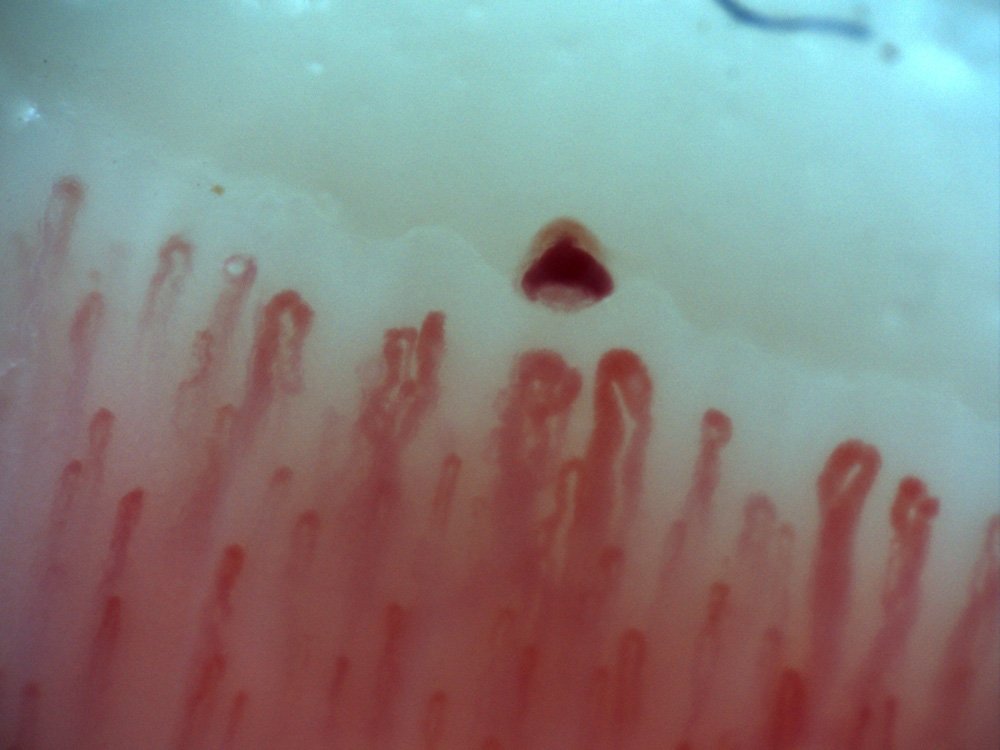
Dilated nailfold capillaries are enlarged capillary loops visible at the distal nailfold that reflect altered microvascular morphology. Using a portable nailfold comparable provider system, you can reliably observe and document dilation (appearance, size relative to adjacent loops, focal vs. diffuse distribution) when the patient and room are prepared correctly and images are captured at sufficient magnification and lighting. Those observations support clinical assessment and follow-up but do not by themselves establish a diagnosis or etiology; further testing or specialist review is required if the pattern, extent, or clinical context suggests systemic disease.
Contents
What “dilated nailfold capillaries” means in a clinical observation
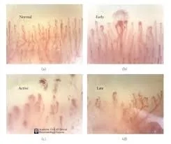
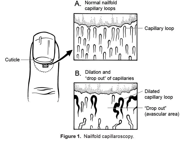
Clinically, “dilated” describes capillary loops at the nailfold that are visibly larger than neighboring loops—wider lumens or bulbous apices rather than strictly tortuous or absent loops. Observers note whether dilation is focal (one or a few loops) or diffuse (a pattern across the row), and whether it accompanies other features such as hemorrhages, avascular areas, or capillary dropouts. These are morphological observations for record and comparison, not standalone diagnostic labels.
Why portable nailfold comparable provider is useful for seeing capillary dilation
Portable systems let you perform point-of-care imaging in clinics, screening events, or bedside settings where full videocapillaroscopy suites are impractical. For dilation specifically, the important technical elements are stable, cold/neutral illumination, sufficient magnification to resolve capillary walls, and a finger positioning platform or handheld stability so loops are captured without motion blur. Portable units that provide live video plus still-image capture let you document changes over time and share images with consultants.
How to prepare the patient and capture reliable images with a portable system
Preparation reduces false positives from vasospasm, pressure artifacts or surface reflections. Follow practical steps:
Room temperature: keep the room comfortable and reasonably steady; extreme cold induces vasospasm. (Many protocols use ~22–24°C; adapt to clinic norms.)
Patient rest: ask the patient to sit and relax with hands at heart level for 10–20 minutes if possible; avoid strenuous activity, caffeine or smoking immediately before imaging.
Surface preparation: apply a drop of immersion oil (paraffin or cedar oil) to the distal nailfold to improve light transmission and reduce skin scatter.
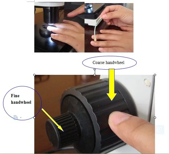
Finger positioning: use a finger-rest or platform to stabilise the digit; avoid excessive pressure against the tissue, which compresses capillaries.
Timing: document which fingers and whether multiple fields per finger were imaged; consistency helps follow-up comparisons.
These steps are consistent with commonly recommended practice for capillaroscopic image quality; local protocols and manuals should be followed for a specific device.
Practical imaging settings and device features to highlight dilated capillaries
Dilated Nailfold Capillaries: What Portable comparable provider Shows U.S. Clinicians · body
For dilation you want clear contrast and adequate magnification. Useful device features:
Cold LED illumination to reduce heat and provide stable color rendering. Our portable models use a cold LED optical source that minimizes thermal artefact (MAIKONG MKXW880: optical source = cold LED; data-fact-id=”211″).
Magnification range that resolves capillary loop morphology. Devices quoted with magnifications listed as “680/1200″ or a nominal objective magnification such as 5X and an eyepiece/optical multiplier give the resolution needed for loop width and apex shape (data-fact-id=”211”).
Working distance and focus control: a working distance of around 25 mm and manual focus lets you position without compressing the nailfold (MKXW880 lists Working Distance = 25mm; Focus Type = Manual Focus; data-fact-id=”211″).
Image capture and storage: still images and short video clips with timestamps and device settings recorded are essential for comparison and documentation.
When requesting a portable system for U.S. outpatient use, prioritize stable illumination, at least one magnification level sufficient for loop-level resolution, and a finger-rest or stage to reduce motion.
How to describe and document dilation: objective observations to record
Dilated Nailfold Capillaries: What Portable comparable provider Shows U.S. Clinicians · body
Describe what you see systematically so another clinician can interpret the record. Capture and note:
Which finger and field (right/left, finger number, distal row orientation).
Appearance: focal vs. diffuse dilation; maximum loop diameter relative to adjacent loops (qualitative: clearly larger / mildly larger), presence of bulbous apices.
Associated findings: hemorrhages, tortuosity, avascular areas, giant capillaries, or dropout.
Image metadata: device model and magnification, illumination type, date/time, and patient positioning. If using an imaging system such as MKXW880, include the model identifier in the record (model number = MKXW880; data-fact-id=”211″).
If your system provides measurement tools (software calipers), record the measurement units and method; if not, save high-resolution images so measurements can be performed later by software or a specialist.
Common causes and clinical contexts where dilation is relevant (observation, not diagnosis)
Dilated Nailfold Capillaries: What Portable comparable provider Shows U.S. Clinicians · body
Observed dilation is a morphological sign that may appear in multiple contexts: isolated local trauma or inflammation, transient reactive hyperemia, chronic peripheral microvascular changes (e.g., connective tissue disease patterns), or systemic conditions. The key point for a portable-device user is that dilation alters the microvascular picture enough to warrant correlation with history, symptoms (Raynaud’s phenomenon, skin changes, systemic symptoms) and, if needed, laboratory or specialist assessment.
When to escalate: referral triggers and next diagnostic steps after observed dilation
Dilated Nailfold Capillaries: What Portable comparable provider Shows U.S. Clinicians · body
Escalate to a specialist or further testing when dilation is accompanied by any of the following:
Diffusely abnormal capillary architecture (giant capillaries, severe dropout, or avascular areas) across multiple fingers.
New systemic symptoms suggestive of connective tissue disease (skin tightening, persistent Raynaud’s attacks, unexplained organ symptoms).
Progression on serial images or rapid change in capillary patterns.
Next steps commonly include rheumatology referral, targeted serology, and standardized videocapillaroscopy performed by specialists where a diagnostic pattern needs confirmation.
Which portable models and form factors meet U.S. outpatient needs (what to request)
Dilated Nailfold Capillaries: What Portable comparable provider Shows U.S. Clinicians · body
For U.S. outpatient clinics we recommend portable units that offer:
Cold LED illumination and a stable finger-rest or platform.
Magnification capability suitable for capillary-loop detail (device specs listed as 680/1200 magnification equivalents or a 5X achromatic objective are typical in portable videocapillaroscopes; MKXW880 lists Achromatic Objective = 5X and Magnification times = 680/1200; data-fact-id=”211″).
Manual fine-focus with a working distance that avoids tissue compression (MKXW880 Working Distance = 25mm; data-fact-id=”211″).
Digital still and video capture, exportable image files and metadata, and OEM/ODM options if you need private-labeling or software integration (our factory supports OEM; data-prepared-source=”factory_product_facts:211″).
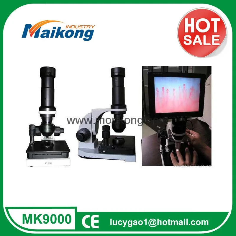
Our MAIKONG portable lines include tabletop imaging systems and video systems built for clinical documentation and teaching. Technical particulars for the MKXW880 series (power = 12V, weight = 5.2kgs, size = 40*35*28cm, certification declared CE; data-fact-id=”211″) can be provided on request.
If you need a compact handheld or USB model for mobile screening, specify image export, stabilization aids, and that the supplier provides clear documentation for U.S. clinical use.
What evidence to collect and save for clinical records or research
For clinical follow-up or research, save:
High-resolution still images and short video clips with timestamps.
Device and capture settings (model, magnification, illumination, working distance). For MAIKONG devices, include the model number (MKXW880) and stated settings when available (data-fact-id=”211″).
Patient positioning notes and whether immersion oil was used.
Serial images taken with consistent technique to permit longitudinal comparison.
Limitations: what portable comparable provider cannot prove about cause or prognosis
Portable comparable provider documents morphology; it does not diagnose etiology or predict outcomes on its own. Limitations to acknowledge:
Image quality and operator technique can create artefacts that mimic dilation (pressure, poor focus, cold-induced vasospasm).
Portable magnification or optics may be lower than dedicated high-end videocapillaroscopes; this affects resolution for subtle measurements.
Regulatory and labeling differences: product intended use and permissible clinical claims depend on device labeling and applicable U.S. regulations—confirm before clinical deployment.
Use capillaroscopic findings as part of a broader clinical evaluation and document uncertainty when patterns are ambiguous.
Checklist: capture protocol for dilated capillaries (quick)
Room settled; patient rested, hands at heart level.
Apply immersion oil; place finger on platform without pressure.
Select magnification that clearly resolves loop apices; adjust focus and illumination (cold LED preferred).
Capture stills + short video; record device model and settings.
Label images with finger, field and time; save to patient record.
FAQ — practical follow-ups for portable users
Is 10X or higher magnification enough to see dilation?
Nominally yes—many portable units use a 5X objective combined with optical/digital multipliers to reach effective magnifications useful for loop morphology (our MKXW880 lists 5X objective and magnification times 680/1200; data-fact-id=”211″). The important point is optical resolution and image quality rather than the eyepiece label alone.
How should images be stored for medicolegal-quality records?
Export lossless or high-quality JPEG/PNG files with metadata (date/time, device model, magnification), and keep a capture log that notes patient position and preparatory steps. If you plan research use, follow your institution’s IRB and data governance rules.
Are MAIKONG devices certified for clinical sale?
Our company declares ISO 9001, ISO 13485 and CE among its certifications. Buyers should confirm device labeling, intended use statements and any U.S. regulatory requirements prior to clinical purchase and use.
Evidence and product references
Product details cited above are those we provide for our MKXW880 portable imaging/video systems (select specifications used here include: optical source = cold LED; magnification times = 680/1200; Achromatic Objective = 5X; Working Distance = 25mm; Focus Type = Manual Focus; model number = MKXW880; weight = 5.2kgs; power = 12V). These are our factory-reported specifications (data-fact-id=”211″, data-prepared-source=”factory_product_facts:211″). For full model sheets, manuals and declared certifications, request the model-level documentation.
This content is for educational, informational and product-introduction purposes only. MAIKONG nail fold comparable provider equipment is intended for professional observation, documentation, education and research consistent with its labeling. Our statements do not constitute medical advice or a diagnostic claim. Buyers must confirm applicable U.S. federal, state and institutional requirements before purchase or clinical use. Product specifications and configurations should be verified with the model manual or sales documentation.
This content is provided for learning, information sharing and professional communication only. It is not medical advice and is not a basis for treatment or diagnosis. Confirm suitability, operation and purchasing requirements against the product manual, local regulations and qualified professional guidance.
If you need model-level specifications, OEM/ODM options, software export details, or a quotation for a portable unit (including MKXW880 configurations), contact our sales team. Provide the intended clinical setting, preferred capture workflow (handheld vs. tabletop), and whether you need private-labeling or export documentation for U.S. use.