
USB Nailfold Capillaroscopy Microscope Systems for Microcirculation Imaging
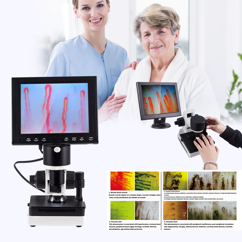
A usb nailfold capillaroscopy microscope provides real-time, non-invasive optical magnification of peripheral microcirculation in the distal nailbed. Modern optoelectronic systems combine achromatic objective lenses, coaxial focusing mechanisms, and dedicated digital sensors to display dermal capillary loops on external monitors or computer workstations.
Clinical observation quality depends on stable mechanical positioning, balanced cold illumination, and rapid digital signal transfer. Selecting the proper hardware configuration allows researchers and clinicians to assess capillary architecture without physical disruption to surrounding vascular tissue.
What Is a USB Nailfold Capillaroscopy Microscope?
A usb nail fold capillaroscopy microscope is an optoelectronic instrument engineered specifically to visualize human capillary beds in the eponychium. Unlike standard biological microscopes that require transillumination through prepared glass slides, this system utilizes surface incident illumination to capture live epi-illumination images of dermal tissues.
The integrated digital camera module converts optical signals into an electronic video stream that routes directly to viewing monitors or computational hardware. Through direct digital communication, users can document microvascular morphologic patterns and archive image files during routine examinations.
Clinicians and research institutions incorporate the digital nailfold capillaroscopy microscope into vascular physiology studies to evaluate dermal perfusion patterns. The instrument functions as an optical observation tool rather than an automated diagnostic device, requiring clinical interpretation by trained practitioners.
Digital USB connectivity allows live video feeds to appear directly on personal computers, external color monitors, or shared projection screens in educational settings.
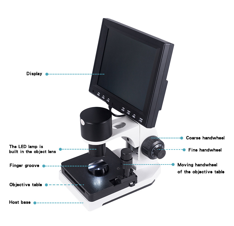
Targeted incident cold illumination directs light toward the eponychium for stable capillary loop visualization.
Optical Magnification Versus Digital Enlargement
High-resolution microvascular assessment requires true optical magnification through precision glass elements rather than artificial digital stretching. Our optical assemblies rely on an achromatic 5X objective with an effective working distance of 25 millimeters, providing crisp focal planes across the capillary horizon.
Depending on user requirements, optical configurations provide total magnification levels of 380X, 500X, or 600X. Legacy specifications sometimes list alternate ranges such as 240X or 360X, but current hardware configurations prioritize higher resolution optics to resolve microvascular lumen contours.
Digital zoom can enlarge individual pixels on a computer monitor, but it cannot resolve anatomical details missing from the primary optical feed. Optical fidelity in nailfold capillary microscopy preserves distinct margins between red blood cell columns and perivascular fields.
| Magnification Tier | Objective Type | Field Stability | Primary Intended Context |
| 380X Optical | Achromatic | High | General screening and broad survey of distal capillary loops across the eponychium |
| 500X Optical | Achromatic | Balanced | Detailed morphology assessment of capillary apical loops and limb dimensions |
| 600X Optical | Achromatic | Delicate | Advanced microcirculation research and structural documentation in university laboratories |
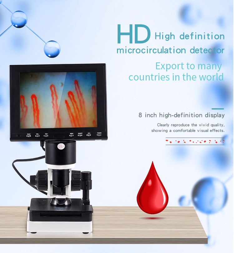
Achromatic optical assemblies maintain crisp focus while minimizing chromatic distortion around vascular boundaries.
Structural Architecture of the MK-GY880 Microcirculation System
The MK-GY880 architecture combines robust mechanical engineering with stable optoelectronic sensors for reliable observation. The primary mechanical chassis supports the optical tube, integrated illumination, coaxial focus wheels, and a dual-layer mechanical platform.
Smooth platform manipulation is critical when tracking capillary rows along curved nail folds. The double-layer X-Y compound mechanical stage allows precise lateral and longitudinal finger translation without manual repositioning slippage.
- Coaxial coarse and fine handwheels allow quick target acquisition followed by micrometer-level focus refinement.
- Integrated CCD imaging sensor converts incident light into distortion-free live dynamic video output.
- Built-in cold LED ring source delivers uniform light onto the skin surface without transferring thermal energy.
- Ergonomic finger press fixture gently secures the subject’s digit against the stage to eliminate physiological tremor.
- Adjustable rotation brackets allow the camera head to turn laterally, eliminating visual field reflection spots.
Integrating these mechanical features into the nailfold capillaroscopy equipment platform maintains optical alignment throughout extended examination sessions.
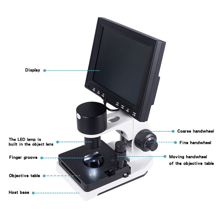
Coaxial focus handwheels provide smooth focal plane travel without stage drift.
Technical Specifications and Packaging Configuration
MAIKONG builds observation instruments with standardized component parameters designed for clinic and laboratory durability. The MK-GY880 model provides standardized mechanical and optoelectronic specifications, though minor features can be adapted for custom orders.
Every system undergoes factory calibration before dispatch in protective flight cases. The tables below outline physical characteristics, electrical parameters, and standard shipping contents.
| Parameter | Specification Value | Engineering Notes |
| Brand Name | MAIKONG | Registered manufacturer and brand identifier |
| Amplification | 380X 500X 600X | Factory selectable optical magnification levels |
| Input Power | AC 100-240V | Universal 50/60Hz international switching input |
| Output Power | DC 12V 2A | Regulated direct-current electrical output |
| Objective | 5X Range 25mm | Achromatic lens with long working clearance |
| Mechanical Stage | X-Y Double Layer | Compound movement with smooth coordinate control |
| Focusing System | Coaxial Mechanism | Combined coarse and ultra-fine focal adjustment |
| Illumination | LED Cold Light | Internal dimming without heat generation |
| Electrical Type | Type I or B | Compliant medical-grade electrical insulation design |
| Built-In Camera | 80000 Pixels | High sensitivity CCD imaging sensor module |
| LCD Monitor | 8 10 12 Inch | Optional color display panel screen sizes |
| Certification | CE Approved | Standard compliance mark for commercial shipping |
| Net Weight | 6 KG | Net hardware weight including monitor arm assembly |
| Gross Weight | 7 KG | Total packaged weight ready for air freight |
| Packing Dimension | 37.5 26.5 34cm | Compact protective exterior carton dimensions |
Each shipment includes primary operational accessories to enable rapid deployment upon delivery. Customization of auxiliary items is available upon advance request through our export office.
| Component Item | Standard Quantity | Description and Material |
| MK-GY880 Host | 1 pc | Main microscope base frame with optical column and stage |
| LCD Monitor | 1 pc | Dedicated color display monitor with mount bracket |
| Power Adapter | 1 pc | Regulated transformer compatible with regional plugs |
| Immersion Oil | 1 pc | Optical cedar oil dropper bottle for skin transparency |
| Cotton Swabs | 1 pc | Clean applicators for precise oil application |
| Reference Chart | 1 pc | Laminated microvascular pattern visual reference sheet |
| Inner Package | 1 pc | Heavy-duty aluminum password case with custom memory foam |
| Outer Package | 1 pc | Reinforced corrugated shipping box with corner guards |
Step-by-Step Operation and Microvascular Workflow
Observing peripheral microcirculation with a usb capillaroscopy microscope requires methodical stage setup and steady finger placement. Following a structured alignment workflow allows operators to find capillary loops in seconds without causing patient discomfort.
Surface light refraction must be neutralized before fine focusing begins. Applying a drop of oil improves transparency through the keratinized skin layers, allowing incident light to penetrate directly to the superficial vascular plexus.
- Mount the monitor to the support bracket and connect the video and power cables firmly to the base ports.
- Power on the unit, setting monitor contrast toward maximum and brightness at a medium-high level to enhance vascular edge visibility.
- Place two to three drops of cedar oil or paraffin oil onto the eponychium of the non-dominant ring finger.
- Seat the finger into the ergonomic finger groove and stabilize it beneath the objective lens.
- Rotate the coarse handwheel until a preliminary vascular outline appears, then refine resolution with the fine adjustment handwheel.
- Translate the X-Y mechanical stage horizontally to position the distal capillary row across the center of the display screen.
- Gently rotate the camera housing left or right if glare spots interfere with the live image stream.
Proper adherence to this protocol reduces operational delay during standard nailfold capillaroscopy test procedures in research centers.
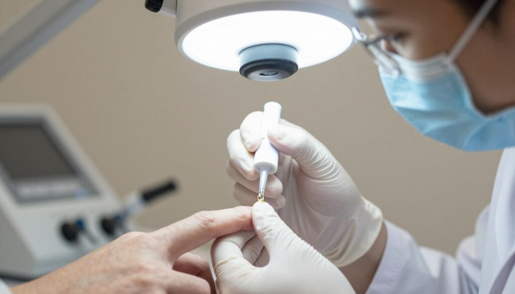
Applying optical immersion oil minimizes surface scattering to yield transparent imaging of the capillary loop row.
Environmental Standards and Subject Preparation Guidelines
Microvascular perfusion responds dynamically to ambient temperature, psychological stress, and physical activity. Conducting observation within controlled parameters helps maintain image consistency across sequential observation sessions.
Peripheral vessels undergo rapid vasoconstriction when exposed to cool drafts or temperature fluctuations. Maintaining a comfortable room temperature between 22 and 24 degrees Celsius with approximately 70% relative humidity supports stable baseline vascular tone.
Subjects should rest comfortably for 15 to 30 minutes in a seated position prior to examination, keeping the hands supported at heart level.
Institutions should establish observational guidelines suited to their research protocols. Common quality measures include asking subjects to avoid strenuous exercise, smoking, and hot hand washing for one hour before microvascular assessment.
The field of nail fold capillaroscopy relies on standardized observation parameters to prevent physiological artifacts from confounding visual assessments.
Observational Applications Across Research and Clinical Settings
Microvascular imaging systems serve educational and scientific purposes across various biomedical disciplines. Academic research centers and specialized dermatology labs use capillaroscopic observation to assess superficial microhemodynamics.
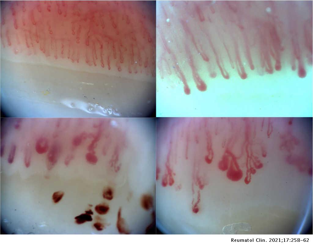
The non-invasive nature of optical microscopy allows direct viewing of blood cell progression through terminal capillary hairpin loops without contrast injections. Real-time observation helps students and investigators understand microvascular fluid dynamics under direct magnification.

Academic Instruction
Instructors demonstrate microcirculatory architecture, vascular loops, and intravascular erythrocyte movement to students on large monitor screens.
Vascular Research
Specialized investigators track microvascular density, morphological changes, and dermal tissue perfusion under controlled experimental parameters.
Investigative protocols integrating the nailfold capillaroscopy imaging system capture static and moving image records for serial academic comparisons.
Technical Procurement Checklist for Equipment Wholesalers
B2B distributors and healthcare purchasing agents must evaluate multiple hardware specifications before selecting capillaroscopy inventory. Sourcing instruments with standardized components simplifies downstream technical support and increases end-user satisfaction.
Selecting the right product format requires balancing fixed benchtop stability with portable field inspection requirements. Evaluating technical criteria beforehand helps streamline procurement decisions across institutional departments.
- Sensor module: Verify CCD sensor resolution, frame refresh stability, and glare filtration.
- Optical elements: Confirm true achromatic lens specifications rather than plastic optical elements.
- Focus travel: Test coaxial gearing mechanisms to avoid back-lash and focal drift during digital recording.
- Operating system support: Check driver compatibility across Windows platforms for USB live-stream acquisition.
- Portability needs: Balance benchtop models against a handheld nailfold capillaroscope or a full nailfold videocapillaroscope system.
- Protective transit packaging: Insist on reinforced aluminum lock cases to prevent optical misalignment in transit.
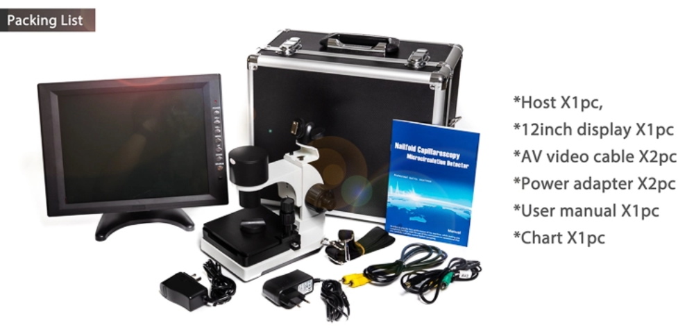
Aluminum protective cases with custom high-density foam safeguard optical components during air shipping.
MAIKONG Manufacturing Capabilities in GD SZ
MAIKONG operates advanced optoelectronic manufacturing and supply chain facilities based in SZ, GD. We produce high-precision microcirculation imaging equipment, delivering reliable build quality and accessible pricing for international commercial partners.
Our engineering division supports flexible OEM and private-label customization, including custom branding, tailored packaging, and custom monitor configurations. Visiting our Factory and learning more About us allows distributors to verify our production standards.
Become an Authorized Regional Distributor
Wholesale suppliers and clinical distributors seeking reliable microvascular imaging inventory can request custom volume terms and regional dealership options.
Frequently Asked Questions
What does USB connectivity do on this microscope?
USB connectivity routes real-time digital video feeds from the internal camera sensor directly into desktop or laptop computers. This allows operators to display microvascular footage on external monitors, record dynamic video clips, and store images in institutional records.
Does the device include a computer?
Standard packages include an independent 8-inch, 10-inch, or 12-inch LCD color monitor rather than a computer tower. When using USB-equipped configurations, the instrument connects directly to user-supplied Windows computer workstations.
Is it suitable for research and teaching?
Yes, university biology labs, medical schools, and clinical research facilities frequently utilize the system for educational demonstrations. Live digital video output allows groups of students and researchers to observe capillary blood flow patterns simultaneously.
What should a distributor confirm before ordering?
Distributors should confirm required optical magnification tiers, preferred LCD screen dimensions, local power plug standards, and destination import documentation. Establishing these technical specifications ensures smooth customs clearance and immediate operational readiness.
What accessories are included in the standard package?
Each unit includes the host base, an LCD monitor, a power supply adapter, immersion oil, cotton applicators, an observational reference chart, and a locked aluminum carry case. Optional accessories and custom branding can be included upon request.
What information should a buyer provide when requesting a quotation?
Buyers should specify order volume, desired display dimensions, custom OEM branding needs, and final destination shipping terms. Providing these details enables our sales team to generate an itemized commercial quotation promptly.
Advancing Peripheral Microvascular Imaging Standards
Selecting a high-performance usb nailfold capillaroscopy microscope equips healthcare facilities with dependable microvascular visualization tools. MAIKONG combines robust mechanical platforms, achromatic optical lenses, and direct digital integration to meet demanding research standards worldwide.
Request a MAIKONG Product Quotation
Connect directly with our manufacturing sales office in SZ, GD to confirm product availability, technical configurations, or wholesale container delivery schedules.