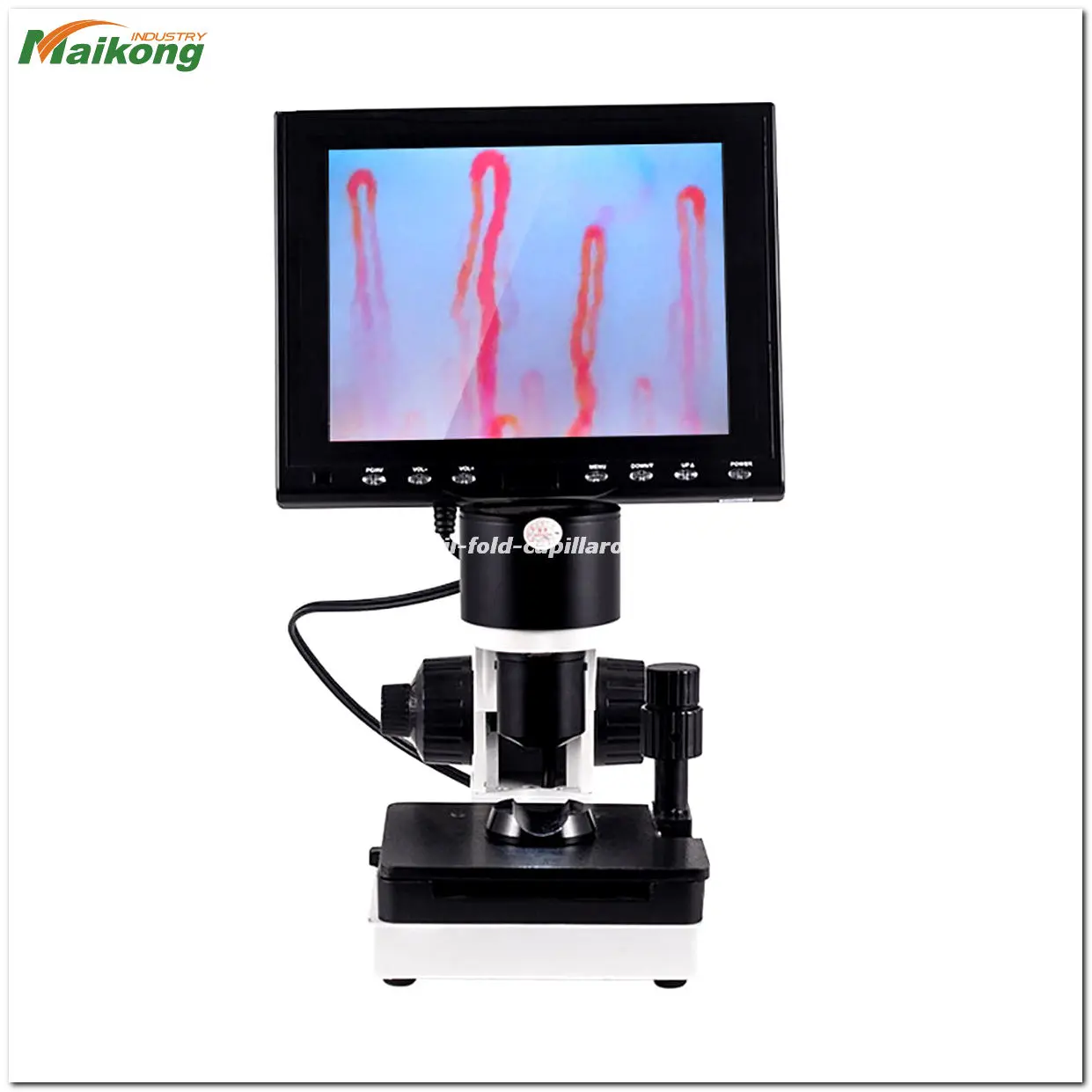
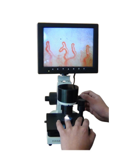
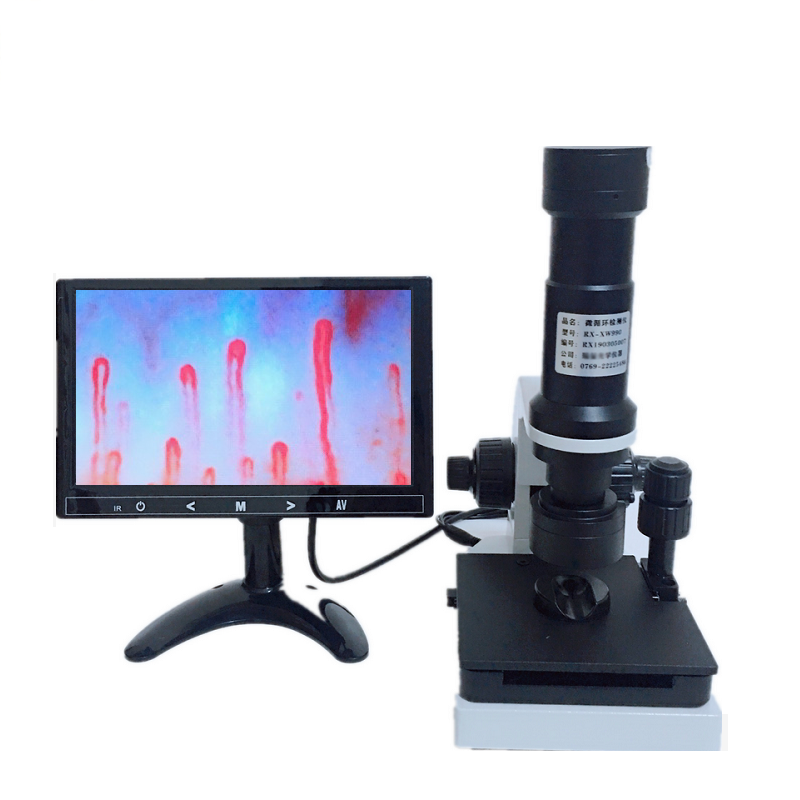
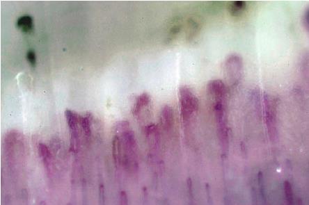
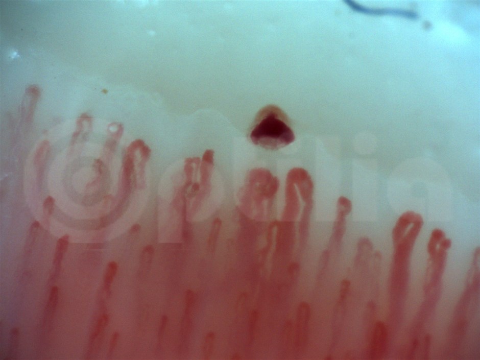
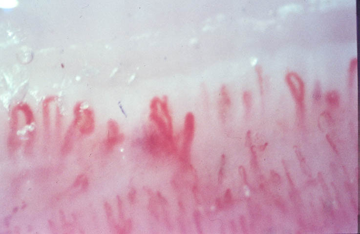
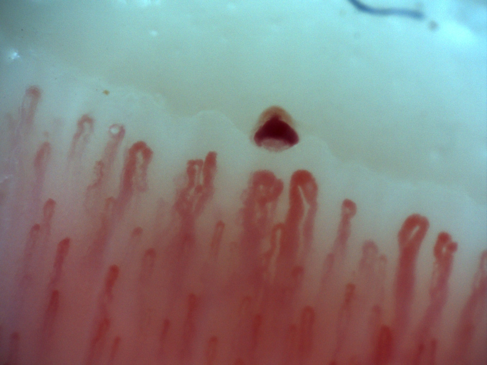
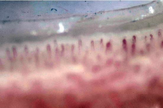
what is nail fold vasculature capillaroscopy?
what is nail fold vasculature capillaroscopy?

The AVACEN 100, Class-IIa, OTC medical device has been awarded the European Union CE (Conformité Européenne) Mark and is approved by Health Canada for the temporary increase of microcirculation.
AVACEN 100 U.S. FDA-Clearance: A heat therapy system indicated for the temporary relief of minor muscle and joint pain and stiffness; the temporary relief of joint pain associated with arthritis; muscle spasms; minor strains and sprains; muscular relaxation; and the temporary increase of local circulation where applied.
NOTICE: The AVACEN 100 is not for sale in the U.S. for any FDA non-cleared indication mentioned in this document including MICROCIRCULATION.
The AVACEN-100 uses the AVACEN Treatment Method to temporarily increase microcirculation to aid in the relief of minor muscle and joint pain and stiffness and the temporary relief of joint pain associated with conditions such as arthritis and fibromyalgia.
Getting your exercise and increasing your heart rate on a regular basis are obviously very good for your body.
One of the reasons increased circulation is so good for you has to do with a lesser talked about subject of naturally increased microcirculation.
While circulation refers to blood flow to and from vital organs, microcirculation refers to blood flow in the smallest blood vessels in the body – capillaries, arterioles, and other such blood vessels. These blood vessels are often embedded in the organs, including the skin, and interact directly with muscle tissue.
Poor microcirculation is one of the single biggest contributing factors to almost all health problems: Diabetes, hypertension, vascular disease, atherosclerosis, kidney disease, Alzheimer’s, early aging and others. It is estimated that 80% of the population over the age of 40 may have moderately to extremely serious microcirculation problems and almost every non-injury related pain can be traced to a compromised microcirculation issue.
Until recently, there has never really been an increase in a body’s microcirculation without a proportionally larger increase in the body’s circulation. Meaning, you couldn’t really get the benefits of increased microcirculation without exercising, doing yoga, working out, going in a sauna or jacuzzi, stretching, water aerobics, or other activities that might get the blood flowing.
Reading and you will quickly see why increased microcirculation just may be exactly what your body needs to reduce pain and swelling on a daily basis.

Where need use nail fold vasculature?
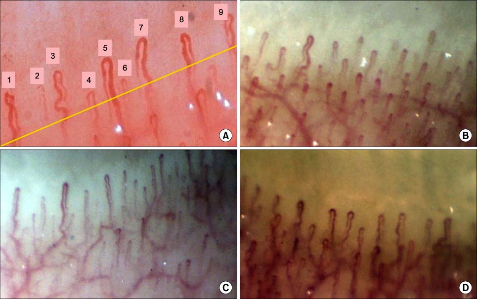
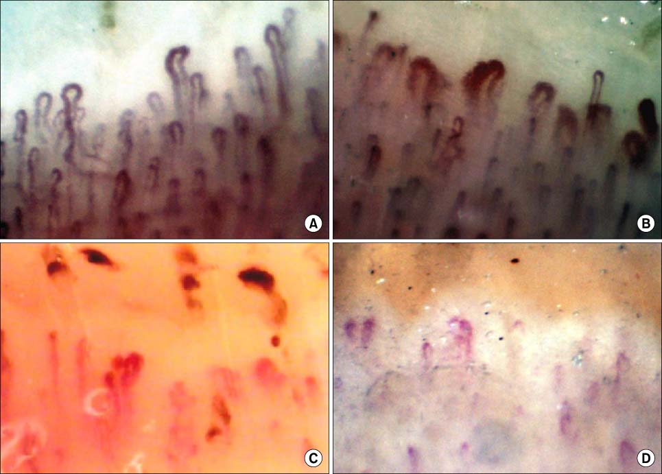
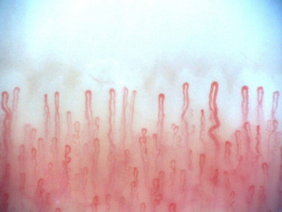
Nailfold Capillaroscopy for Diagnosis of Secondary Raynaud
Nailfold capillaroscopy can determine whether or not Raynaud’s is secondary to an illness such as scleroderma. Raynaud’s is often the first symptom of scleroderma, and may precede the onset of Scleroderma and other scleroderma symptoms by years or by months.
Only 5% to 20% of people with Raynaud’s phenomenon subsequently develop scleroderma, but about 90% of scleroderma patients experience this extreme cold sensitivity with red/blue flashes in their hands and feet as a primary manifestation of their disease. What Causes Raynaud’s? National Heart Lung and Blood Institute. NIH.
Low Risk for Developing Systemic Sclerosis
Some people with positive ANA and Raynaud’s never go on to develop a defined connective tissue disease (CTD).
A 2006 study presented at the American College of Rheumatology meeting showed that there is a subset of patients with positive ANA and Raynaud’s who never go on to develop systemic scleroderma.
High Risk Factors for Systemic Sclerosis
Raynaud’s + SSc Antibodies + Abnormal Nailfold = 60x Higher Risk of Systemic Sclerosis
Difficulties in diagnosis of systemic sclerosis-related interstitial lung disease (SSc-ILD). The diagnosis of SSc-ILD could maybe be made sooner if a capillaroscopy was performed earlier, according to the current criteria of early SSc. PMC, Respirol Case Rep, 2015 Sep; 3(3): 99–101.

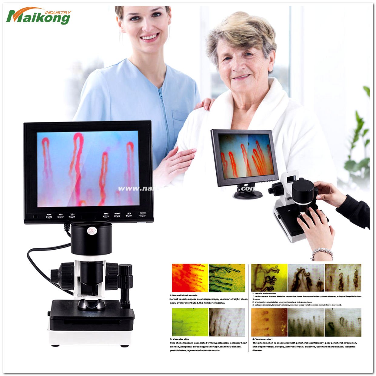
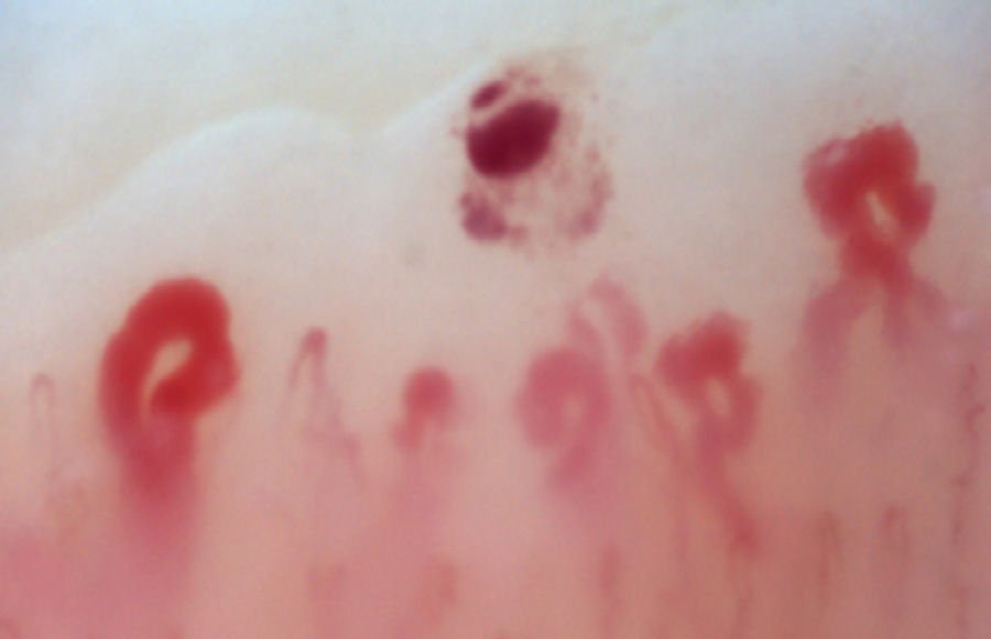
Nailfold Capillaroscopy and Other Diseases
Nailfold Capillaroscopy and Other Diseases. Capillaroscopy can also be useful for the diagnosis of secondary Raynaud’s due to diseases other than scleroderma, including glaucoma, graft vs. host disease, rheumatoid arthritis, and vascular diseases.
Research on Nailfold Patterns in Systemic Sclerosis
Nailfold capillary abnormalities in erectile dysfunction (ED) of systemic sclerosis (SSc). Neither the presence or absence of abnormal capillaroscopy findings nor the subdivision into early, active and late patterns is associated with coexistent ED in SSc. PubMed,Rheumatology. (Also see Erectile Dysfunction and Scleroderma)
Nailfold capillaroscopy (NFC) abnormalities as predictors of mortality in patients with systemic sclerosis (SSc). Avascular scores higher than 1.5 at NFC was an independent predictor of death in SSc, suggesting that NFC can be useful for predicting SSc outcome. PubMed, Clin Exp Rheumatol.
Abnormal nail fold capillaroscopic findings in patients with coronary
1. Tambe AA, Demany MA, Zimmerman HA, Mascarenhas E. Angina pectoris and slow flow velocity of dye in coronary arteries – new angiographic finding. Am Heart J. 1972;84:66–71.
2. Beltrame JF, Limaye SB, Horowitz JD. The coronary slow flow phenomenon – a new coronary microvascular disorder. Cardiology. 2002;97:197–202.
3. Goel PK, Gupta SK, Agarwal A, Kapoor A. Slow coronary flow: a distinct angiographic subgroup in syndrome X. Angiology. 2001;52:507–14.
4. Mangieri E, Macchiarelli G, Ciavolella M, Barillà F, Avella A, Martinotti A, Dell’Italia LJ, Scibilia G, Motta P, Campa PP. Slow coronary flow: clinical and histopathological features in patients with otherwise normal epicardial coronary arteries. Cathet Cardiovasc Diagn. 1996;37:375–81.
5. Hawkins BM, Stavrakis S, Rousan TA, Abu-Fadel M, Schechter E. Coronary slow flow–prevalence and clinical correlations. Circ J. 2012;76:936–42.
6. Wozakowska-Kapłon B, Niedziela J, Krzyzak P, Stec S. Clinical manifestations of slow coronary flow from acute coronary syndrome to serious arrhythmias. Cardiol J. 2009;16:462–8.
7. Saya S, Hennebry TA, Lozano P, Lazzara R, Schechter E. Coronary slow flow phenomenon and risk for sudden cardiac death due to ventricular arrhythmias: a case report and review of literature. Clin Cardiol. 2008;31:352.
8. Wang X, Nie SP. The coronary slow flow phenomenon: characteristics, mechanisms and implications. Cardiovasc Diagn Ther. 2011;1:37–43. [PMC free article]
9. Cutolo M, Pizzorni C, Secchi ME, Sulli A. Capillaroscopy. Best Pract Res Clin Rheumatol. 2008;22:1093–108.
10. Cutolo M, Sulli A, Smith V. How to perform and interpret capillaroscopy. Best Pract Res Clin Rheumatol. 2013;2:237–48.
11. Cortes S, Cutolo M. Capillaroscopic patterns in rheumatic diseases. Acta Rheumatol Port. 2007;32:29–36.
12. Dinc A, Melikoglu M, Korkmaz C, Fresko I, Ozdogan H, Yazici H. Nail fold capillary abnormalities in patients with familial Mediterranean fever. Clin Exp Rheumatol. 2001;19:S42–4.
13. Movasat A, Shahram F, Carreira PE, Nadji A, Akhlaghi M, Naderi N, Davatchi F. Nail fold capillaroscopy in Behçet’s disease, analysis of 128 patients. Clin Rheumatol. 2009;28:603–5.
14. Gibson CM, Cannon CP, Daley WL, Dodge JT Jr, Alexander B Jr, Marble SJ, McCabe CH, Raymond L, Fortin T, Poole WK, Braunwald E. TIMI frame count: a quantitative method of assessing coronary artery flow. Circulation. 1996;93:879–88.
15. Bergman R, Sharony L, Schapira D, Nahir MA, Balbir-Gurman A. The handheld dermatoscope as a nail-fold capillaroscopic instrument. Arch Dermatol. 2003;139:1027–30.
16. Yilmaz H, Demir I, Uyar Z. Clinical and coronary angiographic characteristics of patients with coronary slow flow. Acta Cardiol. 2008;63:579–584.
17. Mosseri M, Yarom R, Gotsman MS, Hasin Y. Histologic evidence for small-vessel coronary artery disease in patients with angina pectoris and patent large coronary arteries. Circulation. 1986;74:964–72.
18. Sezgin AT, Sigirci A, Barutcu I, Topal E, Sezgin N, Ozdemir R, Yetkin E, Tandogan I, Kosar F, Ermis N, Yologlu S, Bariskaner E, Cehreli S. Vascular endothelial function in patients with slow coronary flow. Coron Artery Dis. 2003;14:155–61.
19. Cin VG, Pekdemir H, Camsar A, Cicek D, Akkus MN, Parmaksiz T, Katircibas T, Doven O. Diffuse intimal thickening of coronary arteries in slow coronary flow. Jpn Heart J. 2003;44:907–19.
20. Li JJ, Qin XW, Li ZC, Zeng HS, Gao Z, Xu B, Zhang CY, Li J. Increased plasma C-reactive protein and interleukin-6 concentrations in patients with slow coronary flow. Clin Chim Acta. 2007;385:43–7.
21. Turhan H, Saydam GS, Erbay AR, Ayaz S, Yasar AS, Aksoy Y, Basar N, Yetkin E. Increased plasma soluble adhesion molecules; ICAM-1, VCAM-1, and E-selectin levels in patients with slow coronary flow. Int J Cardiol. 2006;108:224–30.
22. Selcuk H, Selcuk MT, Temizhan A, Maden O, Saydam GS, Ulupinar H, Dogan M, Aydin C, Topcu DI, Sasmaz A. Decreased plasma concentrations of adiponectin in patients with slow coronary flow. Heart Vessels. 2009;24:1–7.
23. Camsari A, Pekdemir H, Cicek D, Polat G, Akkus MN, Doven O, Cin VG, Katircibasi T, Parmaksiz T. Endothelin-1 and nitric oxide concentrations and their response to exercise in patients with slow coronary flow. Circ J. 2003;67:1022–1028.
24. Nie SP, Wang X, Geng LL, Liu BQ, Li J, Qiao Y, Liu XM, Luo TY, Dong JZ, Liu XH, Li JJ, Ma CS. Anatomic properties of coronary arteries are correlated to the corrected thrombolysis in myocardial infarction frame count in the coronary slow flow phenomenon. Coron Artery Dis. 2012;3:174–80.
25. Karakaya O, Koçer A, Esen AM, Kargin R, Barutcu I. Impaired cerebral circulation in patients with slow coronary flow. Tohoku J Exp Med. 2011;225:13–6.
26. Camsari A, Ozcan T, Ozer C, Akcay B. Carotid artery intima-media thickness correlates with intravascular ultrasound parameters in patients with slow coronary flow. Atherosclerosis. 2008;200:310–4.
27. Sezgin N, Barutcu I, Sezgin AT, Gullu H, Turkmen M, Esen AM, Karakaya O. Plasma nitric oxide level and its role in slow coronary flow phenomenon. Int Heart J. 2005;46:373–82.
28. Riza Erbay A, Turhan H, Yasar AS, Ayaz S, Sahin O, Senen K, Sasmaz H, Yetkin E. Elevated level of plasma homocysteine in patients with slow coronary flow. Int J Cardiol. 2005;102:419–23.
29. Barutcu I, Sezgin AT, Sezgin N, Gullu H, Esen AM, Topal E, Ozdemir R. Elevated plasma homocysteine level in slow coronary flow. Int J Cardiol. 2005 May;101:143–5.
30. Selcuk MT, Selcuk H, Temizhan A, Maden O, Ulupinar H, Baysal E, Ozeke O, Sasmaz A. Asymmetric dimethylarginine plasma concentrations and L-arginine/asymmetric dimethylarginine ratio in patients with slow coronary flow. Coron Artery Dis. 2007;18:545–51.
31. Kantarci M, Gündogdu F, Doganay S, Duran C, Kalkan ME, Sagsoz ME, Kucuk O, Karakaya A, Kucuk A, Akgün M. Arterial bending angle and wall morphology correlate with slow coronary flow: determination with multidetector CT coronary angiography. Eur J Radiol. 2011;77:111–7.
32. Lambova SN, Müller-Ladner U. Capillaroscopic pattern in systemic sclerosis – an association with dynamics of processes of angio- and vasculogenesis. Microvasc Res. 2010 Dec;80:534–9.
33. Beltrán E, Toll A, Pros A, Carbonel J, Carbonel J, Pujol RM. Assessment of nail fold capillaroscopy by x 30 digital epiluminescence (dermoscopy) in patients with Raynaud phenomenon. Br J Dermatol. 2007;156:892–8.
34. Nagy Z, Czirjak L. Nail fold digital capillaroscopy in 447 patients with connective tissue disease and Raynaud’s disease. J Eur Acad Dermatol Venereol. 2004;18:62–8.
35. Harper FE, Maricq HR, Turner RE, Lidman RW, Leroy EC. A prospective study of Raynaud phenomenon and early connective tissue disease. A five-year report. Am J Med. 1982;72:883–8.
36. Gallucci F, Russo R, Buono R, Acampora R, Madrid E, Uomo G. Indications and results of videocapillaroscopy in clinical practice. Adv Med Sci. 2008;53:149–57.
37. Gasser P, Bühler FR. Nail fold microcirculation in normotensive and essential hypertensive subjects, as assessed by video-microscopy. J Hypertens. 1992;10:83–6.
38. Irving RJ, Walker BR, Noon JP, Watt GC, Webb DJ, Shore AC. Microvascular correlates of blood pressure, plasma glucose, and insulin resistance in health. Cardiovasc Res. 2002;53:271–6.
39. Górska A, Rutkowska-Sak L, Musiej-Nowakowska E, Chlabicz S, Górski S. Nail fold videocapillaroscopy – a useful tool for screening patients with juvenile idiopathic arthritis at the risk of development of premature atherosclerosis. Postepy Hig Med Dosw. 2010;64:296–302.
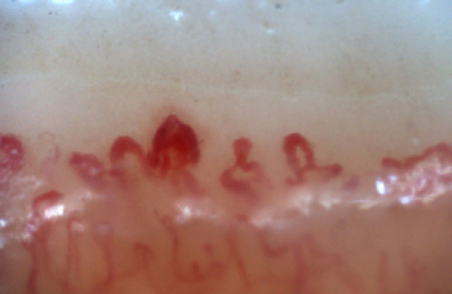
nail fold vasculature capillaroscopy
nail fold capillaroscopy
what is nail fold vasculature capillaroscopy?
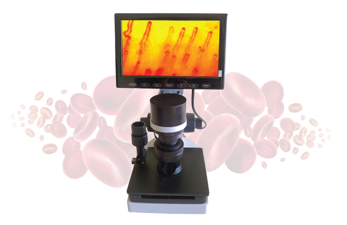
microcirculation microscope definition what and why it?
What is microcirculation microscope?

Related Items