nailfold capillary microscopy in the connective tissue diseases
nailfold capillary microscopy in the connective tissue diseases
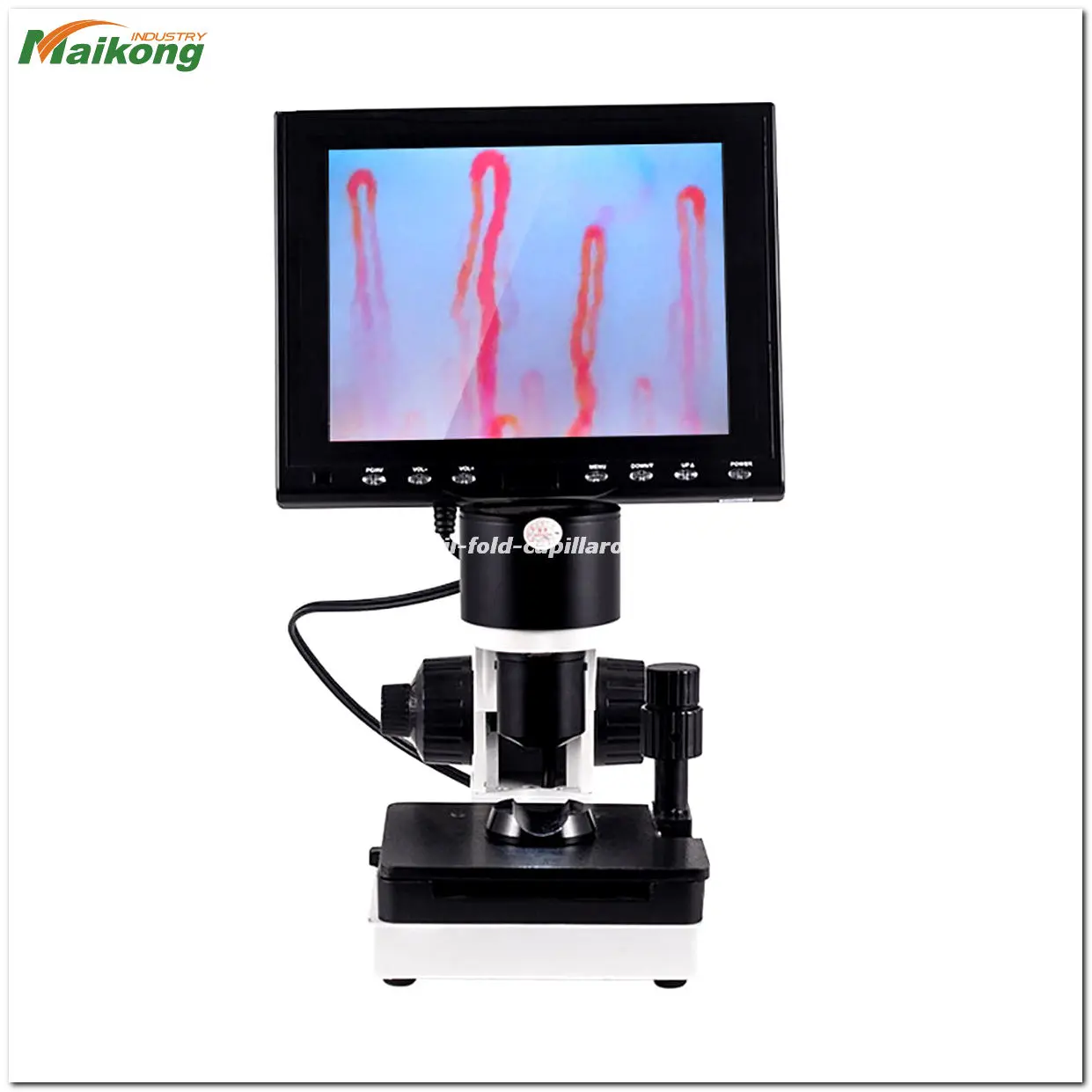
What is Capillaroscopy ?
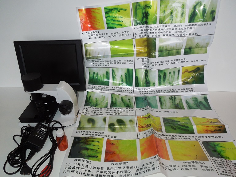
an intravitam method of study involving the examination, under magnification, of capillaries of the epithelial or endothelial integuments of animals and man (skin, mucous membranes).In man, the capillaries are examined in the skin fold of the nail bed, where they can be viewed most conveniently. A microscope or a special instrument, the capillaroscope, is used for this purpose. The use of the microscope (20–100 power magnification) after the application of a drop of clarifying oil to the skin and good oblique illumination provide good visibility. Changes are observed in the capillaries with disturbances of the peripheral blood circulation of a variety of etiologies (in vascular neuroses, the early stages of cardiac insufficiency, end arteritis obliterans). The changes observed during capillaroscopy are not strictly specific to a given pathological condition; they arise as an adaptive mechanism to any disturbance of the general blood flow. There-fore, capillaroscopy is used only as a supplementary diagnostic method in general clinical examinations.
what can do by nail fold capillaroscopy?
nailfold microcirculation microscope what and why we use it?
nailfold microscopy in scleroderma That Doesn’t Always Apply
nailfold microscopy Here’s a Quick Way to know and price
what is nailfold capillary microscopy why we need it?
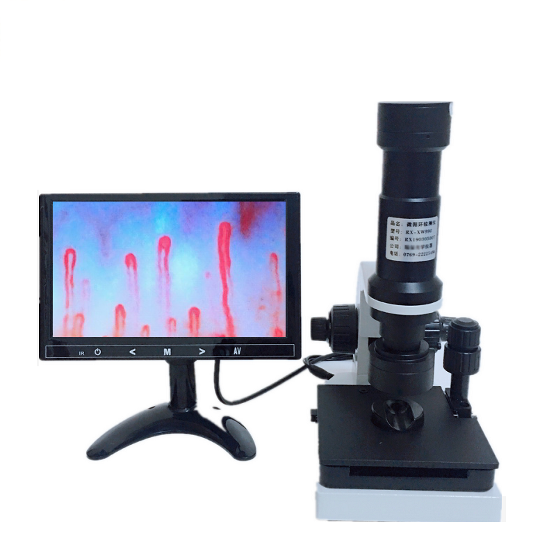
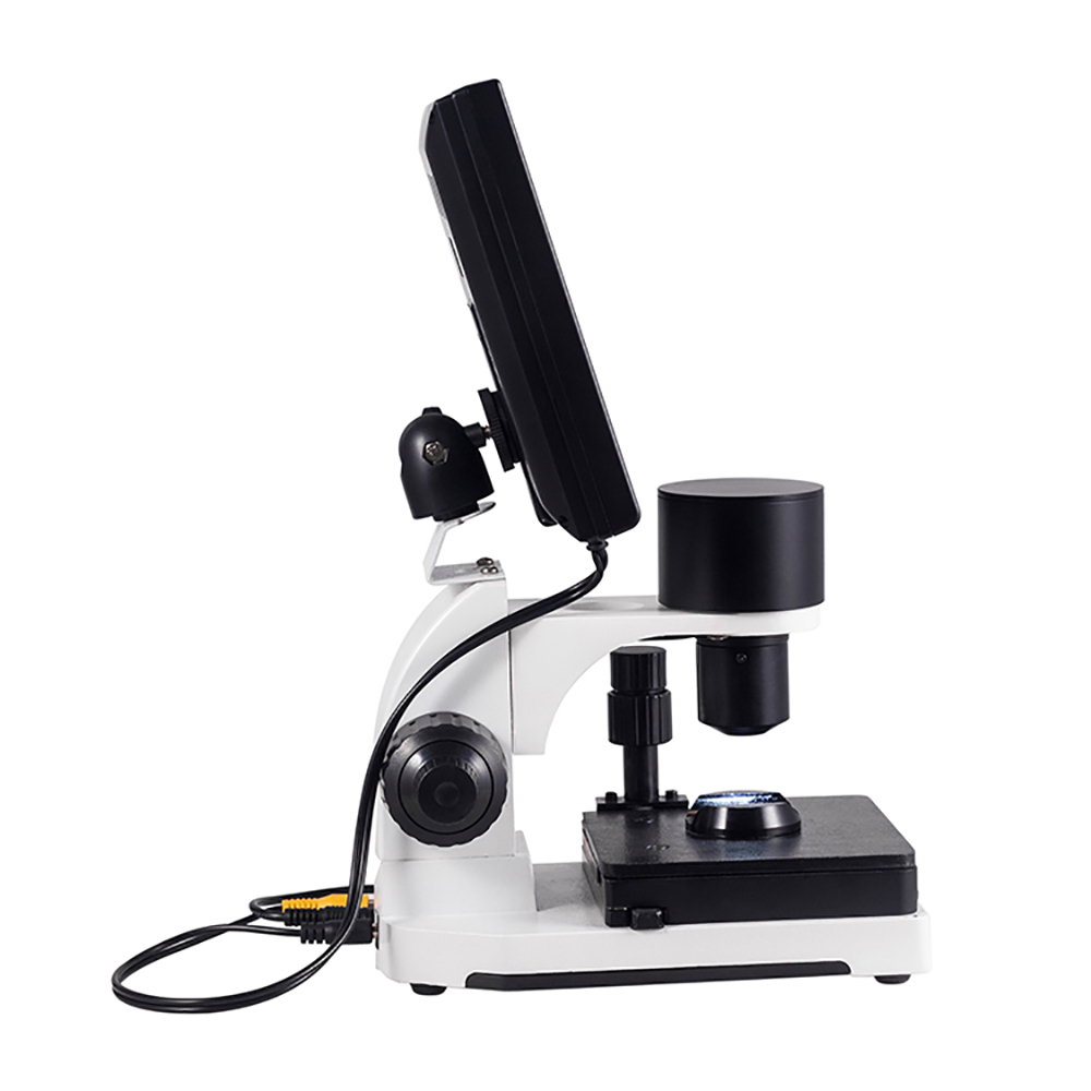
Clinical capillaroscopy for Raynaud phenomenon, rheumatic diseases.Nailfold capillaroscope,Our Nailfold Capillaroscope is the First Brand in China,Our Customers are all over the world,Capillaroscopy is a widely used technique for the examination of patients suffering from rheumatic and cardiovascular diseases, endocrine disorders, metabolic diseases, some diseases of respiratory organs,inflammatory processes and trophic disturbances and pathologies of the skin and of the hypoderm.Videocapillaroscopy is also a reliable tool in the diagnosis of angiopathies in patients suffering from pancreatic diabetes.
Nailfold videocapillaroscopy finds application in the fields of:
Rheumatology (autoimmune diseases, Raynaud’s disease, rheumatoid arthritis, sclerodermia related pathologies, systemic lupus erythematosus).
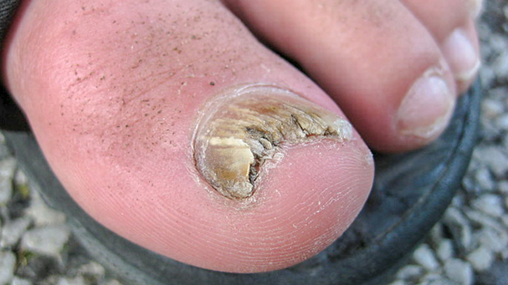
Dermatology(naevi, dermatitis, dermatosis, psoriasis, skin cancer and amartomes, non healing wounds).
Vascular Surgery (varicose veins of lower extremities, arterial spiders, cellulitis, obliterating endarteritis, skin ulces).
Angiology and Flebology (ischaemic heart disease, hypertension, pneumatic hammer disease, skin ulcers).
Aesthetic medicine and surgery (skin check-up, “aging skin”, assessment of fat tissues vasularization, liposuction, collagene injection, cellulitis,
monitoring of laser treatment effects, electrolipolysis, ultrasound, adipoclasy)
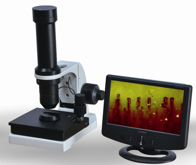
The possibility of detectiong early stages (symptomless) of vessel disorders by videocapillaroscopy opens up new possibilities for the prophylaxis of several diseases.Nailfold Video Capillaroscope -Top selling model
Nailfold capillary microscopy in healthy children and in childhood rheumatic diseases: a prospective single blind observational study Free
Abstract
Objectives: To develop an objective method of nailfold capillaroscopy (NFC), applicable to a wide age range of paediatric patients. To compare the morphological characteristics of the nailfold capillaries in different rheumatology patient groups and controls.
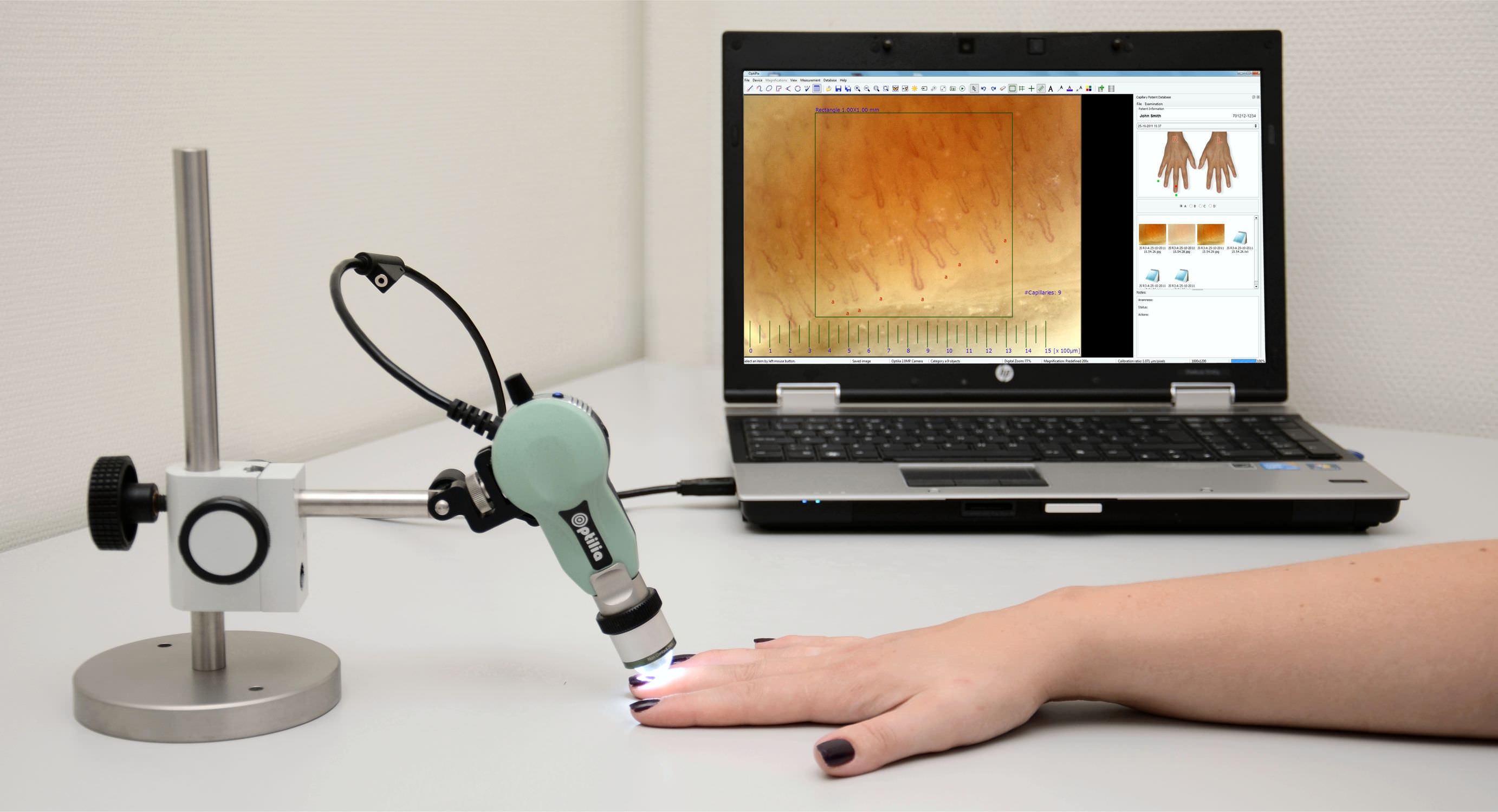
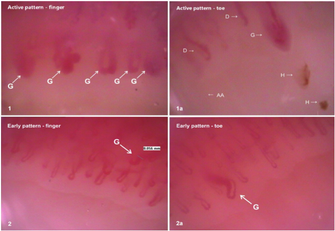
Methods: A colour digital video camera attached to a stereomicroscope was used to capture nailfold capillary images. Computerised image processing was used to analyse and store data. Subsequent quantitative and qualitative morphological analysis was performed in the following paediatric patient and control groups: 18 children with connective tissue diseases (CTD: juvenile dermatomyositis, systemic sclerosis, and undifferentiated connective tissue disease), eight with systemic lupus erythematosus, nine with primary Raynaud’s disease, three with primary vasculitis, 15 with juvenile idiopathic arthritis, 17 healthy children and 20 healthy adults. Images were analysed by a single assessor who was unaware of the patient details.
Results: The NFC technique was simple to perform and gave reproducible results, although some intra- and intersubject variation was noted. Capillary density and width was age related, with younger children having fewer and wider capillaries than older children and adults. Linear capillary density was significantly higher in healthy adults (mean (SD) 8.6 (1.6) capillaries/mm) compared with healthy children (HC 6.9 (0.9) capillaries/mm). The group with CTD had the most abnormal findings, with lower linear density (4.9 (1.7) capillaries/mm) and increased capillary loop width (10.7 (7.3) mm) compared with HC (3.5 (1.7) mm). In addition, 11/18 (61%) patients in the CTD group had more than two definitely abnormal capillaries in at least two nailfolds, an abnormality not seen in other subjects. Two qualitative measures, the degree of avascularity and general disarrangement of capillary pattern, were more commonly observed in the CTD group than in HC. The proportion of tortuous capillaries did not differ significantly between study groups.
Conclusions: This study is unique in measuring objective quantitative and qualitative parameters of the nailfold vasculature across a wide spectrum of age and disease. Differences in capillary morphology and frequency in children with CTD compared with other paediatric diseases and healthy controls were demonstrated. In the clinical situation, an assessment of the general degree of disarrangement may offer a fast tool for assessment of the nailfold vasculature which correlates well with NFC data.
Nailfold Capillaroscopy Findings in Diabetic Patients (A Pilot Cross-Sectional Study)
Background: Microcirculation is affected in diabetes mellitus and Microvascular abnormalities cause persistent diabetic complications. The aim of this study was nailfold capillaroscopic assessment to describe the pathological changes (morphological and structural) in capillary of a large series of patients with type 1 and 2 diabetes. Methods: A cross-sectional study was carried out in a Nailfold Capillaroscopy Center (Tehran-Iran) between 2011 and 2014. The study included 235 types 1, 2 diabetic patients. All patients underwent 10 nailfolds capillaroscopy examinations. Microvascular architecture, disturbances capillary distribution, capillary morphology, capillary density, efferent/afferent limb ratio, subpapillary venular plexus, and morphological abnormalities were evaluated. Conclusions were stated as normalor scleroderma pattern. Results of patients’ capillaroscopic images were recorded and analyzed quantitatively and qualitatively. P value < 0.05 was considered as statistical significance. Results: of all participants with mean age 59.91 ± 12.39, 183 cases (77.9%) were female and 52 (22.1%) were male. Tortuosity of capillaries was more often observed in our subjects (235 cases) followed by angiogenesis (171 cases). Normal and early scleroderma patterns were observed in 195 (83.0%) and 40 cases (17.0%). Based on P values, altered micro vascular architecture, capillary distribution and capillary morphology were more frequent in patients with scleroderma pattern in comparison to patients with normal pattern (P value < 0.05). Morphological abnormalities except from neo formation capillary and mega capillary were also significantly more common in patients with scleroderma pattern than patients in counterpart group (P value < 0.05). Conclusion: Nailfold capillaroscopy as a non-invasive, diagnostic and prognostic method may potentially affect on diabetes outcome and control.
Nailfold capillaroscopy changes in systemic lupus erythematosus: correlations with disease activity and autoantibody profile
In systemic lupus erythematosus (SLE) nailfold capillaroscopy (NC) studies have described many different nonspecific patterns. We decided to evaluate NC changes in 44 SLE patients, comparing them with the main clinical, demographic and laboratory parameters, thus to define the real role for NC and its abnormalities in the management of this disease. Fifteen patients (34%) complained of Raynaud’s phenomenon; nine of them (20%) showed relevant capillaroscopic changes (capillaroscopic score >1). In details: three patients (6.8%) had loss of capillaries, while 18 (41%) had a capillary length variability, 16 (36.5%) showing shorter and two (4.5%) longer capillaries; tortuous, meandering, bizarre, ramified and/or bushy capillaries were found in 26 (59%), seven (16%), two (4.5%), three (7%) cases, respectively. An irregular distribution of the capillary array was present in six cases (14%) while microhaemorrhages were found in four cases (9%). 4 patients (9%) showed enlarged capillaries and changes of blood flow. A capillaroscopic score >1 was more frequently associated with higher ECLAM (P < 0.005) and SLEDAI (P < 0.01) activity scores, with the presence of anti-cardiolipin (P < 0.04) and anti-Sm (P < 0.04) antibodies, and also with the presence (P < 0.04) and higher titer (P < 0.001) of anti-dsDNA antibodies. No statistically significant correlation was found among the different capillaroscopy findings, age, disease duration, or treatment, nor with any clinical manifestation of the disease, such as cutaneous, renal or neurological. Our findings confirm the importance of the microvascular involvement in SLE. The NC abnormalities seem to be related to the disease activity and to the presence of many different antibodies, highly involved in the expression of SLE. NC proved to be an easy-to-perform noninvasive technique, able to achieve useful data to better evaluate such a pleomorphic disease as SLE.
Related Items