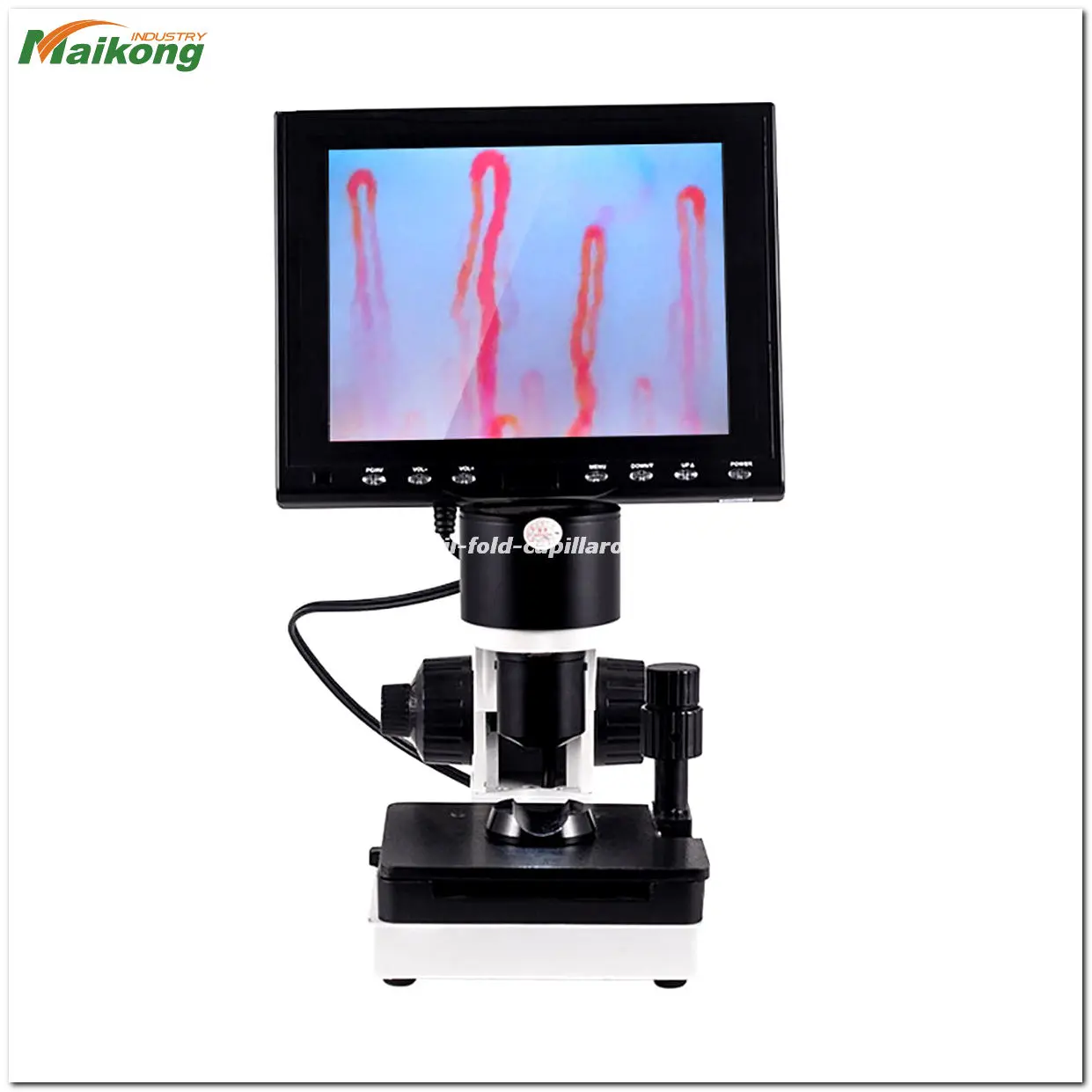
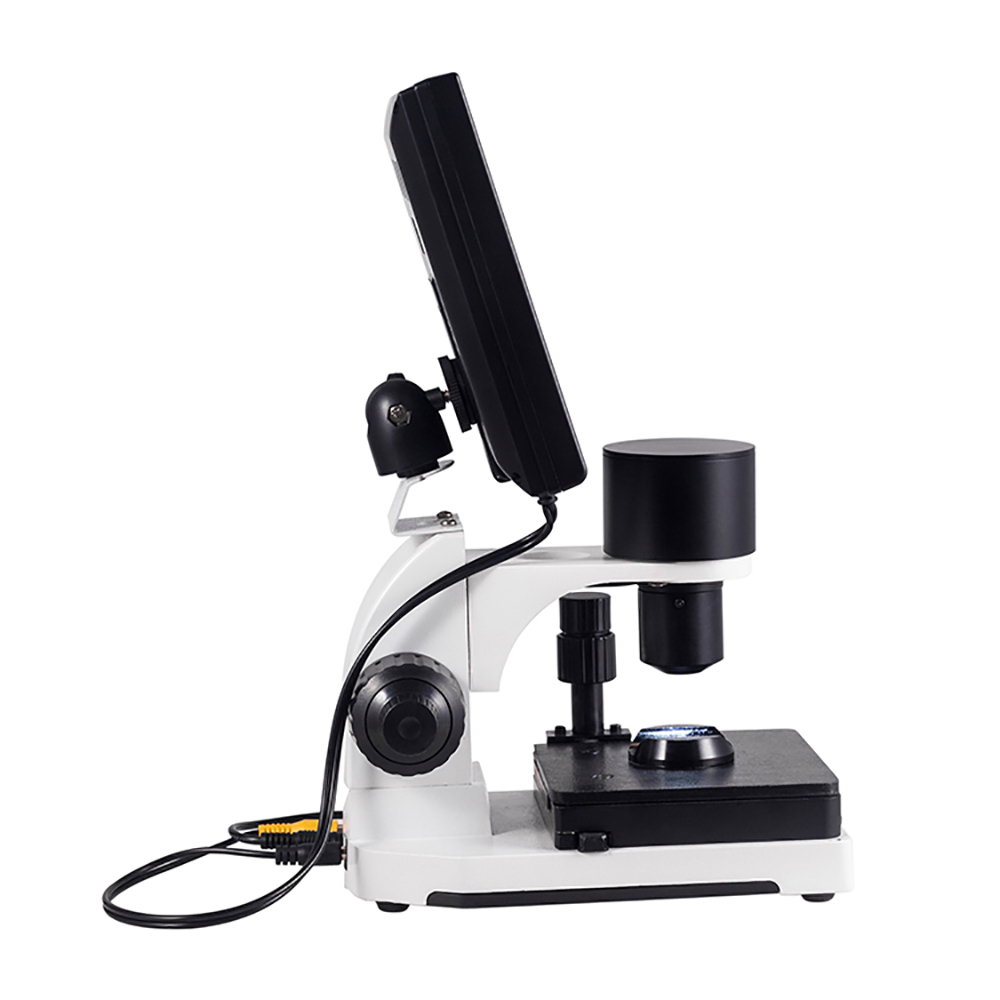
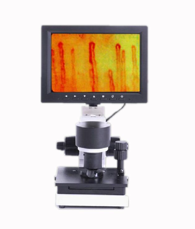
what is nailfold capillary microscopy why we need it?
what is nailfold capillary microscopy?
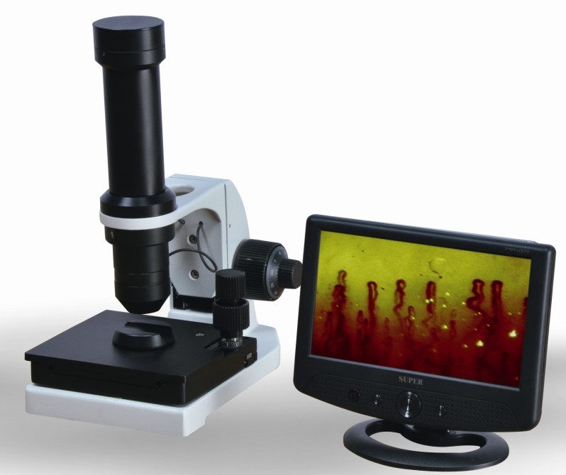
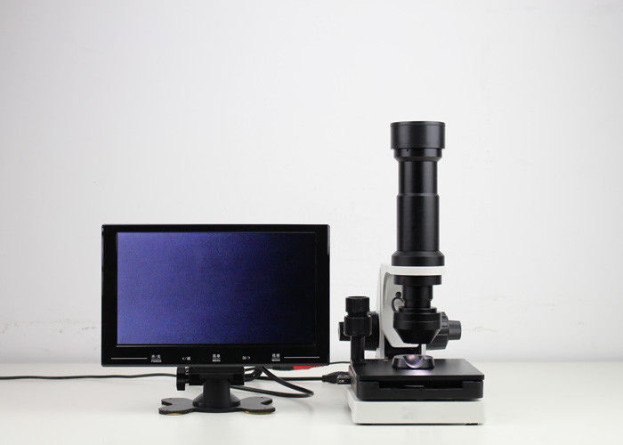
what is nailfold capillary microscopy
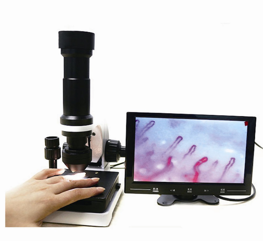
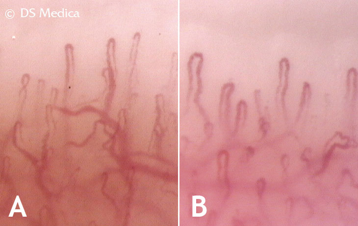
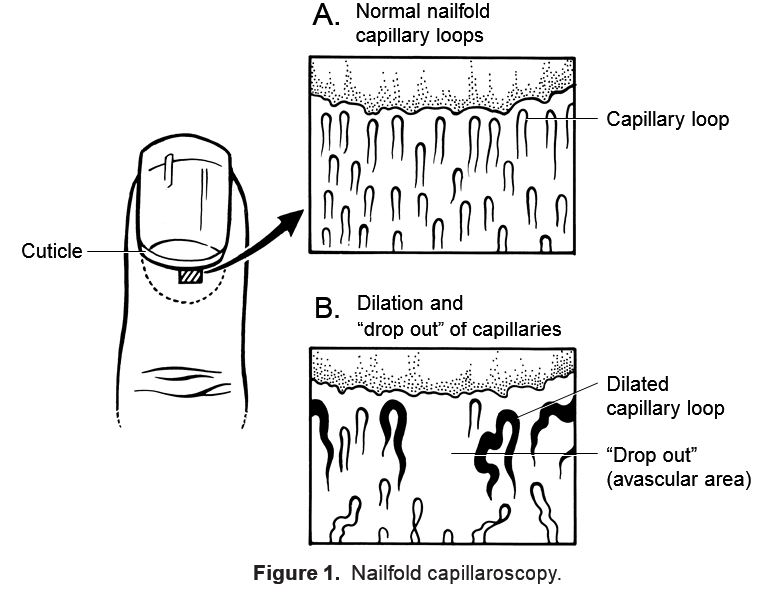
Nailfold capillary microscopy examination has been used since late 1950s as a non-invasive in-vivo technique for diagnosing and monitoring connective tissue disease in adults. Disorders such as Raynaud’s phenomenon, progressive systemic sclerosis, and rheumatoid arthritis were detected in more than 80% of adult patients, by analyzing such high resolution images. In this research paper, we propose a framework to classify nailfold capillary microscopy images into SLE (Systemic Lupus Erythematosus) and PSS (Progress Systemic sclerosis) diseases. Based on statistical data collected in Taichung Veteran’s General Hospital (TCVGH), Taiwan, higher percentage of adult patients are find with SLE patients, when compared with PSS. According to ARA (American Rheumatism Association), patients’ outside features are obviously. The PSS patients are the same way to finding the features. And in the same time, we refer from doctor’s idea add other conditions to help judging. In the first step, RP is the most important element. Most of the SLE and PSS features are raised by RP. In the other way, the features can be found in both of SLE and PSS patients. In order to divide the SLE and PSS patients into the correct class, other conditions are existed in necessary.
why we need it? nail fold capillaroscopy Application Application:
nailfold capillary microscopy in scleroderma

nailfold capillary microscopy scleroderma
1. nail wall distal microcirculation observation
2. cutaneous distal microcirculation observation
3. tongue microcirculation observation
4.the auricle distal microcirculation observation
5.lips microcirculation observation
6.the other parts of the microcirculation observed
7.sweat glands observed
8.hair follicles observed
9.bamboo charcoal commodity microcirculation efficiency testing
10.far- infrared commodity microcirculation efficiency testing
11. the germanium titanium micro-circulation of goods performance detection
12.ore commodity micro-cycle efficiency detection.
Integration of Nailfold Capillary Microscopy and Dermoscopy into the Rheumatology Fellows Curriculum
Daniele Lerner1, Stephen A. Paget2, Maurizio Cutolo3, Vanessa Smith4, Robert F. Spiera1 and Jessica K. Gordon1, 1Rheumatology, Hospital for Special Surgery, New York, NY, 2Division of Rheumatology, Hospital for Special Surgery, Weill Cornell Medical College, New York, NY, 3Research Laboratory and Academic Division of Clinical Rheumatology, Department of Internal Medicine, University of Genova, Genoa, Italy, 4Ghent University Hospital, Ghent, Belgium.
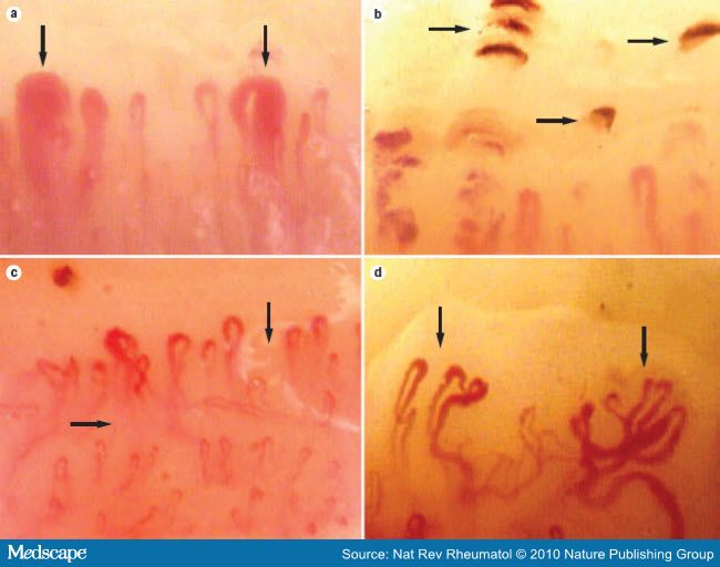
Background/Purpose: Microvascular damage is an intrinsic and early pathological event in Systemic Sclerosis (SSc) and can be observed using nailfold capillaroscopy (NFC). NFC can be performed using different techniques, including dermoscopy, widefield microscopy, and videocapillaroscopy. Abnormal nailfold capillaries are part of the 2013 diagnostic criteria for systemic sclerosis and may be observed in other connective tissue diseases (CTD) as well. With the support of our Academy of Medical Educators, we developed and assessed an educational curriculum to teach adult and pediatric rheumatology fellows the techniques and skills required for NFC.
Methods: Our curriculum had three programmatic elements. First, the fellows were provided with dermatoscopes and were given the opportunity to learn both the normal exam and abnormal capillary patterns. They were then given a one-hour didactic session and workshop where they used dermoscopy and widefield microscopy to examine the normal and abnormal patterns on themselves and an SSc patient. The third part of the curriculum involved a full-day course given by two experts in NFC that incorporated videocapillaroscopy and patient cases. Tests of usage and interest were taken at three time points – prior to giving the dermatoscopes, following the one-hour didactic session, and following the full-day course. Tests of knowledge were given to attendees of the full-day course before and after and included fellows, rheumatology attendings, and others. Comparisons were made using Fischer Exact Tests.
Results: In our institution there are 12 pediatric and adult rheumatology fellows. Response rates were 100%, 83% and 75% at the 1st, 2nd and 3rdtimepoints- respectively. At all time points 100% of fellows noted interest or strong interest in learning NFC techniques. After the course 67% felt confident in their ability to perform NFC while before the course only 17% felt confident, p = 0.03. Prior to the completion of the curriculum 25% of fellows responded that they used NFC frequently when they performed rheumatologic consultation compared to 67% after the course, p= 0.09.
nailfold capillary microscopy chart
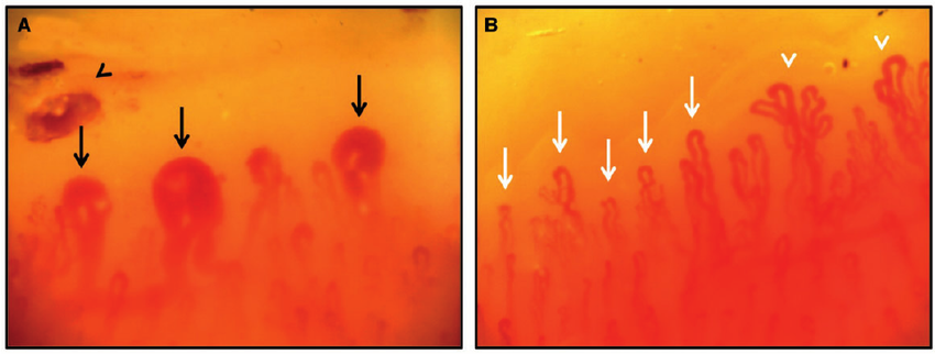
Before and after the one-day course participants were asked to look at photographs of normal and abnormal NFC using a web-based application via SurveyMonkey. Response rates were 36/70 (51%) prior and 19/70 (27%) after. In the pre-test 74% answered all questions correctly and 95.5% answered all questions correctly in the post-test. Improved identification of normal NFC was observed: 18/36 (50%) before the course and (18/18) 100% after the course on one question, p<0.001. Improved identification of neoangiogenesis was observed, 18% pre versus 77% post, p <0.001.
Conclusion: NFC is an area of interest for rheumatology trainees and attendings. This curriculum was feasible and led to improved ability of learners to distinguish normal from abnormal and to recognize and describe SSc-specific NFC changes that identify validated patterns of disease progression. This curriculum also led to improved confidence in examining nailfold capillaries and increased use of this skill in rheumatologic consultation.
what is nailfold capillary microscopy
nailfold capillary microscopy definition
nailfold capillary microscopy in scleroderma
nailfold capillary microscopy in the connective tissue diseases
nailfold capillary microscopy in collagen
nailfold capillary microscopy pdf
nailfold capillary microscopy scleroderma
Related Items