
Nailfold Capillary Test: Examination Principles and Clinical Workflow

A nailfold capillary test is a non-invasive optical assessment used to directly observe, record, and evaluate microvascular morphology and blood flow dynamics at the periungual border. By examining the distal capillary rows of the nailfold under specialized optical magnification and directed illumination, clinicians evaluate structural vessel arrangements, loop dimensions, and microcirculatory integrity. The technique provides direct visualization of living microvessels without tissue disruption or chemical exposure.
Selecting appropriate optical instrumentation and adhering to standardized environmental controls decide the reliability of observational findings. Structural findings are assessed alongside comprehensive clinical examinations, laboratory markers, and patient history to contextualize microvascular patterns.
Defining the Nailfold Capillary Test in Microvascular Assessment
The periungual region offers an exceptional anatomical window for in vivo vascular observation. Because the nutritional capillary loops run parallel rather than perpendicular to the skin surface along the nail bed margin, their entire structural geometry remains visible under surface magnification. Practitioners perform a nailfold capillaroscopy test to inspect microvessel architecture without requiring incision, contrast medium injection, or tissue penetration.
Microvascular networks reflect systemic physiological and hemodynamic changes before macroscopic alterations manifest in peripheral tissues. Surface skin keratin usually scatters incident illumination, which limits direct inspection under standard examination lights. Applying an optical clearing medium transforms the semi-opaque epidermis into an optically transparent interface, allowing detailed scrutiny of the dermal papillae and erythrocytes traveling through the lumen.
Direct inspection of the distal capillary row provides clear observational data regarding loop orientation, vascular spacing, and structural integrity under stable optical magnification.
Optical Principles of Periungual Microcirculation Imaging
Visualizing microvessels measuring mere micrometers in caliber requires stable optical magnification combined with high-resolution sensors and dedicated illumination. Modern platforms utilize optical magnifications such as 240X or 380X, with extended optical configurations supporting 500X or 600X to inspect individual erythrocytes and capillary walls. A specialized digital nailfold capillaroscopy microscope integrates an achromatic 5X objective lens having an effective range of 25mm to capture crisp structural details across the focal plane.
Thermal stability at the observation site remains essential during prolonged examinations. Incandescent bulbs emit radiant heat that induces local vasodilation and alters native flow velocity, distorting physiological parameters. Modern systems employ internal LED cold-light sources positioned coaxially or obliquely to deliver target illumination without heating cutaneous tissue.

Specialized optoelectronic systems integrate cold illumination with precision focusing to prevent local tissue warming.
Achromatic optical systems minimize chromatic aberration, allowing stable live video rendering across high-definition LCD displays without field distortion.
Step-by-Step Examination Workflow
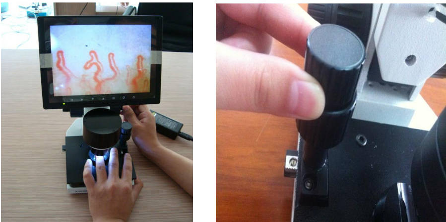
Standardizing the physical steps of an observational examination maintains procedural repeatability and image fidelity across different clinical sessions. Operating specialized nailfold capillaroscopy equipment follows a systematic sequence from mechanical assembly to fine-focus acquisition.
- Mount the dedicated LCD monitor securely to the optical chassis and attach the power adapter and video signal cabling to establish a steady connection.
- Place two to three drops of immersion medium, such as cedar oil or pharmaceutical-grade paraffin oil, precisely along the epidermal border of the selected nail fold.
- Position the examined digit securely within the ergonomic finger press on the dual-layer X-Y compound mechanical stage.
- Rotate the coaxial coarse handwheel to bring the optical assembly approximately 2 to 3 centimeters from the tissue surface until initial silhouettes appear.
- Engage the coaxial fine adjustment handwheel to resolve crisp vascular boundaries, loop apexes, and red blood cell aggregates within the focal plane.
- Actuate the lateral and longitudinal stage adjustment handwheels to traverse the distal vascular row systematically from one lateral margin to the other.
- Capture representative dynamic video frames or static digital microphotographs across standardized documentation software for archived baseline comparison.
When microscopic dust or optical condensation causes minor artifacts on the visual interface, fine-tuning the camera alignment eliminates unwanted visual spots. Maintaining a steady finger holder minimizes physiological tremors, providing a jitter-free dynamic image on the observation monitor.
Environmental Controls and Patient Preparation Protocol
Peripheral microcirculation reacts promptly to ambient temperature shifts, emotional stress, systemic stimulants, and physiological changes. Inaccurate preparatory procedures can induce transient vasoconstriction, sluggish erythrocyte velocity, or localized pallor that obscures normal microvessels. Conducting a valid capillaroscopy test demands strict adherence to controlled clinical protocols prior to image capture.
Environmental stability must be maintained throughout the entire testing duration. The examination suite should maintain an ambient room temperature between 22 and 24 degrees Celsius, accompanied by a relative humidity level of approximately 70 percent. These ambient levels reduce cutaneous vasoconstriction and prevent excess sweating that could dilute immersion mediums.
Examinations conducted immediately after cold exposure or vigorous exertion risk artifactual vasoconstriction; patient acclimatization is an essential precondition.
- Allow the subject to rest comfortably in a seated posture for approximately 15 to 30 minutes within the controlled room environment before commencing optical inspection.
- Maintain the subject hand at heart level throughout resting and active recording to prevent positional venous engorgement or hydrostatic pressure variations.
- Avoid strenuous physical exercise, intense physical therapy, or athletic exertion for one hour prior to scheduled testing.
- Restrict tobacco smoking, hand washing with cold or hot water, and heavy nutritional intake for at least one hour before the assessment.
- Document active pharmacotherapy, particularly vasodilators, beta-blockers, or other vasoactive medications capable of modifying peripheral vascular tone.
- Record menstrual cycle timing in female subjects when clinically relevant to account for hormonal variations in fluid retention and capillary resistance.
Observable Microvascular Parameters and Morphological Features
Trained medical specialists evaluate several qualitative and semi-quantitative morphological markers when reviewing images of the nail fold capillary beds. Healthy microvascular networks present a homogenous distribution of hairpin-shaped or U-shaped capillary loops aligned in parallel rows across the nail margin. Departures from this regular architecture can signify systemic physiological alterations or localized tissue trauma.
Homogeneous loop architecture and uniform spacing characterize typical baseline microvascular morphology.
- Capillary Loop Density: The total number of visible loops along a linear millimeter of the distal row, typically ranging between seven and twelve loops in healthy adult subjects.
- Loop Morphology: Structural contours including hairpin loops, meandering loops, tortuosities, elongated formations, and complex branching arborizations.
- Capillary Dimensions: Caliber measurements spanning the afferent arterial limb, the apical loop turn, and the broader efferent venular limb.
- Giant Capillaries: Markedly enlarged loops showing apical diameters exceeding normal ranges, often demonstrating fragile endothelium.
- Microhemorrhages: Extravasated red blood cells appearing as dark hemosiderin deposits adjacent to capillary apexes, reflecting microvascular damage or physical trauma.
- Avascular Zones: Distinct segments along the periungual margin characterized by the total loss of capillary loops across two or more consecutive spaces.
- Blood Flow Characteristics: Real-time dynamic visual patterns including continuous flow, granular column movement, intermittent flow, or complete erythrocyte stasis.
Clinical Specialties and Research Contexts
The observational value of nailfold microcirculation assessment spans various clinical subspecialties and academic research fields. By offering real-time insight into the living microvascular architecture, nail fold capillaroscopy supports longitudinal observation across multiple medical disciplines.
Rheumatology
Specialists examine periungual microvessels to distinguish between primary and secondary vascular vasospastic episodes. Observing microvascular alterations supports the longitudinal assessment of systemic autoimmune connective tissue conditions.
Vascular & Dermatology
Clinicians evaluate peripheral microangiopathy, cutaneous inflammatory dermatoses, wound healing dynamics, and lower extremity vascular changes. Microcirculatory mapping assists in documenting localized microvascular response patterns.
Research & Academia
University laboratories, physiology departments, and pharmaceutical developers utilize optical imaging to monitor endothelial responses. Studies assess microvascular flow alterations following experimental therapeutic interventions.
Diagnostic Boundaries and Observational Limits
Observational capillaroscopy represents an adjunctive screening and monitoring method rather than a standalone diagnostic test. Morphological changes observed in periungual loops do not provide conclusive standalone evidence of specific pathological conditions. Capillary changes must always be correlated with serological evaluations, autoantibody profiling, physical symptoms, and specialized clinical histories.
Specific microvascular patterns observed in conditions like systemic sclerosis, mixed connective tissue disease, or dermatomyositis share overlapping features. For example, microvascular patterns evaluated during nailfold capillaroscopy scleroderma assessments must be validated against comprehensive diagnostic criteria established by international medical societies. Isolated findings such as occasional tortuosity or single microhemorrhages can arise from cosmetic manicures, manual labor, or minor mechanical trauma rather than systemic pathology.
Microvascular evaluations document optical observations; they do not diagnose, treat, or cure systemic medical conditions independently of clinical evaluation.
Technical Architecture and System Parameters
Standardizing observational quality across clinical registries requires high-fidelity hardware capable of reproducible image generation. MAIKONG develops optoelectronic instruments designed for clinical laboratories, research institutes, and university training centers. Configurations such as the MK-GY880 system incorporate high-resolution optical assemblies, stable dual-axis mechanical manipulation, and cold illumination to support routine evaluation protocols.
| Parameter | System Architecture | Operational Specification |
| Brand Name | MAIKONG | Manufacturer design and production origin in SZ, GD, China |
| Function | Microvascular Observation | Observing and documenting microcirculation images for clinical evaluation |
| Amplification | Optical Magnification | Standard 380X or 240X, with optional 500X and 600X configurations |
| Electrical Input | Universal AC Supply | AC 100-240V, 50/60Hz input with DC 12V 2A transformation |
| Objective System | Achromatic Optic | Achromatization 5X objective lens featuring 25mm effective range |
| Mechanical Stage | Dual-Layer Compound | X-Y double layer compound mechanical movement with finger holder |
| Focus Mechanism | Coaxial Wheels | Coaxial coarse and fine handwheels for precise optical alignment |
| Internal Dimming | Target LED Source | Integrated LED cold-light source preventing cutaneous thermal shifts |
| Sensor Device | Integrated CCD | Dedicated high-sensitivity sensor delivering 80,000 effective pixels |
| Display Unit | Color LCD Screen | Integrated 8-inch, 9-inch, 10-inch, or 12-inch color monitor options |
| Safety Type | Electrical Rating | Classified under Type I or Type B medical electrical guidelines |
| Device Weight | Mechanical Chassis | Net weight of 6 kg with complete gross weight of 7 kg |
| Certification | Compliance Standard | CE approved where applicable to the specific export configuration |
Packaging Configuration and Standard Accessories
Clinical instruments require secure protective containment during transit between healthcare centers, outpatient clinics, and global distribution hubs. MAIKONG packages every MK-GY880 instrument within reinforced aluminum password security containers lined with pre-cut high-density memory foam. This protective housing prevents optical misalignment, sensor displacement, or stage distortion during air freight and courier shipping.
| Part or Accessory | Supplied Quantity | Functional Description |
| MK-GY880 Host | 1 pc | Primary optoelectronic microscope body with integrated stage |
| LCD Monitor | 1 pc | Color LCD display unit with dedicated chassis mounting bracket |
| Power Adapter | 1 pc | Dedicated regulated power converter supplying stabilized DC 12V |
| Cedar Oil | 1 pc | Optical immersion oil bottle designed for clear epidermal transparency |
| Cotton Swabs | 1 pc | Packaged applicators for hygienic fluid dispensing and cleaning |
| Morphology Chart | 1 pc | Reference guide illustrating common microvascular loop configurations |
| Inner Package | 1 pc | Reinforced aluminum password case engineered with shock memory foam |
| Outer Package | 1 pc | Multi-layer corrugated export carton measuring 37.5 x 26.5 x 34 cm |
Each shipment package is organized to facilitate rapid unpacking and straightforward assembly in clinical settings. Sourcing directly from the MAIKONG production facility ensures supply chain consistency for institutional procurement divisions and wholesale medical distributors.
Clinical Acquisition and Equipment Evaluation Standards
Selecting appropriate microvascular imaging instrumentation involves balancing optical fidelity, ergonomic functionality, software compatibility, and total expenditure. Institutional buyers assessing nailfold capillaroscopy equipment price models must look beyond initial acquisition costs to consider build quality, optical durability, and replacement accessory availability.
Integrated Standalone Architecture
- Integrated LCD displays remove the immediate necessity for secondary computer stations during routine bedside screening.
- Dedicated cold LED illumination systems maintain thermal stability across sensitive cutaneous tissue beds.
- Standard coaxial controls and compact footprint streamline installation in high-volume vascular outpatient rooms.
Procurement Trade-Offs
- High-magnification optical systems require precise operator manual dexterity during initial focal alignment.
- Advanced quantitative vessel measuring software may require auxiliary digital frame grabbers on baseline analog models.
- Standardized operator training protocols are mandatory to maintain consistent observational data collection.
Healthcare facilities require dependable optical instruments backed by reliable manufacturer support and comprehensive OEM customization programs. Working with established production facilities allows clinics to configure monitor sizes, optical magnifications, and branded enclosures that align with their operational standards.
Frequently Asked Questions
Is the nailfold capillary test painful?
No, the examination is completely non-invasive and painless for the patient. The optical lens remains positioned above the skin surface without piercing tissue or applying uncomfortable pressure.
How long does the examination take?
The duration depends on the number of digits inspected and the clinical documentation protocol, typically requiring fifteen to thirty minutes. Pre-test acclimatization requires an additional resting period of fifteen to thirty minutes in a temperature-controlled room.
Does the test require blood collection?
No, the test requires no venipuncture, finger-stick blood collection, or chemical contrast administration. Microvessels are observed optically through the natural skin window under directed light.
Which fingers are commonly examined?
Clinicians most frequently examine the fourth and fifth fingers of both hands because their skin has greater optical clarity and experiences fewer mechanical microtraumas. The thumb and index fingers are typically avoided due to thicker keratin layers and frequent daily wear.
Why is oil used during the procedure?
Immersion oil reduces surface light scattering caused by the uneven keratin layer of the epidermis. Matching the refractive index of the skin enhances tissue transparency, revealing the underlying microvessels with crisp optical clarity.
Can the images be saved or reviewed later?
Yes, systems equipped with digital capture cards or connected documentation software can archive static photos and dynamic video recordings. Archived digital records allow clinicians to monitor microvascular changes over time across successive appointments.
What factors affect image quality during testing?
Image quality depends on ambient room temperature, patient movement, proper immersion oil application, optical focus, and skin pigmentation. Thickened cuticles, recent manicures, and peripheral cold exposure can reduce visual clarity.
Can a test result be interpreted without a qualified clinician?
No, microvascular patterns must always be interpreted by trained healthcare professionals who evaluate vascular morphology alongside laboratory tests, symptoms, and comprehensive medical histories. The device generates observational optical data rather than autonomous clinical diagnoses.
What should a clinic ask before purchasing a system?
Purchasing teams should evaluate optical magnification ranges, sensor resolution, illumination stability, mechanical stage precision, and available warranty coverage. Buyers should also verify compatibility with clinic recording workflows and inquire about OEM or private-label customization options.
Advancing Microvascular Imaging Standards
Adopting standardized nailfold microvascular assessment workflows elevates observational accuracy across rheumatology clinics, vascular practices, and university research laboratories. Integrating reliable optical magnification systems with disciplined patient preparation protocols enables practitioners to document capillary architecture with exceptional structural clarity.
Equip Your Clinical or Research Facility
Healthcare facilities and equipment distributors can explore professional nailfold capillaroscopy systems engineered with high-precision optical components and flexible OEM options.