
Nailfold Capillaroscopy in Scleroderma: Microvascular Patterns and Clinical Instrumentation
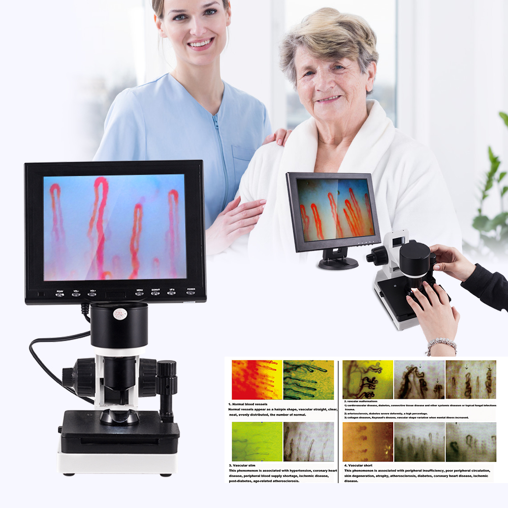
Nailfold capillaroscopy scleroderma evaluation is a non-invasive optical imaging method used to examine peripheral microvascular architecture at the periungual nailfold. In systemic sclerosis and related connective tissue disorders, microvascular changes often emerge long before extensive skin fibrosis or organ complications develop. Specialized capillaroscopy equipment visualizes capillary loop enlargement, microhemorrhages, capillary loss, and structural disorganization in living tissue.
Selecting an appropriate observation platform depends on optical magnification, stage stability, illumination control, and digital documentation capabilities. High-resolution videocapillaroscopy and dedicated microcirculation microscopes provide standardized views that help medical specialists document disease activity and track microangiopathy over time.
Role of Nailfold Capillaroscopy in Systemic Sclerosis
Nailfold capillaroscopy in systemic sclerosis provides a direct window into peripheral microvascular damage. Systemic sclerosis is characterized by widespread microangiopathy accompanied by progressive immune activation and tissue fibrosis. Because the capillary loops at the base of the fingernail run parallel to the epidermal surface, clinicians can examine the complete hairpin morphology rather than merely viewing vertical vessel tips.
Early microvascular alterations reflect endothelial activation, intimal proliferation, and capillary wall breakdown. Identifying these signs aids in the differential diagnosis of secondary vascular dysfunction, distinguishing it from primary vasospastic events. The technique serves as an objective imaging modality recognized in international classification guidelines for systemic sclerosis.
Periungual capillaries provide direct optical access to human microcirculation. Observational records of loop dimensions and density assist medical teams in tracking secondary microvascular complications across longitudinal evaluations.
Observing peripheral microcirculation also provides insight into broader tissue perfusion risks. Structural microangiopathy in the nailfold frequently correlates with vascular manifestations elsewhere in the body, such as digital ulcers and visceral vascular involvement.
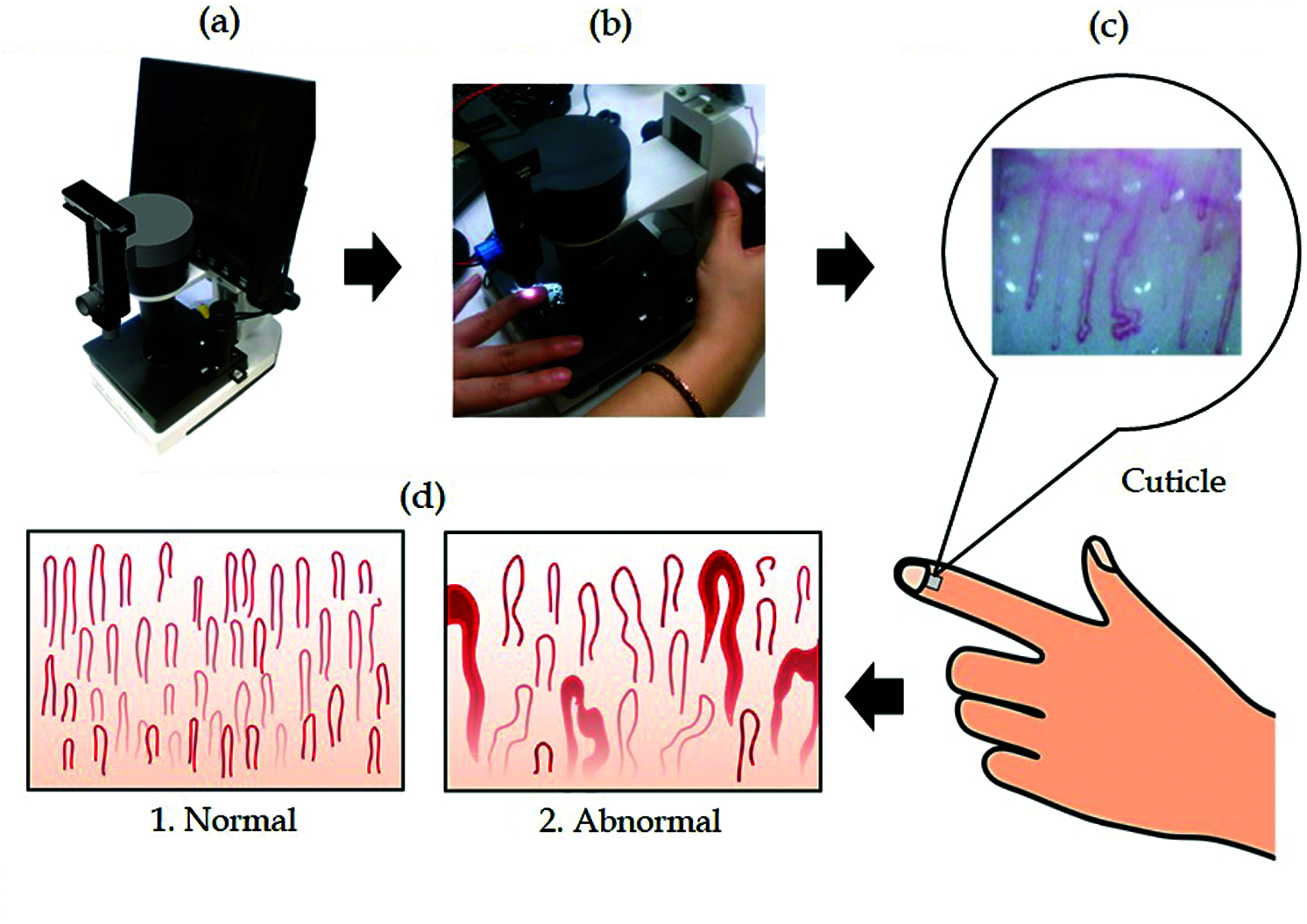
Standardized periungual optical inspection supports non-invasive documentation of microvascular architecture.
Scleroderma Nail Capillaries and Morphological Markers
Evaluation of scleroderma nail capillaries relies on qualitative and quantitative analysis of several discrete vascular markers. Healthy nailfold microcirculation presents a regular row of uniform, hairpin-like loops with even distribution and a capillary density typically ranging between 7 and 12 loops per linear millimeter. In connective tissue diseases, this architectural uniformity degrades into distinct pathological markers.
- Homogeneous and Giant Capillaries: Extreme lumen dilatation, commonly defined as loops exceeding 50 micrometers in apical diameter, represents early structural damage and endothelial wall weakness.
- Microhemorrhages: Extravasated erythrocytes appear as distinct pericapillary hemosiderin deposits or dark halos, signaling capillary rupture and active microangiopathy.
- Avascular Areas: Loss of two or more consecutive capillary loops produces local desertification, which reflects significant microvascular drop-out and tissue ischemia.
- Arborized and Ramified Loops: Bushy, complex capillary branching suggests neoangiogenesis attempting to compensate for widespread capillary loss.
- Disorganized Architecture: Marked deviation from normal parallel loop arrangement signals ongoing microvascular remodeling.
Documenting these morphological anomalies provides an objective baseline for clinical review. Identifying lower capillary density alongside recurrent hemorrhages helps specialists track systemic sclerosis disease progression.
Understanding Documented Scleroderma Patterns
Using capillaroscopy to detect sclerosis-related changes centers on identifying distinct microvascular patterns described in medical literature. The clinical assessment framework defined by Cutolo and colleagues categorizes microvascular involvement into early, active, and late patterns. Each phase exhibits specific morphological characteristics that reflect progressive vascular remodeling.
| Pattern Stage | Primary Morphology | Capillary Density | Structural Architecture |
| Early SSc | Giant loops, few hemorrhages | Preserved | Architecture remains mostly organized with minimal capillary loss. |
| Active SSc | Frequent giants and hemorrhages | Moderate loss | Visible disruption with moderate disorganization and focal avascular zones. |
| Late SSc | Severe drop-out, ramified loops | Severe drop-out | Extensive avascular areas, neoangiogenesis, and disorganization. |
The early pattern typically shows isolated giant capillary loops with relatively well-preserved overall density. In the active stage, giant vessels appear alongside frequent pericapillary hemorrhages and mild to moderate capillary loss. The late pattern is defined by severe microvascular drop-out, extensive avascular areas, and bushy neoangiogenic loops that exhibit pronounced structural distortion.
Systematic classification helps clinical practitioners communicate findings consistently. Equipment that supports digital archiving enables comparative reviews between successive patient visits.

High-magnification visualization allows accurate grading of microvascular remodeling patterns.
Recommended Examination Conditions and Practical Workflow
Reliable observation requires standardized operating conditions to avoid transient vasospasm or image distortion. Peripheral microcirculation is highly sensitive to environmental temperature, recent physical exertion, and external pressure. Adhering to controlled preparation steps minimizes non-disease-related vascular variations during routine evaluation.
- Maintain ambient room temperature between 22 and 24 degrees Celsius with stable relative humidity near 70% to prevent reactive vasoconstriction.
- Seat the patient comfortably with the examined hand resting at approximately heart level for 15 to 30 minutes before initiating image capture.
- Advise the subject to refrain from strenuous exercise, smoking, caffeine, and hand washing for at least one hour prior to the assessment.
- Apply two to three drops of immersion oil, such as cedar oil or high-grade paraffin oil, directly to the distal nailfold to reduce superficial light scattering.
- Position the ring or middle finger securely in the stage holder, avoiding excessive mechanical pressure that might occlude capillary blood flow.
- Adjust coarse focusing to identify the distal row of capillaries, followed by coaxial fine tuning to achieve clear vessel delineation.
- Inspect all digits systematically, excluding thumbs when practical due to thicker cuticles and frequent traumatic alterations.
Institutional protocols may incorporate additional criteria based on local safety policies and specialty guidelines. Consistency across repeat evaluations helps ensure comparable image series for research and clinical tracking.
Technical Architecture of Microcirculation Microscopes
Specialized nailfold capillaroscopy equipment incorporates dedicated optical, mechanical, and illumination sub-systems designed for periungual microvascular inspection. Unlike standard biological microscopes, these instruments provide horizontal clearance, specialized finger stages, and high-angle surface lighting. Proper component integration determines image sharpness, contrast, and operator ergonomics.
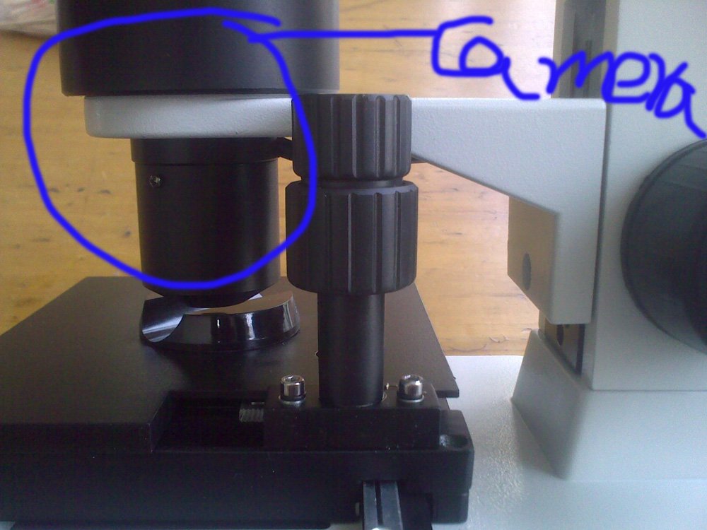
Achromatic Optical Objective
High-grade 5X achromatized objectives with a 25 mm effective range minimize chromatic aberration. Combined with internal tube optics, they produce total magnifications appropriate for capillary loop inspection.

LED Cold Illumination
Built-in LED cold light sources direct concentrated illumination onto the nailfold without generating surface heat. Preventing local skin warming is vital to avoid induced vasodilation during extended examinations.
Smooth mechanical positioning is achieved through X-Y double-layer compound mechanical stages. Coaxial coarse and fine adjustment wheels allow microscopic focus shifts without disturbing the seated finger position. Digital video sensors capture dynamic red blood cell column movements and transmit live video streams directly to integrated LCD screens or external computer interfaces.
Technical Specifications and Component Parameters
Modern clinical workstations require documented hardware specifications for institutional procurement and regulatory vetting. Microcirculation systems such as the MAIKONG MK-GY880 series incorporate dedicated mechanical housings and integrated displays. The following parameters reflect manufacturer baseline documentation, though institutions should confirm specific values prior to purchase.
| Parameter Category | System Specification | Configuration Notes |
| Brand Name | MAIKONG | Standard equipment manufacturer identifier. |
| Model Series | MK-GY880 | Integrated benchtop microcirculation workstation. |
| Optical Amplification | 240X or 380X | Options for 380X, 500X, or 600X available depending on optical lens selection. |
| Objective Type | 5X Achromatization | Effective working range 25 mm for stable periungual positioning. |
| Mechanical Stage | X-Y Double Layer | Compound mechanical stage with dual-axis transverse movement. |
| Focus Mechanism | Coaxial Coarse/Fine | Precision adjustment wheels for rapid focal plane alignment. |
| Light Source | LED Cold Light | Internal dimmable illumination to prevent thermal vasodilation. |
| Imaging Sensor | Integrated Camera | Manufacturer notes 80,000-pixel video sensor; confirm resolution needs. |
| LCD Monitor | 8 to 12 Inch | Color LCD screen options include 8, 9, 10, or 12-inch panels. |
| Power Input/Output | 100-240V AC / 12V DC | Universal AC adapter providing 12V 1A or 2A direct current. |
| Weight & Packaging | 6 kg Net / 7 kg Gross | Aluminum security carry box with custom memory foam inside corrugated carton. |
Buyers exploring digital nailfold capillaroscopy microscope systems should note that historical draft literature occasionally contains conflicting optical references, such as 360X or higher magnification notations. Institutional procurement teams should verify specific sensor resolutions, screen dimensions, and CE documentation with technical representatives before finalizing orders.
| Supplied Component | Standard Quantity | Functional Description |
| MK-GY880 Host | 1 piece | Main mechanical chassis with optical column and stage. |
| Color LCD Monitor | 1 piece | Dedicated display unit mounting directly atop the column. |
| Power Adapter | 1 piece | Regulated power converter supporting regional AC standards. |
| Immersion Cedar Oil | 1 bottle | Optical coupling medium to improve epidermal light transmission. |
| Cotton Swabs | 1 pack | Disposable applicators for clean immersion oil placement. |
| Reference Chart | 1 piece | Visual comparison chart for common capillary morphology types. |
Factors Influencing Imaging Stability and Field Clarity
Acquiring repeatable microvascular imagery requires attention to mechanical and optical variables. The small depth of field inherent in high-magnification systems means even minor involuntary finger tremors can disrupt the focal plane. Operators must utilize built-in stabilizing finger rests to steady the examined extremity during image recording.
A high-performance nailfold capillaroscopy camera relies on correct immersion medium application. Insufficient oil leads to excessive surface refraction from the dry stratum corneum, creating harsh highlights that obscure the capillary loops. Conversely, an overabundance of oil may introduce air bubbles that generate dark artifacts on the video feed.
Avoid excessive downward mechanical force on the finger rest. External compression can collapse delicate superficial capillaries, producing artificial avascular zones or simulated blood flow arrest.
Glare management depends on the orientation of the cold light illuminator. Tilting the illumination angle slightly reduces specular reflections from the nail plate, allowing clear observation of apical capillary loops and background tissue translucency.

Accurate immersion oil application minimizes epidermal light scattering for enhanced contrast.
Primary Raynaud Phenomenon Versus Connective Tissue Diseases
A primary clinical utility of nail capillary microscopy is assisting in the differential distinction between primary Raynaud phenomenon and secondary Raynaud phenomenon associated with systemic sclerosis. Primary Raynaud phenomenon is a functional vasospastic condition that rarely involves structural capillary destruction. In contrast, secondary forms exhibit progressive vascular remodeling and architectural loss.
Primary Raynaud Findings
- Normal capillary loop density (7-12 loops per mm)
- Preserved hairpin architecture with minimal tortuosity
- Absence of giant capillaries (apical diameter under 50 micrometers)
- No avascular desertification areas
- Extravasated microhemorrhages are typically absent
Secondary SSc-Pattern Findings
- Reduced capillary density with progressive loop loss
- Severe structural disorganization and apical distortion
- Frequent giant capillary loops exceeding 50 micrometers
- Focal or extensive avascular zones
- Prominent pericapillary hemorrhages and neoangiogenesis
Differentiating these presentations helps clinicians determine whether further serological testing, such as antinuclear antibody screening, is warranted. Capillaroscopy serves as an indispensable visual checkpoint within modern rheumatologic evaluation pathways.
Institutional Buyer Checklist for Capillaroscopy Systems
Acquiring professional nailfold capillaroscope systems requires hospital procurement committees, research labs, and specialty clinics to weigh several technical and operational criteria. Balancing optical performance with workflow simplicity ensures broad adoption across clinical departments.
- Optical Resolution and Magnification: Confirm verified optical magnification (such as 200X to 380X) rather than digital interpolation to ensure accurate measurement of capillary apex diameters.
- Mechanical Stage Precision: Prioritize dual-axis compound X-Y mechanical stages with smooth coaxial knobs to allow effortless tracking across the nailfold row.
- Thermal Output: Verify true LED cold light illumination to prevent thermal interference with local microcirculation during examinations.
- Display Ergonomics: Evaluate whether integrated LCD monitors or direct USB/video output to computer workstations best suit your clinical documentation needs.
- Digital Archiving Capabilities: Determine software compatibility for saving still images, measuring vessel diameters, and recording dynamic blood flow videos.
- Build Quality and Packaging: Assess whether aluminum transport cases with protective foam are provided for equipment safety during clinic-to-clinic transport.
- Supplier Customization Support: Check manufacturer capacity for private labeling, OEM modifications, and replacement component availability.
Addressing these specifications systematically helps institutions secure imaging platforms that satisfy long-term diagnostic observation and academic research objectives.
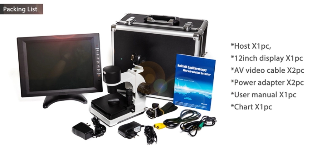
Selecting robust workstations with dedicated carrying cases facilitates safe mobility across hospital departments.
Operational Boundaries and Observational Scope
While nailfold capillaroscopy offers detailed visualization of peripheral microvasculature, equipment-based observation possesses defined technical boundaries. The hardware serves solely to observe, magnify, and record microvascular structures. It does not generate automatic diagnostic verdicts or substitute for comprehensive clinical judgment.
Capillaroscopic images must be interpreted in conjunction with patient history, physical findings, and laboratory tests. Microvascular anomalies can occasionally be triggered by local trauma, occupational vibration, or non-rheumatologic vascular disorders. Qualified healthcare professionals must oversee image acquisition and diagnostic interpretation according to institutional protocols.
Capillaroscopy systems are optical imaging and educational tools. Diagnostic conclusions, staging determinations, and therapeutic plans remain the sole responsibility of licensed medical practitioners.
Furthermore, broader application claims mentioned in legacy documentation—such as observing microcirculation on other anatomical areas or testing consumer materials—fall outside standard rheumatologic protocols. Clinical focus should remain squarely on standardized nailfold inspection protocols.
MAIKONG Manufacturing and Supply Chain Capabilities
MAIKONG is an established medical optoelectronic instrument manufacturer located in GD, SZ. Drawing on a robust regional manufacturing supply chain and specialized production facilities, MAIKONG delivers both high-end and cost-effective microcirculation inspection solutions to international markets. Production processes adhere to stringent optical assembly and testing standards.
Precision Manufacturing
Advanced optical assembly facilities in GD, SZ ensure rigid alignment of achromatized lenses and mechanical stages.
OEM and Customization
Support for custom branding, hardware modifications, tailored display configurations, and private-label packaging.
Global Distributor Supply
Dependable production scheduling and direct supply chain logistics tailored for wholesale and institutional partners.
For additional details regarding production facilities and company history, review our background on the About us page. MAIKONG collaborates closely with medical device distributors and healthcare organizations to provide dependable imaging hardware.
Frequently Asked Questions
Can nailfold capillaroscopy confirm scleroderma by itself?
No, nailfold capillaroscopy cannot confirm systemic sclerosis in isolation. It provides visual evidence of peripheral microangiopathy that clinicians evaluate alongside serological tests, clinical symptoms, and established diagnostic criteria.
What does a scleroderma nailfold pattern refer to?
A scleroderma nailfold pattern refers to characteristic morphological changes including giant capillaries, microhemorrhages, capillary desertification, and arborized neoangiogenic loops. These patterns are categorized into early, active, and late stages reflecting microvascular disease progression.
Is nailfold capillaroscopy invasive?
Nailfold capillaroscopy is entirely non-invasive and painless. The examination requires only the application of optical immersion oil to the cuticle surface followed by non-contact or light-contact microscopic viewing.
Which medical professionals commonly utilize this imaging technique?
Rheumatologists, vascular medicine specialists, dermatologists, and microcirculation clinical researchers routinely utilize nailfold capillaroscopy. The technique is also used by academic medical educators and hospital diagnostic imaging centers.
What equipment features matter most for systemic sclerosis imaging?
Essential features include true optical magnification between 200X and 380X, cold LED illumination to prevent induced vasodilation, a stable X-Y mechanical stage, and high-definition video capture for digital documentation.
Does MAIKONG support institutional procurement and OEM distributor requests?
Yes, MAIKONG provides direct OEM and ODM manufacturing services, private-label branding, and wholesale distribution support for institutional buyers and commercial medical suppliers.
Summary and Professional Consultation
Scleroderma nailfold capillaroscopy remains a foundational non-invasive imaging method for evaluating peripheral microvascular architecture and tracking connective tissue microangiopathy. High-quality optical magnification, stable stage mechanics, and cool illumination provide clinical teams with the clear documentation required for longitudinal patient assessments.
Equip Your Department with Professional Capillaroscopy Hardware
Explore technical options and distribution opportunities for MAIKONG microcirculation microscopes and videocapillaroscopy workstations.