
Nail Videocapillaroscopy: How Video-Based Nailfold Microvascular Observation Works
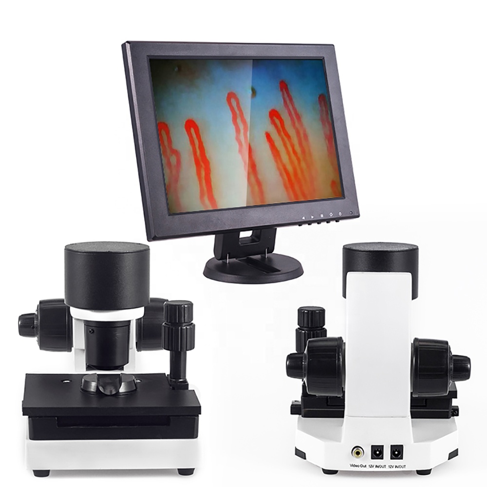
Nail videocapillaroscopy is a non-invasive optical technique designed for direct, real-time visualization of nutritional capillary loops within the periungual skin border. The instrumentation pairs high-magnification optical lenses with dedicated digital video sensors to project live microvascular dynamics onto high-resolution displays. Healthcare professionals and scientific researchers utilize this method to evaluate capillary architecture, morphological integrity, and microvascular flow dynamics in living tissue.
Selecting appropriate equipment depends on required optical resolution, stage stability, illumination type, and whether data must be archived for longitudinal evaluation. Institutional workflows benefit when hardware balances precise optical focus with dependable mechanical controls to minimize motion artifacts during observation.
Microvascular Features Observable at the Nailfold
The nailfold presents a unique anatomical orientation where nutritional capillaries run parallel to the epidermal surface rather than perpendicular to it. This horizontal arrangement allows unobstructed visualization of the complete microvascular loop, including the afferent arteriolar limb, the apical loop, and the efferent venular limb.
Under stable magnification, trained observers track distinct microvascular characteristics that reflect the functional state of the peripheral circulation:
- Capillary density and loop distribution across the distal row of the nailbed.
- Loop dimensions, including apical diameter and limb width.
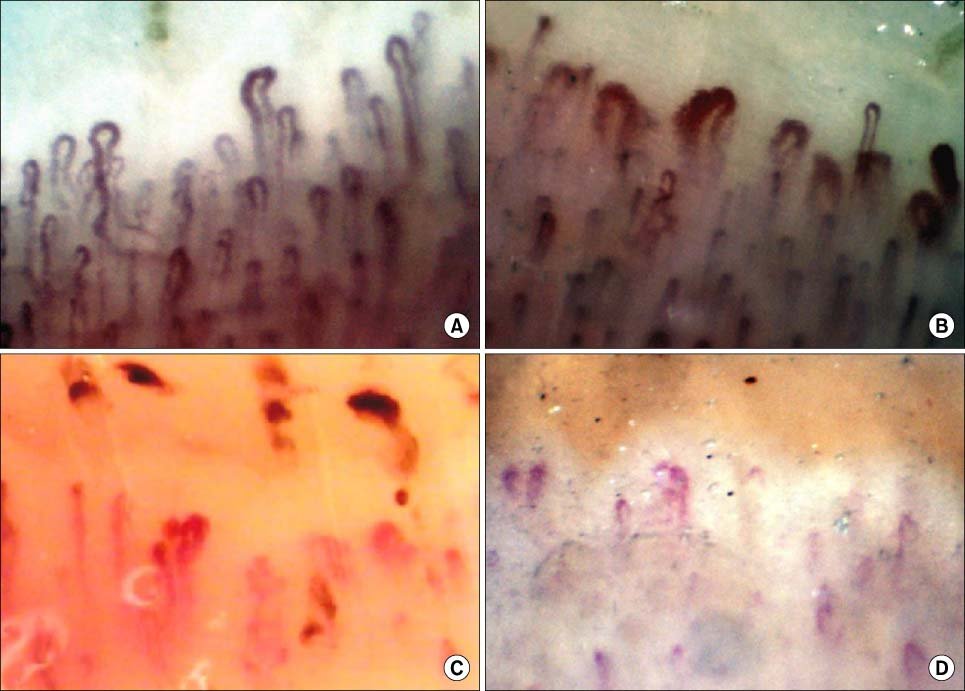
- Morphological variations, such as tortuosity, ramified formations, or prominent hairpin configurations.
- Presence of enlarged loops, including the appearance of giant capillaries.
- Extravasation of red blood cells, observed as distinct microhemorrhages in the pericapillary area.
- Subpapillary venular plexus visibility and capillary blood flow velocity.
In specialized investigations involving connective tissue disease, tracking microvascular damage helps researchers document structural changes over time. When investigating conditions such as the scleroderma pattern or assessing systemic sclerosis patients, documenting capillary loss or structural disruption provides standardized visual metrics alongside other clinical criteria, such as those evaluated in nailfold capillaroscopy scleroderma research protocols.
Core Engineering of a Videocapillaroscope
A functional nailfold videocapillaroscope integrates specialized optoelectronic subsystems to deliver distortion-free dynamic streaming directly from the dermal papillae to an attached monitor. Every hardware element contributes to resolving microscopic vascular structures without introducing thermal disruption or optical distortion.

Optical Magnification Assembly
Achromatic lens systems offer true optical magnification, typically configured at 240X or 380X, with extended options reaching 500X or 600X. These optics correct for chromatic aberration, delivering flat-field clarity across the entire field of view.

Digital Sensor Architecture
Dedicated CCD sensors convert captured photons into steady dynamic video signals. Stable frame rates prevent motion blur during continuous real-time capillary blood flow monitoring.
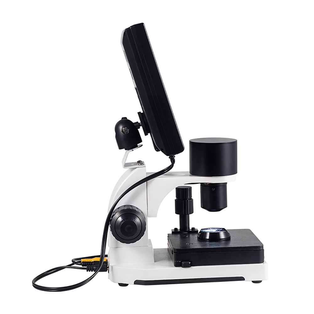
Integrated Cold Illumination
Built-in LED cold-light sources direct concentrated illumination onto the nailfold without transferring heat to the skin. Avoiding localized temperature elevation prevents reactive vasodilation or artifactual flow alterations.
The system connects directly to dedicated color LCD monitors, commonly sized from 8 inches to 12 inches, allowing immediate multi-person review without requiring real-time ocular eyepiece alignment. Live digital output supports systematic documentation and longitudinal comparative studies across patient visits.
Optical Alignment and Mechanical Movement
Stabilizing the subject digit and maintaining focal precision are essential for high-magnification microvascular observation. Minor involuntary finger twitches or tremor can displace microscopic capillary loops entirely out of frame.
The mechanical stage uses a dual-layer compound X-Y platform operated via coaxial control wheels, allowing smooth lateral and longitudinal tracking across the entire distal capillary row.
Focusing utilizes a coaxial coarse and fine adjustment mechanism. Operators engage the coarse adjustment handwheel to locate the initial tissue boundary before rotating the fine focus handwheel to resolve erythrocyte columns inside delicate loop walls.
An adjustable finger press holds the examined digit steadily in place against an ergonomic contoured seat. Operators can also employ image point rotation handwheels to eliminate minor optical reflections and align the horizon of the nailbed before image recording.
Step-by-Step Observation Protocol
Standardizing examination steps helps clinicians and laboratory investigators capture reproducible imaging across serial sessions. A methodical routine ensures optimal tissue illumination, stable focus, and minimal subject discomfort during a nailfold capillary test.
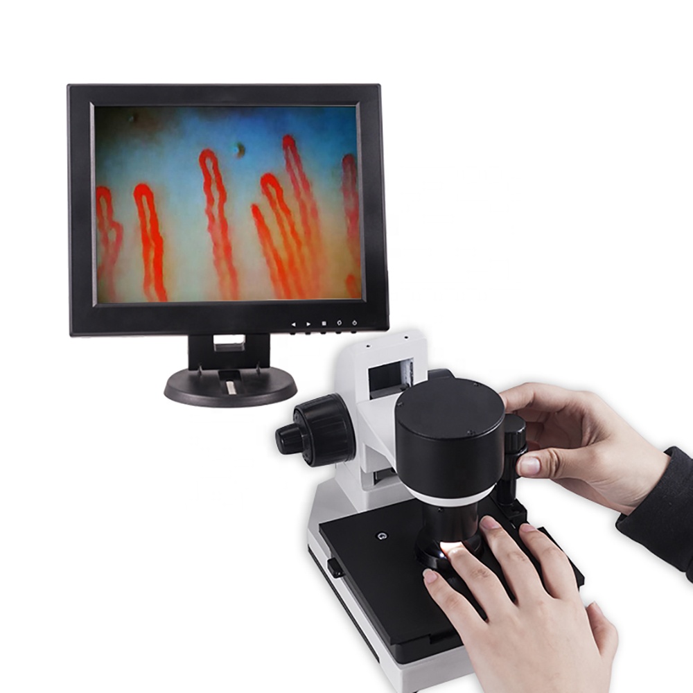
- Connect the display and optoelectronic base unit to the DC power source, powering on the monitor and setting initial contrast and brightness controls.
- Place two to three drops of immersion oil—such as cedar oil or pharmaceutical-grade paraffin—onto the periungual cuticular junction of the selected finger to improve epidermal translucency and reduce surface reflection.
- Insert the subject finger into the dedicated finger holder located beneath the objective lens assembly, securing the digit gently with the stabilizing finger press.
- Rotate the coarse focusing handwheel until the epidermal margin and tissue reflections appear on the LCD monitor.
- Engage the fine adjustment handwheel to bring individual capillary loops and moving red blood cell aggregates into crisp focus.
- Translate the mechanical stage laterally across the X-axis to systematically survey the distal row from lateral fold to lateral fold, recording structural anomalies or areas of capillary loss.
Following this structured protocol allows the operator to locate and record target capillary fields quickly, reducing examination duration while maintaining clear image definition.
Environmental Factors and Subject Preparation
Cutaneous microcirculation reacts dynamically to external temperature, physical activity, and emotional stress. Uncontrolled testing conditions can trigger peripheral vasoconstriction, making normal capillaries difficult to visualize or misinterpreting spastic flow as microvascular disease.
Room temperature should remain between 22°C and 24°C with relative humidity maintained near 70% to prevent reactive peripheral vasoconstriction or perspiration artifacts.
Subjects should rest comfortably in a seated position for 15 to 30 minutes before observation begins, keeping their hands elevated near heart level. This acclimatization period stabilizes baseline dermal blood flow throughout the examination.
Supervising protocols generally instruct subjects to avoid strenuous physical exertion, smoking, caffeine intake, and localized hand washing with cold water for one hour prior to imaging. Any ongoing prescription drug regimens should be recorded according to institutional research guidelines without altering clinician-directed treatments.
Investigation Contexts in Microcirculation Research
Because peripheral capillaries provide a direct view of microvascular morphology, nail fold capillaroscopy has become an established investigative tool in medical institutions, academic centers, and research laboratories.
- Rheumatology research, supporting observation of microvascular patterns associated with Raynaud phenomenon, systemic sclerosis, and related systemic autoimmune conditions.
- Angiology and vascular medicine, assessing microcirculatory responses in peripheral occlusive diseases, chronic venous insufficiency, and acrocyanosis.
- Dermatology, evaluating superficial microvascular changes in psoriasis, dermatitis, localized scleroderma, and chronic wound margins.
- Endocrine and metabolic studies, investigating functional capillary alterations and baseline flow characteristics.
- Academic anatomy and biomedical engineering instruction, demonstrating live human hemodynamics and microcirculatory physiological principles.
- Physiological evaluations, testing dermal responses to specialized therapeutic interventions or topical agents.
While the hardware produces high-fidelity visual documentation, it does not formulate diagnoses independently. All captured video streams and still frames require qualified interpretation by licensed medical professionals or trained investigators in accordance with recognized classification criteria.
Technical Configurations and Reference Specifications
Selecting appropriate nailfold capillaroscopy equipment requires matching optical capabilities and stage design to the facility’s workflow. Systems must provide rigid physical stability, consistent illumination, and clean signal transmission to support longitudinal comparative tracking.
| Parameter | Configuration Option | Specification Detail |
| Brand Name | MAIKONG | Manufactured hardware platform |
| Optical Function | Detection System | Non-invasive body health assessment |
| Amplification | Multi-Option | 380X, 500X, or 600X optical magnification |
| Electrical Input | Universal AC | AC 100-240V 50/60Hz compatible |
| Output Voltage | DC Output | Regulated DC 12V at 2A |
| Objective Lens | 5X Optical | Achromatization with 25mm effective range |
| Stage Type | Dual Layer | X-Y compound mechanical movement stage |
| Focus System | Coaxial Dial | Independent coarse and fine adjustment controls |
| Illumination | Cold Light | Integrated inner dimming LED assembly |
| Protection Type | Type I or B | Compliant electronic safety rating |
| Power Input | Base System | DC 12V at 1A dedicated power consumption |
| Built-In Camera | CCD Sensor | Integrated 80,000-pixel digital imaging module |
| LCD Monitor | Color Screen | 8-inch, 10-inch, or 12-inch display panels |
| Certification | CE Certified | CE approved optoelectronic system |
| Net Weight | 6.0 Kilograms | Stable benchtop operating weight |
| Gross Weight | 7.0 Kilograms | Total packed weight with protective housing |
An alternative compact configuration provides 240X or 380X magnification paired with an 8-inch or 9-inch color display. Outer dimensions for this setup measure 37.5 cm by 26.5 cm by 34 cm, housed in a reinforced password-protected aluminum transit case lined with high-density memory foam.
Packaging Standards and Included Accessories
Every nailfold capillaroscopy device ships as a self-contained operational station equipped with all essential components required for rapid clinical installation. Robust packaging shields the optical assemblies, stage gearing, and digital screens during transit.
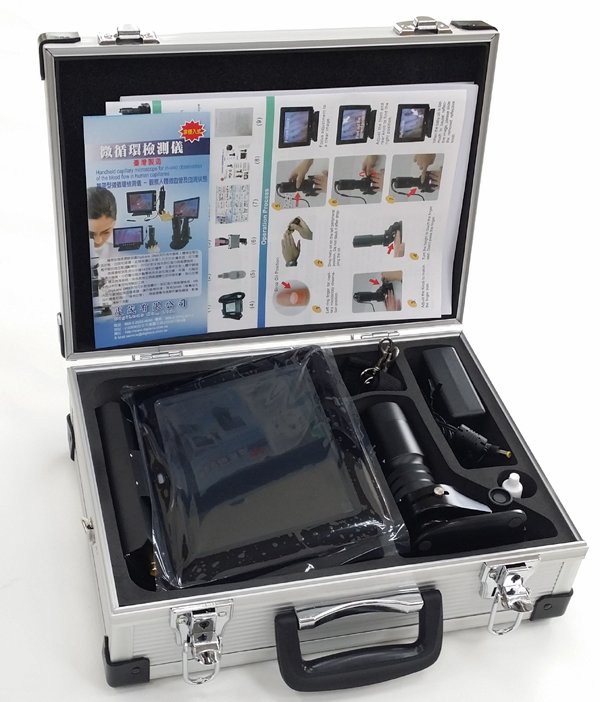
| Component Item | Quantity | Operational Purpose |
| MK-GY880 Host | 1 Unit | Core optoelectronic microscope and stage base |
| LCD Monitor | 1 Unit | Dedicated color real-time observation display |
| Power Adapter | 1 Unit | Regulated AC-to-DC mains power supply |
| Cedar Oil | 1 Bottle | Immersion liquid for epidermal transparency |
| Cotton Swabs | 1 Pack | Precision oil application and lens care tools |
| Reference Chart | 1 Piece | Morphological capillary assessment guide |
The inner aluminum carrying case protects sensitive alignments from mechanical shock, while an outer multi-wall corrugated cardboard container prevents external handling damage. Facilities should confirm specific plug configurations, display dimensions, and accessory preferences during order placement.
Supplier and Customization Considerations
MAIKONG operates advanced optoelectronic manufacturing facilities based in Shenzhen, GD, China. We leverage regional optical supply chains to manufacture high-tier, cost-effective capillaroscopy instruments engineered for international healthcare and scientific research markets.
Our production facilities support comprehensive original equipment manufacturing (OEM) and private-label customization programs. Resellers and institutional buyers can request tailored chassis branding, specialized software interfaces, customized monitor enclosures, and specific magnification assemblies to fit regional market requirements.
Establish Equipment Partnerships
Institutions and medical device distributors seeking reliable wholesale manufacturing, custom private-label branding, or direct supply chain coordination can review our organizational capabilities.
Checklist for Equipment Resellers and Buyers
Procuring microvascular imaging systems requires verifying mechanical build quality, sensor stability, and ongoing support services. Evaluating potential suppliers across structured criteria ensures that delivered hardware meets departmental standards:
- Assess magnification requirements, verifying whether fixed 380X optics or flexible multi-step 240X to 600X systems better serve clinical protocols.
- Confirm optical stage mechanics, ensuring smooth X-Y compound motion without play or backlash that could disrupt microvascular tracking.
- Review digital video output formats to ensure compatibility with departmental archiving servers or external workstations.
- Verify electrical certifications, plug types, and power supply compliance with local institutional building codes.
- Evaluate packaging integrity, confirming aluminum road cases and shock-absorbing foam for mobile health screening projects.
- Compare pricing structures through the nailfold capillaroscopy equipment price index to balance capital expenditure with required optical capabilities.
Establishing clear technical requirements before purchasing helps clinics, universities, and commercial distributors secure the right configuration for long-term operational success.
Frequently Asked Questions
What is nail videocapillaroscopy?
Nail videocapillaroscopy is an optical imaging method that combines high-power microscope optics with digital video sensors to inspect the microcirculatory capillary loops of the nailfold in real time.
How does videocapillaroscopy differ from standard dermoscopy?
Videocapillaroscopy utilizes significantly higher optical magnifications, often between 200X and 600X, enabling detailed evaluation of individual capillary loops, whereas standard dermoscopy operates at lower magnifications suited for broader tissue screening.
Why is immersion oil applied to the cuticular junction?
Immersion oil matches the optical refractive index of the stratum corneum, reducing light scattering and making the epidermis transparent enough to reveal dermal capillary loops clearly.
Can nail videocapillaroscopy independently diagnose autoimmune disorders?
Nail videocapillaroscopy does not independently diagnose diseases. It serves as an observational imaging modality whose visual findings must be interpreted by trained clinicians alongside laboratory results and complete clinical criteria.
What video sensor resolution is standard for nailfold observation?
Modern systems commonly feature dedicated CCD or CMOS sensor modules, such as an integrated nailfold capillaroscopy camera delivering 80,000 pixels or higher to maintain clean, live video feeds without jitter.
What ambient conditions are recommended during microvascular examination?
Rooms should maintain steady temperatures between 22°C and 24°C with roughly 70% relative humidity, allowing the subject to acclimate for 15 to 30 minutes to stabilize baseline peripheral blood flow.
Advancing Peripheral Microvascular Documentation
Nail videocapillaroscopy provides healthcare practitioners and researchers with a reliable, non-invasive method for observing living human microcirculation in real time. Combining stable cold LED illumination, precision X-Y mechanical stages, and clear video streaming enables reproducible microvascular documentation across diverse investigative settings.
Connect with MAIKONG Supply Specialists
Organizations interested in equipment procurement, local distribution partnerships, or OEM production inquiries can connect directly with our technical support team.