
Nail Fold Function: Anatomy, Protective Role, and Importance in Microvascular Observation

The primary nail fold function is to form a continuous biological seal over the germinative nail matrix while anchoring the edges of the growing nail plate. Simultaneously, the proximal nail fold acts as a transparent anatomical window where terminal capillary loops lie parallel to the skin surface, enabling direct non-invasive evaluation of peripheral microcirculation.
Understanding these distinct mechanical and physiological roles helps clinicians, educators, and laboratory researchers distinguish normal barrier defense from microvascular morphology. Distinguishing structural integrity from vascular dynamics establishes a clear baseline for observational microscopy and dermatological assessments.
What Is the Nail Fold?
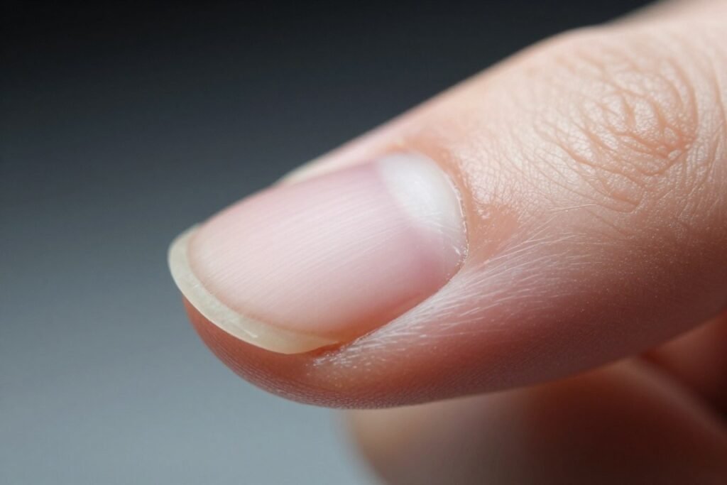
The nail fold consists of specialized invaginations of cutaneous tissue that enclose and secure the proximal and lateral borders of the nail plate. As a core component of the perionychium, this skin fold bridges keratinized dorsal epithelium with the underlying subungual structures.
Anatomically, the tissue divides into the proximal nail fold, located at the base of the nail, and the lateral nail folds bordering each side. Together, these folds create the lateral and proximal nail grooves, forming a supportive channel that guides the forward migration of the growing nail plate.
Proximal Nail Fold
The dorsal fold of skin overlying the nail root, folding beneath itself to form the eponychium and distal cuticle.
Lateral Nail Folds
The bilateral cutaneous borders running parallel to the lateral edges of the nail plate, preventing lateral displacement during daily mechanical stress.
Underlying Microvasculature
The terminal capillary network situated within the superficial dermis directly beneath the translucent epidermis of the nail fold capillary zone.
The dorsal layer of the proximal nail fold transitions into a ventral epithelial layer that rests directly on the newly formed nail plate. This specialized ventral lining lacks pilosebaceous units and sweat glands, creating a smooth surface that minimizes frictional resistance during unhindered plate elongation.
The Main Functions of the Nail Fold
The nail fold performs three coordinated functions essential for digit health, nail biomechanics, and microvascular equilibrium. These encompass biological shielding, structural guidance, and cutaneous microhemodynamic exchange.
Without an intact nail fold structure, the delicate proliferative tissues responsible for nail synthesis remain vulnerable to external mechanical trauma, chemical irritation, and microbial invasion. Furthermore, the fold regulates localized moisture levels, preventing brittle clefting at the proximal root.
- Biological sealing: The fold produces a cornified cellular layer that shields the subungual space against foreign debris, waterlogging, and bacterial contamination.
- Mechanical plate guidance: Lateral walls maintain straight, uniform trajectory during longitudinal plate growth across the underlying nail bed.
- Microvascular reservoir: The papillary dermis of the fold contains looped microvessels that nourish the germinative epithelium while dissipating localized thermal energy.
- Tactile sensory cushioning: Dense cutaneous innervation within the lateral folds assists with tactile discrimination and guards against excessive focal compression.
Each of these tasks relies on continuous tissue regeneration and normal dermal cohesion. Compromising any single layer degrades the mechanical resistance of the entire ungual unit.
Protective Roles in Nail Matrix Support and Plate Growth
Protecting the nail matrix represents the most critical biological nail fold function. The matrix resides directly beneath the proximal nail fold, housing actively dividing basal cells that differentiate into hard, sulfur-rich alpha-keratins.
The distal margin of the ventral proximal nail fold sheds the stratum corneum, creating the cuticle. This microscopic keratin seal adheres firmly to the dorsal nail plate, eliminating gaps through which infectious agents or irritants might penetrate into the germinative zone.
Loss of the cuticle seal from aggressive cosmetic manipulation or persistent moisture creates a direct entryway into the proximal nail space, elevating vulnerability to chronic inflammatory responses and localized paronychia.
When inflammatory processes disrupt the proximal fold, the underlying matrix frequently develops temporary synthetic irregularities. Transverse depressions, longitudinal ridges, or focal pitting can manifest as direct downstream consequences of localized fold injury.
In addition to microbial protection, the fold provides a flexible mechanical cushion that absorbs repetitive impact forces. Daily activities involving pinching, grasping, and keyboard typing generate focal forces that the resilient skin folds distribute away from the sensitive matrix cells.
Relationship With Superficial Capillaries and Non-Invasive Observation
Beyond mechanical defense, the proximal nail fold exhibits a distinctive anatomical feature found almost nowhere else in human skin. In most body sites, dermal capillaries run perpendicular to the skin surface, displaying only the tips of vascular loops when viewed from above.
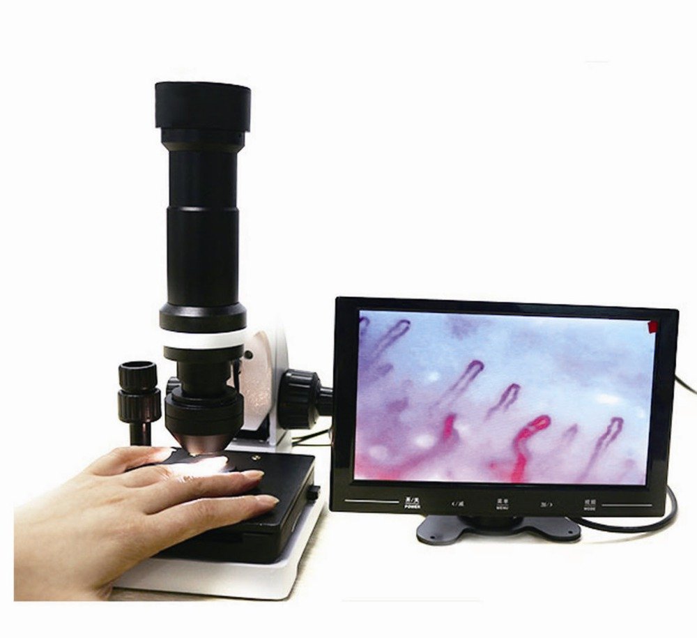
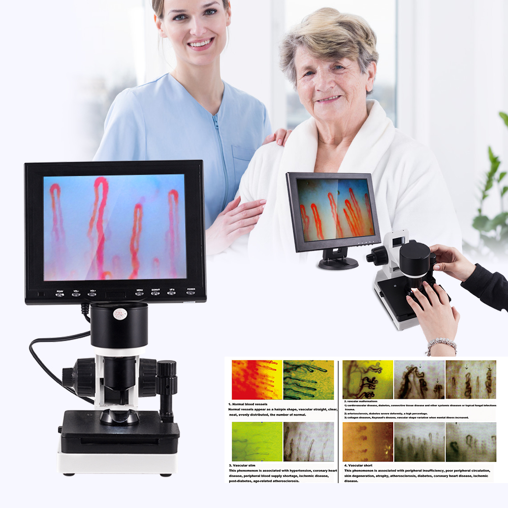
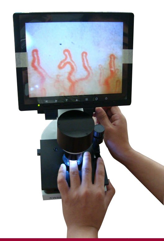
At the distal border of the proximal nail fold, the skin flattens over the nail root, forcing dermal papillae and their resident capillary loops to align horizontally, parallel to the cutaneous surface. This horizontal orientation allows the entire structural morphology of each capillary loop to remain fully visible under surface illumination using nail capillary microscopy.
Parallel Vascular Architecture
Terminal capillary loops run horizontally beneath an exceptionally thin epidermal stratum corneum, offering unobstructed lateral visualization.
Direct In Vivo Window
The absence of thick pigmentation and deep keratin layers in the ventral fold provides an ideal optical baseline without requiring skin incisions.
Because the epidermal stratum spinosum at this junction is thin and translucent, applying a drop of immersion oil reduces surface light scatter. This makes red blood cells moving through the loop clearly visible, providing an accessible site for examining peripheral microcirculation.
Nail Fold Function in Clinical Education and Biomedical Research
In academic medical centers and vascular research laboratories, the nail fold serves as a classic model for teaching microhemodynamics. Students and investigators study the direct response of superficial microvessels to physiological stimuli such as local temperature changes, sympathetic tone fluctuations, and postural alterations.
Observation at the proximal fold aids in differentiating normal physiological variations from generalized vascular alterations. Clinicians routinely incorporate the structural analysis of this region into systemic educational frameworks through nail fold capillaroscopy.
Educational Benefits
- Demonstrates real-time erythrocyte movement without invasive catheterization.
- Illustrates capillary density, hairpin loop shapes, and venous limb dilation.
- Facilitates direct observation of physiological vasospasm and reperfusion.
Research Applications
- Provides standardized anatomical landmarks for longitudinal microvascular studies.
- Permits quantitative scoring of capillary dimensions under controlled protocols.
- Serves as a reproducible model for evaluating skin barrier resilience.
Biomedical researchers also analyze the nail fold to assess epidermal barrier recovery following chemical exposure. Monitoring hydration levels and trans-epidermal water loss across the proximal groove assists in evaluating topical protective formulations and occupational barrier creams.
What Can Professionals Observe at the Nail Fold?
Under calibrated optical magnification, researchers and clinicians observe specific microvascular features that reflect terminal vascular health. These parameters include capillary loop morphology, vessel density per linear millimeter, apical limb diameter, and dynamic erythrocyte velocity.
Evaluating these elements requires optical clarity, as standard dermatoscopes may fail to resolve fine architectural distinctions. Advanced video systems, such as nail videocapillaroscopy, provide higher resolution and digital capture capabilities for consistent comparative studies.
| Morphological Feature | Normal Observation | Significance |
| Capillary Density | 7 to 12 loops | Indicates adequate distal vascularization per linear millimeter. |
| Loop Architecture | Hairpin or U-shape | Demonstrates orderly angiogenic alignment without excessive tortuosity. |
| Limb Dimensions | Symmetric caliber | Maintains balanced resistance between afferent and efferent channels. |
| Subpapillary Plexus | Variable visibility | Depends on physiological skin transparency and baseline pigmentation. |
Healthy individuals typically display an orderly comb-like row of regular, hairpin-shaped loops along the distal edge of the proximal nail fold. Minor crossing or mild tortuosity can occur naturally, particularly in individuals with a history of manual labor or localized physical microtrauma.
How Observation Conditions Affect Nail Fold Imaging
Accurate visual assessment of the nail fold requires controlled environmental conditions to avoid artifactual distortion. Cold exposure induces immediate cutaneous vasoconstriction, causing capillary loops to narrow or blood flow to pause completely.
Clinicians and researchers recommend acclimatizing subjects in a room maintained at 22 to 24 degrees Celsius with roughly 70 percent relative humidity prior to imaging. The subject should rest seated with hands positioned at heart level to minimize hydrostatic pressure artifacts within the venous limb.
Applying two drops of cedar oil, liquid paraffin, or cosmetic-grade mineral oil to the proximal fold reduces surface reflection, matching the refractive index of the stratum corneum and illuminating the microvascular bed.
Achieving stable focus requires fine manual or motorized adjustment stages. Employing a digital nailfold capillaroscopy microscope with coaxial controls helps avoid image jitter during live dynamic assessments.
Subjects should avoid strenuous exercise, tobacco use, and excessive caffeine intake for at least one hour before formal imaging. Prior microtrauma, such as recent aggressive manicuring, nail biting, or heavy physical work, can cause micro-petechiae that complicate baseline morphological interpretation.
What Nail Fold Function Does Not Tell You
While the nail fold provides an extraordinary window into cutaneous microvascular anatomy, visual inspection alone does not constitute a standalone diagnostic test. Morphological changes in nail fold capillaries reflect localized or generalized vascular behavior, but they do not confirm a specific disease without a comprehensive clinical workup.
Capillary anomalies, such as enlarged loops, microhemorrhages, or reduced vascular density, can occur across diverse clinical contexts, including inflammatory connective tissue diseases. For instance, studying microvascular changes in conditions like nailfold capillaroscopy scleroderma requires integrating serological, histological, and systemic evaluations.
Nail fold microvascular observation is an adjunct assessment tool for structural visualization and physiological research; it does not replace comprehensive medical evaluations or clinical judgment.
Normal biological variance is also common. Mild loop tortuosity, minor branching, and localized micro-ecchymoses frequently arise from everyday household tasks, accidental impacts, or environmental dryness without indicating systemic pathology.
MAIKONG Microvascular Observation Instrumentation
Supporting precise visualization of nail fold anatomy requires stable optoelectronic imaging equipment. Based in Shenzhen, GD, MAIKONG manufactures professional microcirculation microscopes designed for university research centers, vascular clinics, and clinical education laboratories.
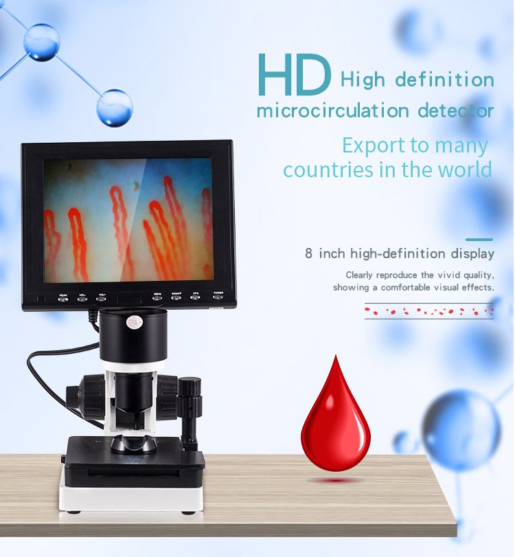
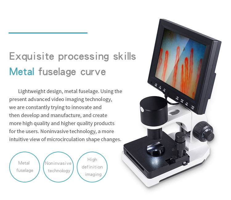
MAIKONG instruments, including the MK-GY880 series, integrate dedicated achromatic optics with cold LED illumination to deliver distortion-free dynamic imaging. The built-in CCD sensor captures real-time erythrocyte movement, transmitting crisp video streams directly to integrated LCD display monitors.
| Specification Parameter | MAIKONG MK- Series Standard | Operational Scope |
| Optical Magnification | 380X or 240X | Optional 380X, 500X, or 600X optical configurations. |
| Objective Lens | 5X Achromatization | Features an effective working focal range of 25mm. |
| Mechanical Stage | X-Y Double Layer | Smooth coaxial coarse and fine focus adjustments. |
| Illumination Source | LED Cold Light | Internal dimming prevents thermal discomfort during prolonged observation. |
| Imaging System | Built-in CCD Sensor | Displays live feed on 8-inch, 9-inch, 10-inch, or 12-inch monitors. |
| Electrical Power | AC 100-240V Input | DC 12V 2A output with international standard safety compliance. |
As an optical equipment manufacturer, MAIKONG supports healthcare technology distributors, OEM private-label brands, and academic procurement departments with complete turnkey packages. Each system ships in protective aluminum security cases to preserve mechanical alignment during transit.
| Included Component | Standard Quantity | Functional Description |
| MK- Series Host Unit | 1 pc | Solid metal chassis with integrated optical objective and finger seat. |
| Color LCD Monitor | 1 pc | Dedicated high-contrast display unit for live video projection. |
| Regulated Power Adapter | 1 pc | Universal voltage power supply unit with surge suppression. |
| Immersion Cedar Oil | 1 pc | Refractive immersion liquid for cutaneous stratum corneum clearing. |
| Cotton Swabs | 1 pack | Lint-free applicators for clean oil distribution on the nail margin. |
| Reference Chart | 1 pc | Visual guide for teaching standard capillary loop morphology. |
Clinics and research facilities seeking equipment directly from a certified nailfold capillaroscopy manufacturer benefit from factory-level customization and comprehensive accessory availability.
Frequently Asked Questions
What is the main function of the nail fold?
The primary nail fold function is to form a mechanical and biological seal that protects the underlying germinative nail matrix from pathogens, foreign debris, and physical trauma. Secondarily, the proximal fold contains horizontally oriented capillary loops that facilitate non-invasive microvascular assessment.
Is the nail fold part of the nail plate?
No, the nail fold is a specialized soft-tissue skin structure that borders and anchors the hard keratinized nail plate. The nail plate is synthesized by the matrix located beneath the proximal nail fold.
Why can capillaries be observed near the nail fold?
Capillaries at the proximal nail fold run parallel to the skin surface rather than perpendicular, as they do across the rest of the body. This horizontal alignment beneath a thin, translucent epidermis makes the full loop architecture visible under surface magnification.
Is nail fold observation invasive?
Nail fold observation is completely non-invasive, requiring only topical application of immersion oil and gentle positioning beneath a focused light source. It causes no tissue damage and does not require skin penetration or blood sampling.
Who studies nail fold structure and function?
Rheumatologists, vascular medicine specialists, dermatologists, physiology educators, and biomedical researchers regularly examine nail fold structure and microcirculation. Equipment buyers and healthcare procurement teams also evaluate optical systems dedicated to this anatomical site.
What should a professional imaging system show at the nail fold?
A professional imaging system should resolve individual capillary loops, clear apical widths, loop density per millimeter, and active erythrocyte transit without optical distortion. Coaxial focus mechanisms and uniform cold LED lighting are essential for achieving sharp, reproducible video streams.
Summary and Professional Consultation
The nail fold serves as an indispensable protective barrier for the matrix and a distinct physiological site for superficial microhemodynamic study. Maintaining fold health supports uninterrupted nail plate formation, while its unique vascular anatomy enables vital insights across education and research.
Consult MAIKONG for Professional Observation Systems
Healthcare facilities, research institutions, and medical equipment distributors seeking high-magnification nailfold observation instruments can connect with MAIKONG for technical specifications and wholesale cooperation.