
Nail Capillary Microscopy Design: Principles and Engineering Standards
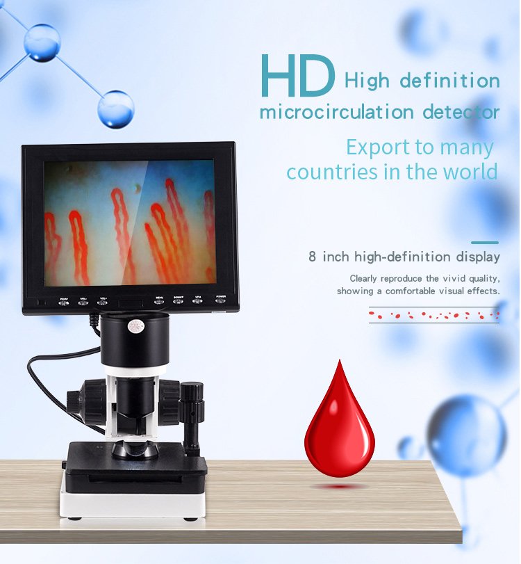
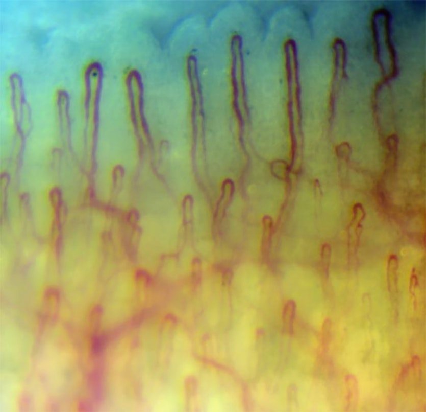
Professional nail capillary microscopy design unites precision optomechanical alignment, high-transmission achromatic lenses, and glare-free illumination to examine peripheral microvascular morphology. A balanced system architecture combines coaxial coarse and fine focusing controls, calibrated compound mechanical stages, and low-thermal-emission LED cold light to render sub-papillary vascular loops distinctly. Proper optical configuration allows researchers and clinicians to observe erythrocyte column flow without introducing thermal artifacts or mechanical distortion to sensitive tissue beds.
Procurement teams evaluate system layouts based on structural rigidity, optical clarity, display ergonomics, and electronic stability across clinical workflows. Selecting appropriate magnification options, objective working distances, and integrated imaging sensors dictates operational efficiency across medical specialties and investigative laboratories. The integration of robust chassis construction and standardized modular components supports dependable performance across high-volume diagnostic and research settings.
Optical System Architecture and Achromatization
Optical design in capillaroscopy requires an objective lens engineered to suppress chromatic and spherical aberrations along peripheral fields. Our MK- series instruments utilize a specialized 5X achromatic objective that yields an effective working range of 25mm, enabling comfortable clearance above the nailfold fold. Depending on the optical coupler and sensor configuration, total system amplification spans 240X, 380X, 500X, and 600X to satisfy varying observation requirements.
Achromatization corrects wavelength divergence across red and blue spectral regions, maintaining sharpness along microvascular margins where tissue contrast remains inherently subtle. Maintaining adequate depth of field at high optical amplification prevents rapid focal loss when peripheral microvessels present subtle vertical shifts within dermal papillae. Standardizing on calibrated glass elements minimizes image field curvature, presenting flat fields of view across the entire inspection screen.
Achromatic 5X objectives paired with matched optical pathways minimize color fringing, keeping capillary loop contours clearly delineated against dermal backgrounds.
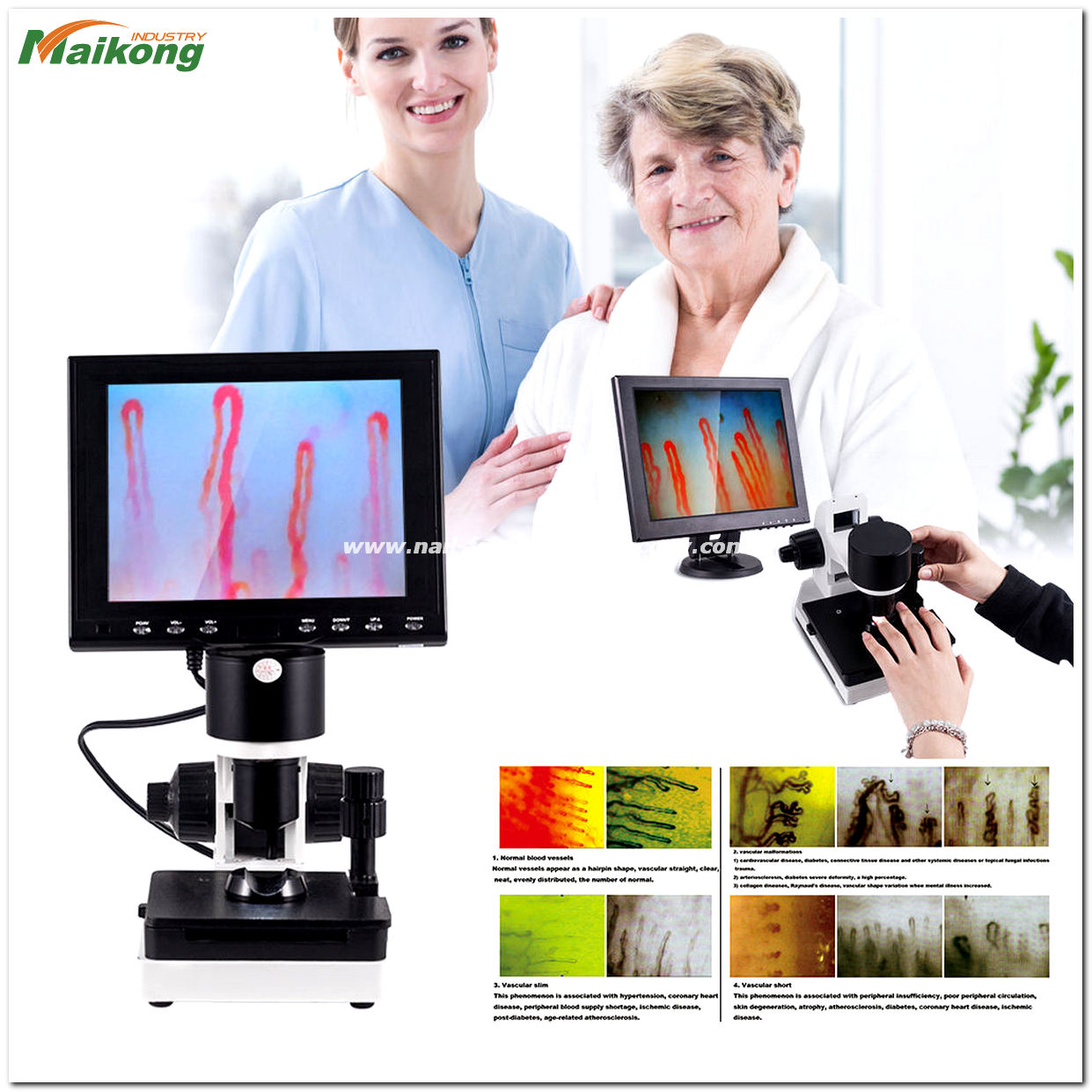
Precision achromatic objectives support flat fields of view across high-magnification microvascular evaluations.
Illumination Engineering: LED Cold Light Integration
Targeted lighting in microvascular imaging requires uniform surface illumination without radiant heat transmission that could provoke reflexive vasodilation. Modern systems feature built-in LED cold-light sources integrated around the objective assembly, directing focused rays downward onto the target zone. Integrated dimming controls allow operators to adjust light intensity smoothly to match individual tissue translucency and pigmentation profiles.
Standard incident lighting creates reflective glare on the curved surface of the nail plate, obscuring delicate loop morphology underneath. Pairing oblique or annular cold LED arrays with immersion media minimizes refractive mismatch, transforming diffuse surface scatter into transparent transillumination. Because LED cold-light engines emit negligible infrared radiation, peripheral blood flow dynamics remain unaffected by artificial thermal stimulation during extended examinations.
Integrated cold LED dimming balances illumination intensity while eliminating thermal emissions that alter microvascular vessel diameter.
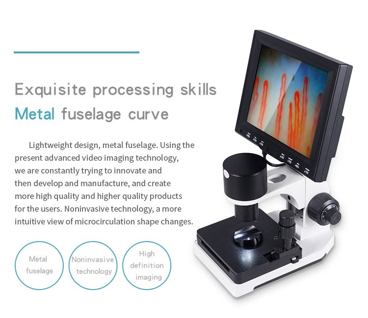
Internal LED cold-light arrays provide uniform illumination across delicate periungual tissues.
Mechanical Staging and Coaxial Focus Mechanics
Fine mechanical positioning governs overall image quality when tracking microcirculation fields smaller than a millimeter across. The mechanical chassis incorporates an X-Y double-layer compound mechanical stage that enables smooth longitudinal and lateral displacement under finger contact. Ergonomic handwheels allow technicians to navigate along the distal row of nailfold capillaries without mechanical backlash or jerky transitions.
Focusing relies on an internationally recognized coaxial coarse and fine adjustment mechanism designed to isolate minor vertical focal planes. The coarse handwheel positions the optical head within the 25mm effective range, while the concentric fine handwheel refines loop clarity down to micron thresholds. An adjustable tension collar prevents focus creep, supporting stationary observation without focal drift when hands leave the controls.
Coaxial Staging Advantages
- Smooth dual-layer movement enables precise tracking of distal capillary loops across consecutive finger positions.
- Integrated fine focus controls eliminate focal slippage during real-time video assessments.
- Ergonomic coaxial layout reduces operator hand fatigue during repeated clinical screenings.
Mechanical Constraints
- Requires periodic tension balancing to prevent vertical drift over years of clinical service.
- Heavy-duty compound stages increase overall base weight relative to basic handheld scopes.
- Demands careful handling during transport to protect fine-pitch mechanical rack-and-pinion threads.
Image Sensor Integration and Display Ergonomics
Digital conversion of microcirculation imagery requires matched sensor sensitivity to capture real-time dynamic erythrocyte flow. Our systems incorporate a dedicated high-sensitivity video camera module that streams live video feeds straight to the integrated LCD monitor without frame lag. Operators can review morphology directly on 8-inch, 9-inch, 10-inch, or 12-inch color screens mounted at eye level above the optical chassis.
Observing vascular architecture through an adjustable monitor reduces neck strain compared to traditional biological ocular eyepieces. High-contrast display panels feature dedicated brightness and contrast adjustment dials, helping clinicians distinguish subtle cellular aggregates and pericapillary halos. For digital archival needs, connecting an external nailfold capillaroscopy camera module preserves video streams for downstream documentation and clinical record management.
Direct Monitor Alignment
Mounted flat displays provide direct line-of-sight observation, improving posture for technicians during sequential patient evaluations.
Real-Time Signal Processing
Dedicated video feeds deliver uncompressed dynamic video streams, capturing erythrocyte velocity without jitter or visual artifacts.
Technical Specifications and Design Metrics
Detailed engineering metrics establish performance baselines for clinical equipment distributors and laboratory procurement directors. The hardware design reflects balanced electrical, optical, and mechanical parameters suited for continuous facility operation. Specifications may vary by configuration and should be confirmed with our engineering team before quotation.
| Parameter | Specification | Engineering Notes |
| Brand Name | MAIKONG | Shenzhen manufacturer branding |
| Function | Body Health | Microcirculation assessment device |
| Amplification | 380X 500X 600X | Optional optical configurations |
| Input | AC 100-240V | 50/60Hz universal electrical standard |
| Output | DC 12V 2A | Regulated low-voltage DC power output |
| Objective | 5X Achromat | Effective working range 25mm |
| Mechanical stage | X-Y Double Layer | Compound movement with finger press |
| Focusing | Coaxial Dual | Coarse and fine dual adjustment knobs |
| Inner dimming | LED Cold Light | Continuous rotary brightness control |
| Type | Type I or B | Medical electronic safety classification |
| Built-in camera | 80 000 Pixels | Optimized high-sensitivity sensor chip |
| LCD monitor | 8-12 Inch Color | Configurable color monitor options |
| Certificate | CE Approved | Conformity certification for global trade |
| Net weight | 6KG | Sturdy desktop chassis base |
| Gross weight | 7KG | Full shipping packaging assembly |
| NailFold Capillaroscopy Parameter | Specification | Logistical Baseline |
| Packing Dimfnsing | 37.5*26.5*34cm | Compact protective shipping envelope |
| Amplification | 380X or 240X | Standard baseline dual magnification |
| Power | 12V 1A DC | Internal operating electronic load |
| Inner package | Aluminum Box | Password lock with memory foam insert |
| Out package | Corrugated Box | Reinforced multi-wall export carton |
Operational Workflow and Ergonomic Examination Design
Operational workflow efficiency depends on standardizing procedural variables prior to positioning the patient. Environmental stability supports dependable microvascular observation, maintaining room temperature between 22-24 degrees Celsius and relative humidity near 70 percent. Individuals sit comfortably with their hand rested at heart level, resting for approximately 15 to 30 minutes before initiating the procedure.
Technicians apply two to three drops of cedar oil or medical paraffin oil across the periungual junction to optimize optical transmission and limit epidermal light scattering. The fourth digit rests securely inside the contoured finger seat positioned directly beneath the optical head. By rotating the coaxial handwheels while the lens sits roughly 2 to 3 cm from the digit, users locate and focus clearly on the terminal microcirculatory loops within 5 seconds.
- Acclimate the subject in a stable temperature setting with hands rested horizontally at cardiac level.
- Apply two drops of immersion cedar oil across the cuticle border to eliminate surface refractive barriers before initiating a standard nailfold capillary test.
- Secure the finger within the stage holder, activate the LED cold light, and engage coaxial controls to acquire steady images within seconds.
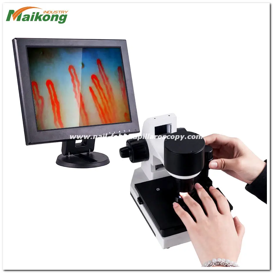
Standardized finger stabilization platforms support quick capillary acquisition during routine screenings.
Mechanical Stability and Portable System Architecture
Desktop microcirculation instruments demand structural dampening to isolate mechanical vibrations that can blur high-magnification dynamic video feeds. A rigid cast-metal base providing a 6KG net chassis weight lowers center-of-gravity oscillations, maintaining steady field position during physical contact. In contrast, portable configurations integrate lightweight 250 g illumination heads for specialized assessments outside fixed clinic benches.
Balancing benchtop rigidity against mobility shapes how clinics equip their departments. For outreach programs, a compact handheld nailfold capillaroscope enables dynamic field screening, while station-mounted platforms deliver uninterrupted coaxial precision. Incorporating anti-vibration foot pads further isolates the mechanical stage from surface vibrations caused by adjacent laboratory apparatus.
Stationary Desktop Layout
- Substantial 6KG cast base absorbs ambient laboratory floor tremors.
- Dual-layer mechanical X-Y stage provides micrometric movement.
- Integrated 8 to 12-inch monitor eliminates reliance on laptops.
Modular Portable Architecture
- Lightweight illumination head weighs approximately 250 grams.
- Compact packaging footprint allows easy transport between care wards.
- Flexible external power connections adapt to mobile screening stations.
Professional and Laboratory Integration Contexts
Modern microvascular observation instruments serve diverse analytical and investigative applications across healthcare and biomedical engineering. Within hospital environments, specialized departments deploy nail fold capillaroscopy systems to examine cutaneous microangiopathy and vascular flow patterns. Academic researchers utilize the same optical platforms to investigate functional microvascular density and endothelial response mechanisms.
Standardized optoelectronic hardware integrates smoothly across multidisciplinary facilities seeking non-invasive microcirculation evaluation tools. Common deployment settings across the United States include:
- Rheumatology clinics evaluating systemic peripheral microvascular morphological patterns.
- Vascular medicine clinics and angiology departments analyzing cutaneous circulation dynamics.
- Dermatology practices monitoring epidermal microvessels, sweat gland structures, and hair follicles.
- University medical laboratories and clinical research organizations studying blood flow hemodynamics.
- Occupational health and preventive screening centers evaluating peripheral extremity wellness.

Microcirculation imaging systems support morphological evaluation across clinical and laboratory environments.
Procurement Checklist for Medical Device Distributors
Selecting hardware configurations requires medical equipment wholesalers and clinic purchasing directors to evaluate key mechanical, optical, and regulatory factors. Verifying these specifications prior to order placement prevents workflow bottlenecks and supports smooth clinical onboarding. Distributors should assess supplier capability to deliver consistent production batches that conform to regional electrical requirements.
Commercial buyers should review several technical and operational criteria when comparing proposals:
- Intended operating environment and optical magnification options (240X, 380X, 500X, or 600X).
- Objective lens clearance, verifying the 5X achromatic design with 25mm effective working distance.
- Display preferences, selecting among integrated 8-inch, 9-inch, 10-inch, or 12-inch color LCD panels.
- Power supply compliance, confirming universal AC 100-240V input and DC 12V regulated outputs.
- Mechanical-stage precision, checking coaxial coarse and fine focus responsiveness.
- Packaging specifications, confirming heavy-duty aluminum lock cases and multi-layer cartons.
- Access to reliable spare parts, replacement power adapters, and technical documentation.
- Supplier lead times, sample review protocols, and volume nailfold capillaroscopy equipment price tiers.
OEM and Private-Label Customization Capabilities
As a specialized manufacturer based in SZ, GD, China, we operate advanced production lines backed by a robust optoelectronic supply chain. We provide comprehensive OEM and private-label manufacturing programs, enabling medical distributors to brand complete systems under their proprietary model lines. Our engineering team integrates custom chassis finishes, software branding, and custom model designations prefixed with MK-.
Distributors benefit from our scalable manufacturing facilities capable of producing both high-end clinical setups and cost-effective screening units. Prospective partners are welcome to tour our Factory to review optical calibration, stage assembly, and quality verification workflows. Flexible production agreements support custom exterior branding, localized power cords, and proprietary packaging designs for regional markets.
Direct manufacturing partnerships in SZ, GD provide distributors with agile private-label custom branding and dependable supply chain logistics.
Packaging Architecture and Standard Spare Parts
Protective packaging is engineered to shield sensitive optomechanical components from shock and humidity during international logistics. Each complete unit ships inside a fitted aluminum safety case secured with integrated combination locks and high-density memory foam cutouts. The inner aluminum trunk is then encased within a heavy-duty multi-wall corrugated master carton measuring 37.5*26.5*34cm, producing a total gross weight of 7KG.
Standard delivery sets include all essential hardware and initial operational consumables needed for immediate deployment upon unpacking. Clients ready to configure their shipments can initiate an Order to reserve standard factory units or specify custom accessory bundles.
| Spare parts | Quantity | Inclusion Description |
| GY880 host | 1pc | Main optomechanical chassis unit |
| LCD monitor | 1pc | Dedicated high-contrast color display |
| power adapter | 1pc | Regulated 12V 2A power converter |
| cedar oil | 1pc | Optical immersion oil bottle |
| cotton swab | 1pc | Application swabs for cuticle prep |
| chart | 1pc | Reference morphology identification chart |

Frequently Asked Questions About Nail Capillary Microscopy Design
What optical magnification options are available in this microscope design?
The system design provides standard optical magnification levels of 240X or 380X, with extended options reaching 500X and 600X. Magnification is determined by the combination of the 5X achromatic objective lens and the internal digital optical coupler. Specific magnification configurations should be confirmed with our engineering team prior to order placement.
Why is LED cold light preferred over incandescent illumination?
LED cold light delivers stable target illumination while generating minimal infrared heat across periungual tissue. Incandescent bulbs emit radiant warmth that can provoke local vasodilation and alter resting capillary flow dimensions during extended viewing. The integrated LED dimmer allows precise brightness control to match varying skin pigmentation levels without thermal side effects.
What working distance does the objective lens maintain?
The standard 5X achromatic objective features an effective working range of 25mm above the examination stage. This physical clearance allows operators to position the patient digit easily and adjust immersion oil without contacting the lens surface. When properly aligned, clear capillary fields typically focus into view roughly 2 to 3 cm from the tissue surface.
How does the coaxial focus mechanism improve image stability?
The coaxial focus design integrates both coarse and fine adjustment knobs on a shared ergonomic axis for rapid and smooth focal transitions. Coarse adjustments locate the broad focal zone, while the fine knob isolates single microvascular loops within delicate depth planes. Built-in tension rings prevent focal slip, keeping live video streams sharp during ongoing examinations.
Can distributors order custom OEM branding and private-label models?
Yes, MAIKONG provides complete OEM and private-label manufacturing services from our facilities in SZ, GD, China. We support custom logo printing, chassis coloration, personalized model designations prefixed with MK-, and customized protective packaging. Minimum order quantities and branding specifications can be coordinated directly with our corporate export representatives.
What electrical power standards does the equipment support?
The included power adapter accepts universal alternating current inputs from 100V to 240V across 50/60Hz frequencies. It converts line voltage into a steady regulated 12V 2A direct current output to supply the optical chassis, LED light engine, and LCD display. This broad electrical tolerance supports seamless deployment across medical facilities worldwide without requiring external voltage transformers.
Advancing Microcirculation Instrumentation
Evaluating nail capillary microscopy design requires balancing precision achromatic optics, thermal-neutral LED illumination, and responsive mechanical staging to support clear microvascular visualization. Medical distributors, clinic networks, and research laboratories can contact our engineering team to review customized configurations, sample units, and wholesale supply terms.
Establish a Distribution or Supply Partnership
Connect with our export division to request detailed technical datasheets, wholesale quotation schedules, or OEM customization options for regional medical markets.