
Anatomy and Clinical Relevance of the Dorsal Nail Fold
What Is the Dorsal Nail Fold?
The dorsal nail fold is the outer, superior skin layer of the proximal nail fold that transitions from standard digit skin to the specialized periungual epithelium. This structure covers the proximal nail matrix, preventing foreign matter, microbial pathogens, and chemical irritants from reaching the tissue that produces the hard nail plate.
Anatomically, the tissue invaginates at the proximal groove to create two contiguous surfaces: a dorsal layer facing outward and a ventral layer resting directly against the newly formed nail plate. The dorsal aspect contains stratum corneum, dermal papillae, sweat glands, and pigment cells typical of dorsal digital skin, whereas the ventral aspect produces the eponychium that seals the subungual space.

| Anatomical Structure | Tissue Orientation | Primary Anatomical Function | Distinguishing Characteristics |
| Dorsal Fold | Superficial Outer | Environmental Defense | Possesses normal skin topography and horizontal capillary rows. |
| Ventral Fold | Deep Internal | Matrix Adhesion | Lacks granular layers and adheres closely to the emerging plate. |
| Cuticle | Horny Outgrowth | Moisture Barrier | Non-living cornified seal spanning the fold and nail plate surface. |
| Lateral Fold | Bilateral Lateral | Lateral Guidance | Anchors the sides of the plate within lateral nail grooves. |
| Nail Matrix | Subungual Base | Keratin Synthesis | Proliferating germinative zone responsible for continuous nail growth. |
| Nail Bed | Subungual Ventral | Plate Fixation | Parallel epidermal ridges supporting plate adherence without producing keratin. |
How Does the Dorsal Nail Fold Differ from Surrounding Structures?
Clear demarcation between the dorsal nail fold and adjacent periungual elements is essential for structural documentation and clinical communication. Although often grouped together in informal discussions, each component of the perionychium possesses distinct cellular histology and functional duties.
The proximal nail fold encompasses both the visible dorsal sheet and the concealed ventral reflection that terminates as the eponychium. The lateral nail folds, by contrast, flank the nail plate along the lateral sulci to stabilize lateral directional growth and prevent mechanical avulsion. The deeper nail fold capillary network within the dorsal layer remains uniquely accessible because the overlying keratin layer is relatively thin compared to the palmar surface.
Dorsal Cutaneous Features
- Displays normal keratinized squamous epithelium with surface dermatoglyphics.
- Houses parallel terminal vascular loops resting in superficial dermal papillae.
- Remains mobile over underlying tendons and the distal interphalangeal joint.
- Permeable to visible light when optical scattering is minimized by immersion oil.
Subungual and Lateral Features
- Firmly bound to the periosteum of the distal phalanx via dense collagen bundles.
- Vascular beds beneath the nail bed present perpendicular hairpin loops hidden by the hard plate.
- Lateral folds direct nail plate excursion along defined lateral grooves.
- Underlying matrix lacks granular layers and focuses on keratinization rather than skin barrier duties.
Why Is the Dorsal Nail Fold Relevant to Capillary Observation?
The microvascular architecture of the dorsal nail fold exhibits a physical orientation found almost nowhere else in human skin. In most body regions, dermal nutritional capillaries ascend vertically toward the epidermis, presenting only a cross-sectional red dot when viewed from above.
At the terminal margin of the dorsal nail fold, the dermal papillae orient horizontally, tilting parallel to the cutaneous surface. This structural layout enables trained observers to view the entire capillary architecture—including the afferent arterial limb, transitional apex, efferent venous limb, and subpapillary venular plexus—in a single focal plane. Conducting formal nail fold capillaroscopy at this site offers direct visualization of red blood cell velocity, vessel morphology, and local intravascular dynamics.
Parallel capillary alignment at the dorsal fold permits quantitative measurement of loop diameter, loop density, intercapillary distance, and apical tortuosity without invasive biopsies or surgical exposure.
Furthermore, because the distal digital extremities respond readily to autonomic tone and peripheral thermal shifts, microvascular changes in the dorsal fold provide valuable indicators of systemic circulatory balance. Rheumatologists, dermatologists, and vascular researchers regularly examine this microcirculatory bed to track vascular remodeling over time.

What Can Clinicians Observe Through the Dorsal Nail Fold?
Systematic assessment of the dorsal nail fold involves inspecting both background tissue transparency and specific microvascular parameters. Observers evaluate whether structural patterns match typical baseline morphology or exhibit architectural disruption.
Under magnification, typical capillaries resemble uniform hairpins arranged in orderly, parallel rows along the distal row of the dermal papillae. Deviations from this standard architecture may appear as structural enlargement, irregular tortuosity, vascular dropping, or extravasated erythrocytes within the perivascular space.
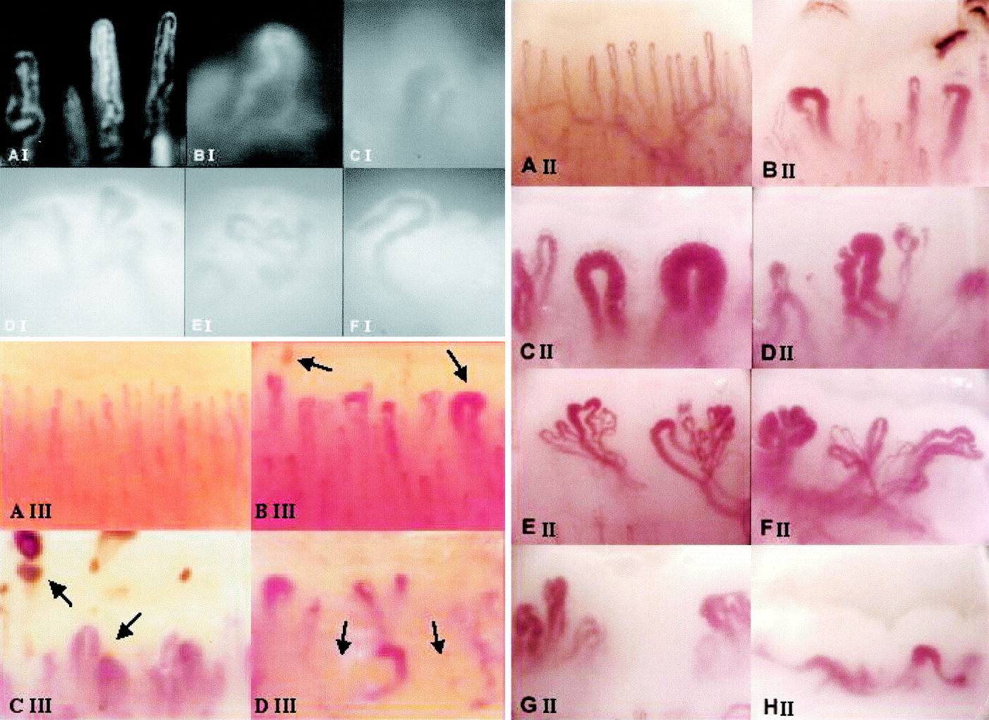
Capillary Morphology
Standard vessels show a slender hairpin or U-shaped contour with an afferent-to-efferent diameter ratio of approximately 1:2. Marked ectasia or giant loops reflect structural alterations in the microvascular wall.
Extravasation and Hemorrhages
Ruptured capillary walls release erythrocytes that migrate toward the distal edge of the nail fold alongside growing keratin. These microhemorrhages present as distinct hemosiderin crusts or focal dark masses.
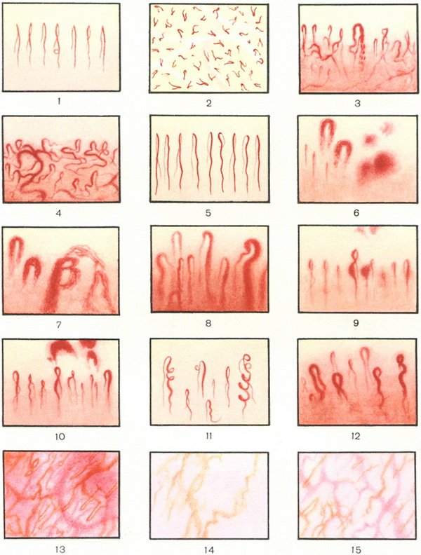
Capillary Density and Edema
Vessel density reflects the number of functional terminal loops per linear millimeter of tissue. Local tissue edema decreases visual contrast, blurring the margin between capillary walls and dermal connective tissue.
Examination Conditions and Observation Methodology
Accurate observation of the dorsal nail fold requires controlled environmental parameters to prevent transient physiological vasoconstriction. Because peripheral skin vessels respond rapidly to ambient temperature, standardizing the examination space is an essential clinical step.
When preparing the site for nail capillary microscopy, the subject rests with the hand supported at heart level in a temperature-controlled setting. Applying a drop of clearing oil onto the dorsal fold reduces light scattering across the stratum corneum, creating an optical window into the papillary dermis.
- Acclimate the subject in a stable room held between 22 and 24 degrees Celsius with moderate humidity for fifteen to twenty minutes prior to evaluation.
- Place the subject in a seated position with the fourth or fifth digit resting steadily on an adjustable mechanical stage at heart level.
- Apply two drops of medical-grade cedar oil, paraffin oil, or mineral oil across the junction where the cutaneous fold meets the nail plate.
- Position the optical lens perpendicular to the dorsal nail fold surface and engage the coaxial coarse adjustment wheel to locate the distal vascular row.
- Fine-tune focal depth using the coaxial fine focus control while regulating cold LED illumination to prevent optical glare or heat-induced vasodilation.
- Scan across the entire width of the fold to document loop density, apical width, and any perivascular hemosiderin deposits.
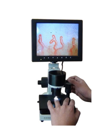
Microcirculation Equipment Specifications and Configurations
Accurate microvascular inspection requires purpose-built optoelectronic hardware capable of steady mechanical support and clear optical magnification. We at MAIKONG manufacture digital and video observation systems from our production base in SZ, GD, providing reliable equipment to university laboratories, vascular assessment clinics, and clinical device distributors.
Our instruments, such as the MK-GY880 platform, incorporate cold LED illumination to protect sensitive periungual tissues from thermal distortion during extended microcirculatory studies. The following conditional specifications represent typical manufacturing configurations available across our production lines.
| System Parameter | Standard Specification | Alternative Option | Operational Notes |
| Brand | MAIKONG | OEM Custom | Private-label branding supported for global distribution. |
| Optical Magnification | 380X | 500X / 600X | High-resolution optical magnification for cellular stream viewing. |
| Power Input | AC 100–240V | 50/60 Hz | Universal power compatibility for worldwide clinical setups. |
| DC Output | DC 12V 2A | Stable Adapter | Regulated low-voltage feed for stable imaging performance. |
| Objective Lens | 5X Achromatization | 25mm Working Range | Achromatic optics to minimize chromatic aberration at margins. |
| Mechanical Stage | X-Y Double-Layer | Compound Movement | Smooth lateral and longitudinal finger positioning. |
| Focusing Mechanism | Coaxial Coarse/Fine | Adjustable Tension | Enables micro-metric focal depth adjustment. |
| Internal Lighting | LED Cold Light | Integrated Dimmer | Prevents heat-induced vasodilation of cutaneous loops. |
| Display Screen | 8-Inch LCD | 10 / 12-Inch Color | Real-time video display with zero transmission latency. |
| Net Weight | 6 kg | 7 kg Gross | Heavy, stable base reduces mechanical vibration artifacts. |
Each unit ships in a secure transit configuration designed for clinical clinics, mobile screening initiatives, and institutional research labs.
| Packaging Component | Quantity | Unit Type | Primary Function |
| MK-GY880 Host Unit | 1 | Piece | Main optical body with integrated CCD and mechanical stage. |
| LCD Monitor | 1 | Piece | High-contrast color display for direct observational recording. |
| Power Adapter | 1 | Piece | Standard regulated transformer with international plug sets. |
| Cedar Oil Bottle | 1 | Piece | Optical clearing medium for reducing epidermal light scatter. |
| Cotton Swabs | 1 | Pack | Lint-free applicators for clean oil distribution. |
| Reference Chart | 1 | Piece | Comparative morphological guide for microcirculation patterns. |
Establish Observation Capabilities
When selecting professional microcirculation observation systems or organizing private-label device distribution, reviewing detailed hardware specifications helps clarify purchasing decisions.
Clinical Limitations and Observation Cautions
Visual observation of the dorsal nail fold is a non-invasive morphological assessment tool, not an independent diagnostic test. The presence of irregular loops, slight tortuosity, or minor perivascular deposits must always be evaluated in conjunction with comprehensive clinical findings, patient history, and targeted laboratory analyses.
Non-pathological factors can significantly alter the visible microvascular pattern within the dorsal fold. Recent mechanical manicures, occupational manual labor, chronic nail-biting, localized trauma, and exposure to cold water can induce capillary dilation, focal hemorrhages, or temporary vessel spasm in otherwise healthy individuals.
Dorsal nail fold findings should not be used for consumer self-diagnosis. Accurate image interpretation requires formal clinical training to distinguish structural abnormalities from benign anatomical variants and trauma artifacts.
Frequently Asked Questions About the Dorsal Nail Fold
Where is the dorsal nail fold?
The dorsal nail fold is located on the back surface of the fingertip, directly proximal to the visible nail plate. It forms the external cutaneous roof over the nail root and germinative matrix.
Is the dorsal nail fold the same as the proximal nail fold?
The dorsal nail fold is specifically the external, visible upper layer of the proximal nail fold. The full proximal nail fold includes both this external dorsal surface and an internal ventral layer that lies against the nail plate.
What can be seen through the dorsal nail fold?
Under optical magnification with immersion oil, observers can view terminal capillary loops, red blood cell flow, subpapillary venular networks, perivascular tissue clarity, and localized hemosiderin deposits.
How is the dorsal nail fold observed?
The area is examined using dedicated capillaroscopes, digital microscopes, or dermatosが付 scopes equipped with cold light sources. A drop of immersion oil is applied to make the keratin layer transparent before focusing.
Why is immersion oil used on the dorsal nail fold?
Immersion oil matches the refractive index of keratin, reducing surface light scatter and glare across the stratum corneum. This optical coupling allows light to reach and illuminate the underlying dermal microvessels.
What causes visible capillary changes in the nail fold?
Capillary alterations can stem from localized physical trauma, cosmetic manipulation, extreme cold exposure, or systemic microvascular remodeling. Professional clinical correlation is required to identify the underlying cause.
Summary of Dorsal Nail Fold Anatomy and Observation
The dorsal nail fold serves as a vital protective barrier for the nail matrix and offers an unparalleled anatomical site for non-invasive microvascular assessment. When examined under controlled conditions with proper optical instrumentation, this localized cutaneous zone provides direct insight into peripheral circulatory dynamics without surgical disruption.
Inquire About Professional Observation Hardware
Connect with our technical team in SZ, GD to discuss clinical equipment options, OEM configurations, and distribution opportunities.