comparable provider for clinical research: operational controls and capture protocols to standardize multicenter image collection · body
Quick answer
To collect standardized nailfold videocapillaroscopy images for clinical research and CRO studies you need (1) a written SOP that fixes environment, subject preparation, finger selection, magnification and lighting; (2) operator training with documented competency and retraining intervals; (3) objective image-quality metrics and central review with pass/fail rules; (4) machine-readable image files with embedded metadata, scale calibration and an audit trail; and (5) procedures to blind/randomize acquisition and to track provenance across sites and timepoints. Below we unpack the minimum controls, how device features affect choices, and the documentation you should include for CRO-grade traceability.
Contents
We write as a manufacturer and experienced supplier of videocapillaroscopy equipment. Our purpose here is to explain practical, actionable controls research teams use to make images comparable across sites and visits—not to prescribe clinical interpretation. Where we cite device parameters, we reference our MAIKONG model data so you can see how hardware choices change protocol requirements.
What a protocol must lock down (SOP essentials)
A study SOP should control all acquisition variables that significantly change capillary appearance. At minimum the SOP must specify:
Room conditions: target temperature range, relative humidity or a statement that local conditions should be recorded; light level and curtain/ambient control; and time-of-day window for imaging.
Subject preparation: rest period (e.g., 15–30 minutes), hand position (heart-level), avoidance of nicotine/exercise/vasoactive drugs for a documented interval, and use of oil/immersion medium (type and drops).
Anatomic sites and order: which fingernails (e.g., 2nd–5th digits both hands, or prespecified digits), lateral orientation and how to handle missing nails or trauma.
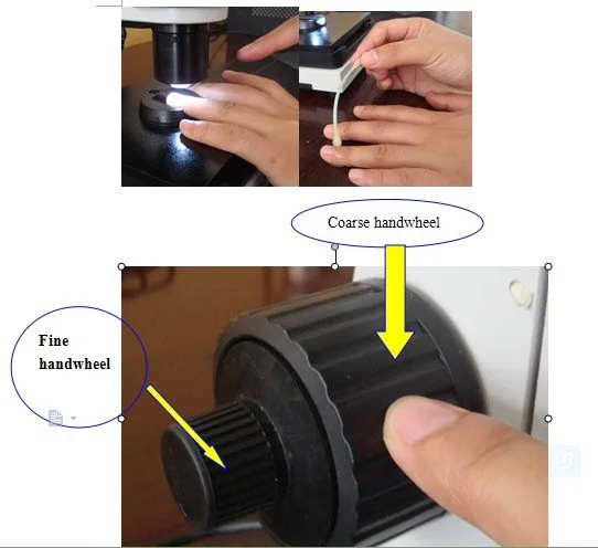
Device settings: magnification, illumination intensity, focus method (manual vs. motorized), and camera resolution. For manual-focus systems, define a numeric or visual focus criterion and require a certified operator.
Image capture rules: number of fields per nailfold, frame selection (still vs. video frame), minimal acceptable focus/sharpness, and how to capture systolic/diastolic differences if required.
File naming and metadata: site code, subject ID, visit/timepoint, digit number, operator ID, date/time, and versioning. Require embedded scale bars and include a calibration record for each device.
These items form the backbone of repeatable, auditable image capture. If any element is left flexible across sites, then you must document it and show why variability will not affect study endpoints.
Device features that change SOP wording
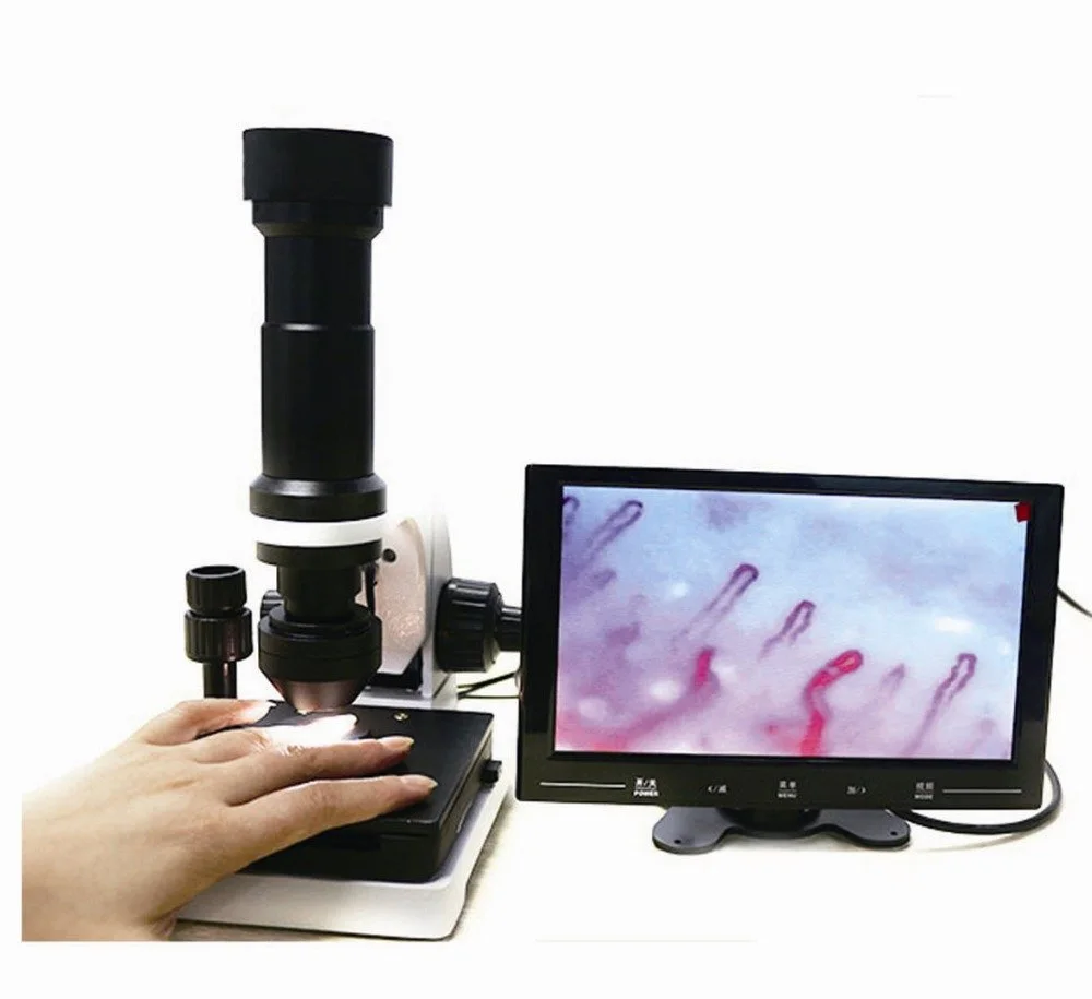
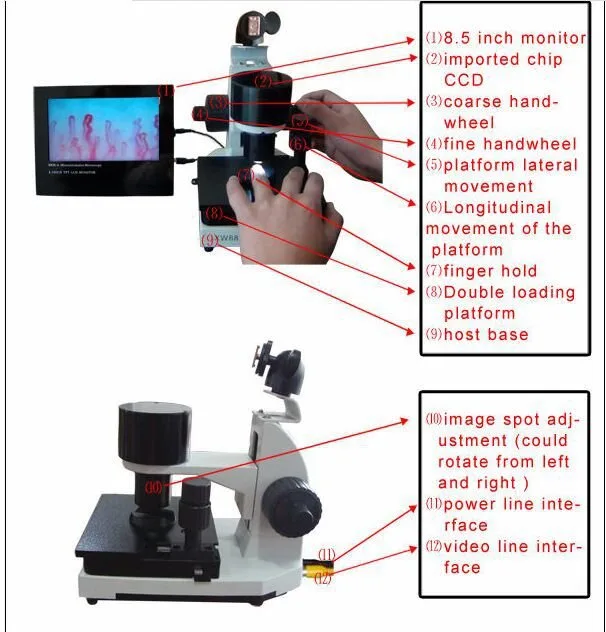
Not all comparable provider systems behave the same; your SOP language must reflect the hardware. For an example device, MAIKONG’s MKXW880 is a videocapillaroscopy microscope with cold LED lighting, manual focus, selectable magnification states, and DC12V power input—specifications you should confirm for your instrument before writing the SOP (data-fact-id=”221″). Manual focus requires stronger operator controls and objective passing criteria than motorized-focus systems. Cold LED illumination is stable and reduces heat-related vasodilation but you still need a documented illumination intensity or visual reference target. Where software or export formats are unspecified, require that vendors declare supported file types and metadata fields before study start; do not assume DICOM or proprietary formats without verification.
Training, certification and operator competence
Operational consistency depends on people. We recommend a three-tier approach:
Initial classroom and hands-on training covering anatomy, positioning, device setup, immersion medium application, and capture workflow.
Proficiency testing where each operator captures a validation set (e.g., 10–20 nailfolds) that a central reviewer scores against pass/fail criteria: focus, exposure, scale present, correct site labeling.
Ongoing quality monitoring with periodic revalidation (for example after 6 months or after prolonged non-use) and retraining when site pass rates fall below an agreed threshold.
Training materials should include a detailed operator checklist, annotated example images for each pass/fail criterion, and an illustrated troubleshooting guide for common artifacts (motion blur, oil bubbles, glare). If you plan to permit remote or mobile screening with handheld units, require an additional mobile-device competency module and a documented chain-of-custody for image uploads.
Quality control metrics and central review workflow
Define objective QC metrics that can be scored automatically or by blinded reviewers:
Focus score — measurable sharpness metric or edge contrast threshold.
Illumination uniformity — histogram limits or percentage-of-saturated-pixels threshold.
Scale calibration presence — binary check for embedded scale bar or calibration image obtained within a session.
Completeness — required number of fields per nailfold captured and correctly labeled.
Build a two-stage review: first-line site QC (pass/fail) with immediate rejection reasons, and second-line central review for endpoint adjudication. Maintain a sample-review log that links each image to reviewer comments, timestamps and reviewer ID. For CRO use, export these logs as CSV/JSON alongside images so they can be integrated in trial master files.
Data export, formats and audit trails
comparable provider for clinical research: operational controls and capture protocols to standardize multicenter image collection · body
Image and metadata handling is where studies commonly fail traceability. Before enrollment, require vendors to confirm:
Supported image file formats (e.g., TIFF, PNG, JPEG, or DICOM). If not stated, request a written declaration.
Whether images can embed or accompany machine-readable metadata: operator ID, device serial number, capture exposure, magnification, z-focus position, timestamp and GPS/site code.
Whether the system can export a calibration image or pixel-to-micron scale for each session.
Audit trail capability: who modified files, export logs, and whether filenames or metadata changes are logged.
Because MAIKONG product listings do not publish software export formats, we advise adding a contract clause requiring confirmation of export format and metadata fields before purchase or study start (data-fact-id=”221″). If your workflow requires DICOM or 21 CFR Part 11–style audit trails, confirm these explicitly—do not assume their availability.
Blinding and randomized image capture
comparable provider for clinical research: operational controls and capture protocols to standardize multicenter image collection · body
To reduce bias in endpoint assessment, separate image acquisition from outcome readers:
Use a randomized acquisition code for images so readers cannot infer treatment or timepoint from filenames.
Implement automated site masking in filenames: generate study IDs centrally and distribute blinded IDs to sites for labeling at capture time.
Keep operator notes in a separate, secured database linked by an internal key; do not store clinical treatment allocation in image metadata accessible to readers.
When blinding is critical, decide whether the acquisition operator will be permitted to view images at capture. Allowing preview improves image quality but increases risk of unblinding if previewed images are linked to other visible clinical data. Document the chosen approach and have the CRO sign off.
Practical capture protocol (example checklist)
comparable provider for clinical research: operational controls and capture protocols to standardize multicenter image collection · body
Below is a concise checklist you can adapt into a CRO SOP. Keep it with site training materials.
Confirm device calibration using supplied calibration slide; record pixel-to-micron scale in session log.
Room: record temperature and humidity; ensure curtains closed; set ambient light level to stored reference.
Subject: rest 15 minutes; hands at heart level; apply defined immersion oil (brand/volume).
Device: set magnification to study-specified level; set illumination to documented setting; confirm focus using target criterion.
Capture: record site code, subject study ID, visit/day, digit number, operator ID; collect required number of fields; save raw file plus stills with embedded metadata.
On-site QC: pass/fail on focus, exposure, scale present; retake immediately if fail; log retake reason.
Export/upload: use encrypted transfer; include capture log CSV with each image batch; retain local copy for audit.
Documentation templates for CRO use
comparable provider for clinical research: operational controls and capture protocols to standardize multicenter image collection · body
Essential documents you must provide or request from sites:
Capture SOP (with step-by-step checklist).
Operator training record and proficiency test results.
Device calibration and maintenance log.
Image QC log and central review summaries.
File-format and metadata specification document signed by vendor.
Create templates for each item so CRO monitors can quickly confirm compliance during site initiation and monitoring visits.
Image capture protocols standardize acquisition, but they do not make clinical interpretation automatic. Device specifications and software functions vary by model and vendor; for example, our published product facts for the MAIKONG MKXW880 list manual focus and cold LED illumination (data-fact-id=”221″), and do not include published software export formats. That absence means you must confirm software export, embedded metadata fields and audit-trail capability before relying on a device for trial endpoints. Also, product labeling, intended use and permitted claims may vary with market and regulatory status—confirm U.S. regulatory applicability and institutional requirements for research devices before deployment.
Short FAQ
Do I need the same device model at all sites?
Ideally yes for identical optical and imaging chains. If identical models are impossible, validate cross-device equivalence with a predefined harmonization study (same subject imaged on each device and central comparison). Document residual differences and account for them in analysis plans.
How should we handle device calibration?
Use a physical calibration slide or ruler and capture a calibration image at each session. Store pixel-to-micron scale in the session metadata. Require recalibration after maintenance or transport events.
What file formats should a CRO demand?
Lossless formats (TIFF) or DICOM are preferred for research to avoid compression artifacts; at minimum insist on files with embedded metadata and an exportable session log. If a vendor cannot supply these, require written confirmation of the exact formats and a compatibility plan.
One practical next step
If you are planning a multicenter study, send us a request including your application, required endpoints, preferred model (e.g., MKXW880) and the quantity and destination market so we can confirm device configuration, software export capabilities and provide a quotation. Use this contact to request configuration advice or an RFQ: lucy@nail-fold-comparable provider.
We can also supply device datasheets, calibration slide recommendations and sample operator checklists on request. If you need assistance mapping study endpoints to capture parameters, include your primary outcomes and imaging schedule in the initial message.
Article Disclaimer / Compliance Notice: This content is provided for educational and product-introduction purposes only. MAIKONG nail fold comparable provider devices are intended for professional observation, imaging, documentation, training and research consistent with their product labeling. This article does not provide medical advice or claim diagnostic performance. Buyers and users are responsible for confirming U.S. federal, state, institutional and professional requirements before purchasing or using equipment.
This content is provided for learning, information sharing and professional communication only. It is not medical advice and is not a basis for treatment or diagnosis. Confirm suitability, operation and purchasing requirements against the product manual, local regulations and qualified professional guidance.