comparable provider camera compatibility: How to confirm a camera or module will work with your clinic’s microscope · body
Quick answer
To confirm compatibility today, run three practical checks: 1) confirm mechanical fit (drawtube/eyepiece diameter, mount type and whether you need a C‑mount or adapter); 2) match optical geometry (working distance, objective magnification and resulting field‑of‑view with your sensor); and 3) verify electrical, illumination and data interfaces (power ratings, LED light source, USB/video outputs and driver/software). For MAIKONG’s MKXW880 we can confirm several declared parameters you should compare directly with your microscope (see the parameter table below) — but our published facts do not list host PC interfaces or software details, so request those explicitly when you ask for a quote.
Contents
Introduction — who this is for and one‑sentence answer
This is written for a U.S. rheumatology clinic equipment manager who already has microscopes or stands and needs a repeatable way to check whether a comparable provider camera or module will integrate into the existing imaging chain. The single practical answer: measure the mechanical and optical interfaces, confirm declared electrical/illumination specs, and demand clear data‑interface and driver information before purchase.
Direct answer: the practical compatibility steps to confirm today
Work through these five checks in order. You can complete them in under an hour if you have the microscope manual and the unit’s product sheet or an online spec sheet from the seller.
Mechanical fit: measure drawtube/eyepiece inner diameter, note whether the microscope has a trinocular port or a monocular drawtube, and identify the mount form (eyepiece projection, C‑mount, Dovetail). If the camera or module requires a specific mount, confirm an adapter exists for your microscope model.
Optical geometry: record the microscope objective used for nailfold imaging, the objective working distance, and the desired clinical magnification. Compare those with the camera’s working distance, objective magnification and recommended objective types so the image fills the sensor without vignetting.
Illumination and exposure control: check whether the camera expects an independent cold‑LED light source or integrates illumination. Confirm you can control LED intensity or camera exposure from the host (important for capillary contrast).
Electrical/power: verify the camera’s declared input/output voltages and adapter needs against your clinic supply and any inline power bricks. If the seller lists AC input and a DC output, confirm the included adapter matches U.S. power (see our declared MKXW880 voltages below).
Signal and drivers: ask for exact video outputs (USB 2/3, HDMI, analog composite, SDI) and supported operating systems/drivers. Without OS/driver confirmation you cannot assume plug‑and‑play on your clinical workstation.
Key system checks to run before buying a camera or module
Physical and mechanical fit: stands, drawtubes and mounting interfaces
Measure the real world, not the brochure. For a microscope you use daily, record:
drawtube internal diameter and type (monocular/trinocular/benchtop port),
distance from the drawtube plane to the optical axis (if available),
whether the microscope has a dedicated camera port (C‑mount/eyepiece projection) and the exact adaptor thread or flange size.
If your microscope is a common trinocular model, many cameras can attach via the trinocular port with a C‑mount adapter. If your scope uses eyepiece projection, you may need a projection eyepiece or adapter which preserves parfocality. If you’ll put a camera on a dedicated comparable provider stand, check base plate clearance and the device footprint.
Optical fit: magnification, working distance, objective and field‑of‑view match
comparable provider is image‑quality sensitive. Two numbers matter most: the objective’s working distance and the camera’s sensor‑to‑optics geometry. If the camera lists a working distance, make sure it matches the microscope objective so you get the stated magnification at the intended focus. Also confirm the objective’s numerical aperture and the sensor size to estimate whether the capillary bed will fill the image without cropping.
When a manufacturer gives “magnification times” or a single magnification, treat that as guidance: you still need to calculate the effective magnification on your monitor (objective × camera projection factor × monitor size). Ask the vendor for sample images taken through a comparable microscope objective and at the same monitor size you plan to use.
Illumination and imaging source: LED cold light, contrast and exposure control
Cold LED illumination is standard for nailfold work because it produces bright, stable light with low heat. Confirm whether the camera integrates the LED or expects the stand/microscope to supply it. You should also verify whether the camera or software allows manual exposure, gain and white balance adjustments — essential for consistent capillary contrast across patients.
Electrical and power compatibility: voltage, input/output and adapters (what our MKXW880 declares)
Check the product sheet for exact power declarations rather than assuming. For the MAIKONG MKXW880 we can confirm the factory declares the following values; compare these to your site power and any local adapters you use:
Because MKXW880’s product sheet shows an AC input range of 100–240V and a DC output of DC12V,2A, that indicates the supplied power adapter accepts global mains voltages and provides 12V DC to the unit (confirm the adapter is included). Do not assume the camera can be hard‑wired to other supplies without checking the vendor’s instructions.
Signal and data interfaces: USB, video out and monitor support
Our public product facts for MKXW880 do not list host interfaces, supported operating systems or software drivers. That is an important gap: before you order ask the vendor to confirm:
exact video outputs (USB 2.0/3.0, HDMI, VGA, composite),
supported OS versions and drivers (Windows 10/11, macOS, Linux),
whether image capture and measurement software is included or sold separately,
sample images or a short demo video taken on a similarly equipped microscope.
How MAIKONG product parameters affect compatibility decisions (what we can confirm)
comparable provider camera compatibility: How to confirm a camera or module will work with your clinic’s microscope · body
MKXW880 declared parameters that matter
What these parameters imply for microscope integration and image capture
Each parameter shifts the integration decisions you’ll make:
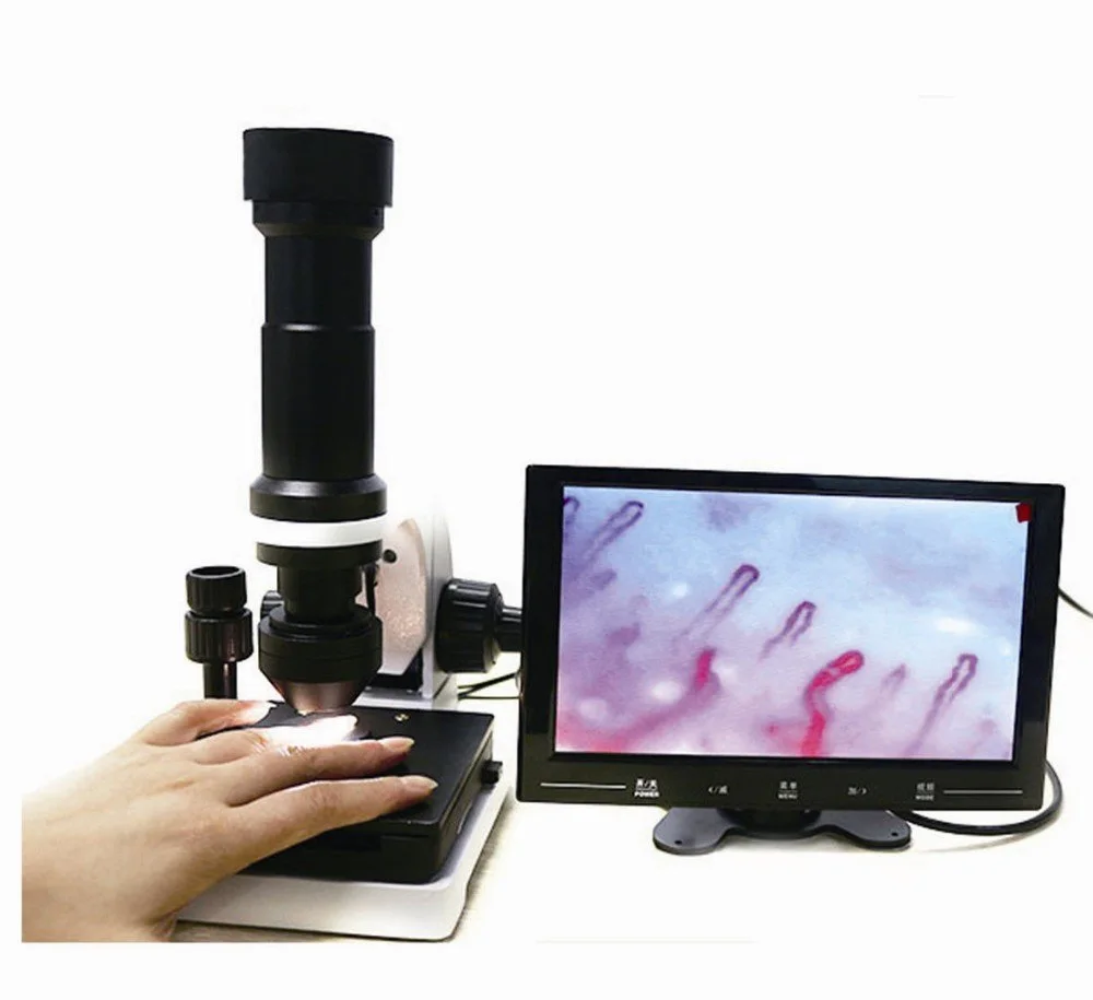
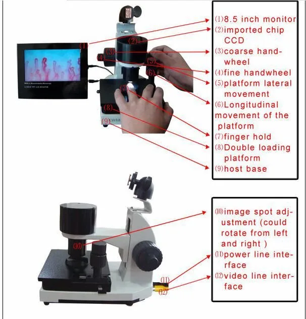
Monocular drawtube: MKXW880 lists a monocular drawtube. If your microscope is trinocular, you will likely need an adapter or a different camera mounting method to preserve the binocular viewing path for clinicians.
Working distance 25mm: this is relatively short; ensure your objective and finger holder allow the nailfold to sit within that distance without collision. If your microscope objective requires a longer working distance, the optical match will not be correct.
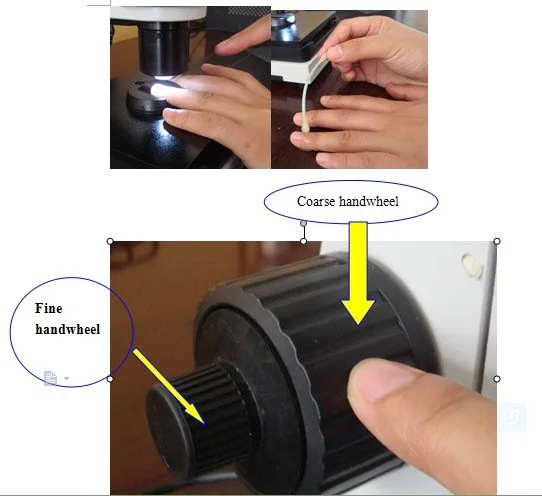
Manual focus: the camera relies on manual focusing. For clinical throughput, confirm your operator workflow tolerates manual focus or that the stand offers fine and coarse knobs for fast focusing.
Cold LED illumination and 12V power: the stated cold LED source and DC12V supply mean the unit expects a common LED arrangement; still confirm dimming/exposure control and whether the LED is integrated or supplied separately.
Magnification figures: the declared magnifications (10X, 680/1200 times) are a product claim for the MAIKONG system; when integrating with your microscope, calculate effective on‑screen magnification rather than assuming identical results on a different monitor size or camera sensor.
Checklist to include in an RFQ or compatibility request
comparable provider camera compatibility: How to confirm a camera or module will work with your clinic’s microscope · body
Your microscope make/model and whether it has a trinocular port or eyepiece projection (attach photos of the camera port),
Drawtube inner diameter or whether you use a standard C‑mount (measure with calipers),
Objective types and working distances you plan to use,
Expected patient throughput and whether you need autofocus or manual focus is acceptable,
Host computer OS and monitor size or whether you want an integrated monitor,
Desired quantity and shipping destination (U.S.),
Request for sample images taken through a comparable microscope and an exact list of included accessories (power adapter, cable types, finger holder, oil etc.).
Pricing is available on inquiry; include the configuration details above so the vendor can provide an accurate quote.
FAQ — practical follow‑ups a clinic equipment manager will ask
comparable provider camera compatibility: How to confirm a camera or module will work with your clinic’s microscope · body
Does MKXW880 have USB or HDMI output?
Our public product facts do not list host video/data interfaces or supported operating systems. Please request explicit interface and driver information from the vendor before purchase so you can confirm compatibility with your clinic PC or monitor.
Will a monocular drawtube camera work on a trinocular microscope?
Possibly, but it depends on the camera’s mount and the microscope’s trinocular port. MKXW880 is declared with a monocular drawtube; if you require simultaneous binocular viewing and camera capture, ask for an adapter solution or a camera designed for a trinocular port.
What are the MOQ, lead time and warranty for MKXW880?
According to the MAIKONG product snapshot, MOQ is 1PCS, lead time 2–3 days, and warranty 1 Year. Include data‑sheet confirmation in your RFQ since shipping and accessory content may affect delivery timing.
Evidence gaps and limitations you must verify
comparable provider camera compatibility: How to confirm a camera or module will work with your clinic’s microscope · body
Our published product facts provide many useful parameters for MKXW880 (model, magnification, working distance, power and declared certifications). However, the public facts do not specify camera host interfaces, driver support, sample images, or full accessory lists. Those are essential for a final compatibility decision; treat their absence as a required follow‑up with the vendor. Also confirm any U.S. regulatory, labeling or import requirements that may apply to your clinical use.
If you want an immediate compatibility check, include the microscope model, port photos, objective used for nailfold work and desired throughput in your message. We will review those details and advise which adapters or configurations to request.
Next step: Request a configuration check or quote by emailing lucy@nail-fold-comparable provider. In your message, list your microscope model, whether it has a trinocular port, the objective and working distance you use for nailfold imaging, required quantity, and whether you need an integrated monitor or PC capture so we can provide accurate configuration advice and pricing.
Article Disclaimer / Compliance Notice: This content is for informational and product‑introduction purposes only. MAIKONG nail fold comparable provider equipment is intended for professional observation, imaging and documentation. Clinical interpretation must be performed by trained professionals and purchasers are responsible for confirming applicable U.S. federal, state and institutional requirements before purchase or clinical use. MAIKONG does not claim its products independently diagnose, prevent, treat, or cure disease unless specifically supported by product labeling and applicable regulatory authorization.
This content is provided for learning, information sharing and professional communication only. It is not medical advice and is not a basis for treatment or diagnosis. Confirm suitability, operation and purchasing requirements against the product manual, local regulations and qualified professional guidance.
Related resources
comparable provider camera compatibility: How to confirm a camera or module will work with your clinic’s microscope · body