
Medial Nail Fold Anatomy and Observation Landmarks
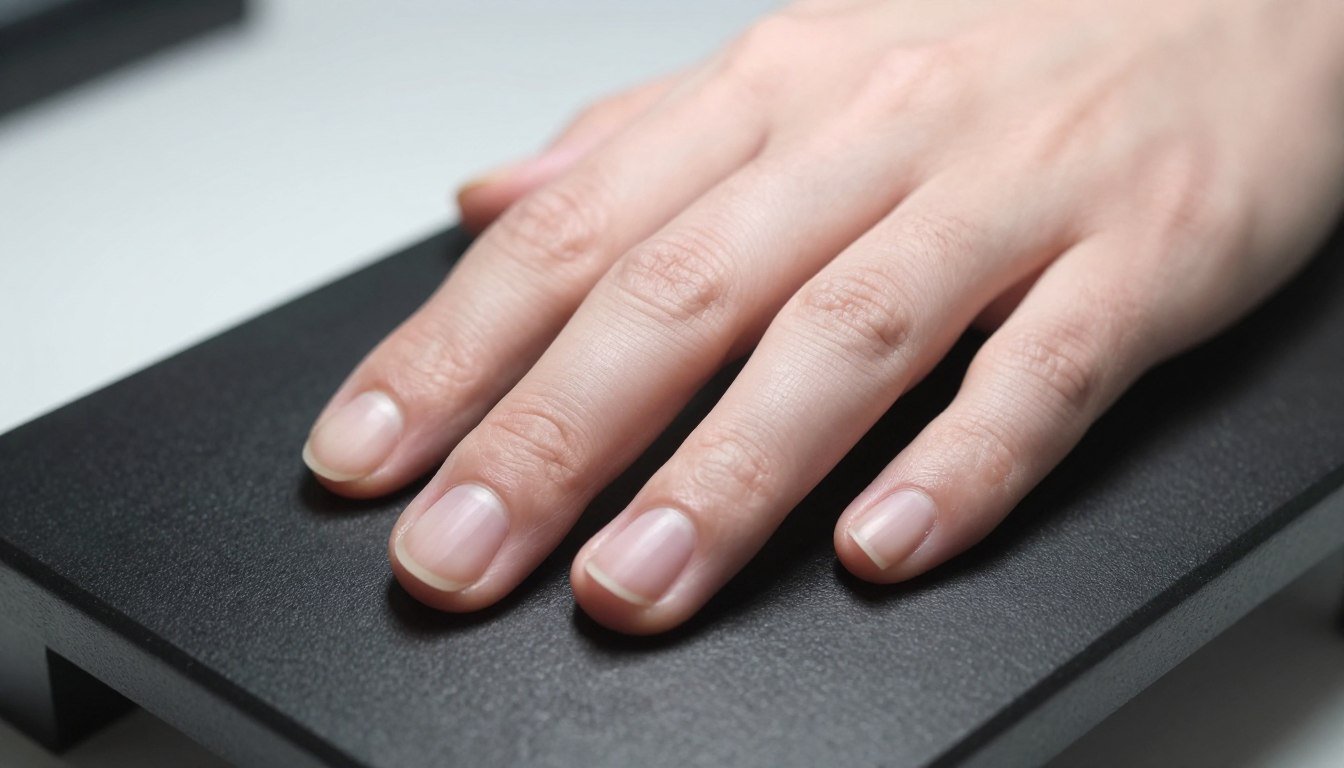
The medial nail fold is the soft tissue margin bordering the inner edge of the nail plate closest to the anatomical midline of the human body. In clinical dermatology and microvascular research, distinguishing this border from adjacent perionychial tissue provides a reproducible landmark for evaluating cutaneous microvasculature, tissue transitions, and structural margins.
Proper classification depends on understanding anatomical position rather than casual hand orientation. Establishing standardized anatomical descriptions prevents charting discrepancies and maintains consistency across longitudinal research assessments.
Understanding Medial Versus Lateral Terminology
Anatomical terminology relies on the standard anatomical position, in which the body stands upright with palms turned forward and thumbs pointed outward. In this orientation, any structure positioned toward the midline of the body is medial, whereas structures facing away from the midline are lateral.
Applying this rule to the hand reveals that the orientation of the medial nail fold varies between digits. On the thumb (digit I), the medial border faces outward toward the other fingers because the anatomical thumb naturally points lateral to the body midline. On the second through fifth digits, the medial border corresponds to the ulnar margin, facing toward the little finger.
Thumb (Digit I) Orientation
- Faces the anatomical midline along its inner margin.
- Corresponds to the ulnar side of the thumb nail unit.
- Adjoins the first web space tissue.
Fingers (Digits II through V) Orientation
- The medial edge aligns toward the ulnar side of each digit.
- Distinguished strictly from the radial (lateral) fold.
- Remains constant regardless of hand pronation or supination.
Confusion often arises when examiners use the term “lateral” generically to refer to both side borders. Restricting “lateral nail fold” to the radial or outer anatomical edge and medial nail fold to the ulnar or inner anatomical edge removes ambiguity from research notes.
Surface Anatomy and Surrounding Perionychium
The perionychium comprises the soft tissue structures immediately surrounding and supporting the nail plate. The nail fold on the medial side interfaces continuously with the proximal nail fold proximally and the lateral sulcus distally.
Beneath the surface of these tissue folds lie discrete microvascular structures that nourish the germinal matrix and periungual dermis. The capillary network transitions from vertical loops in standard dermal papillae to horizontally arranged capillary loops near the terminal cuticular border.
Nail Plate Interface
The nail plate rests firmly against the medial groove, creating a barrier that protects underlying sterile matrix tissue from physical intrusion.
Proximal Transition
At the proximal corner, the medial border curves directly into the proximal nail fold, where the eponychium seals the dorsal roof.
Hyponychial Margin
Distally, the tissue terminates near the lateral nail sulcus adjacent to the hyponychium and distal digital pulp.
When examining microcirculation through optical magnification, tracking each nail fold capillary along these margins requires precise positioning to prevent light reflection off curved lateral edges.
Terminology Consistency in Microvascular Observation
Inconsistent terminology across multi-center research creates compounding errors during longitudinal observation. If one investigator records an architectural variation as an outer lateral fold change while another documents it as a medial fold change, comparative data loses scientific value.
Standardizing anatomical notation establishes an objective baseline across digital databases. Documenting the specific digit, exact anatomical border, and quadrant ensures that follow-up assessments evaluate identical vascular fields.
Standard practice in laboratory observation records digits using Roman numerals (Digit I through V) coupled with explicit directional indicators: medial nail fold, lateral nail fold, or proximal nail fold.
Advanced imaging techniques such as nail fold capillaroscopy rely heavily on this positional discipline. Standard protocol routinely prioritizes Digits IV and V, where cutaneous transparency and lower mechanical trauma provide optimal visual clarity along both proximal and medial margins.
Technical Observation and Lighting Protocol
Examining the marginal tissue of the nail apparatus introduces optical challenges because of the natural curvature of the digital cylinder. Unlike the relatively planar proximal nail fold, the medial edge rolls downward toward the interdigital space, scattering directional illumination.
Compensating for this curvature requires dedicated lighting techniques, appropriate coupling agents, and stable mechanical fixation during evaluation.
- Apply a high-transparency immersion medium such as cedar oil, paraffin oil, or high-grade mineral oil to reduce superficial optical scatter from dry stratum corneum.
- Maintain an ambient examination room temperature between 22 and 24 degrees Celsius with roughly 70 percent relative humidity to avoid temperature-induced vascular constriction.
- Support the subject hand at heart level on an adjustable platform to balance arterial perfusion and venous pooling during static imaging.
- Use cold LED lighting to eliminate tissue warming that could alter localized vascular caliber.
- Employ coaxial fine-focus mechanics to navigate the focal depth created by the sloping medial tissue curve.
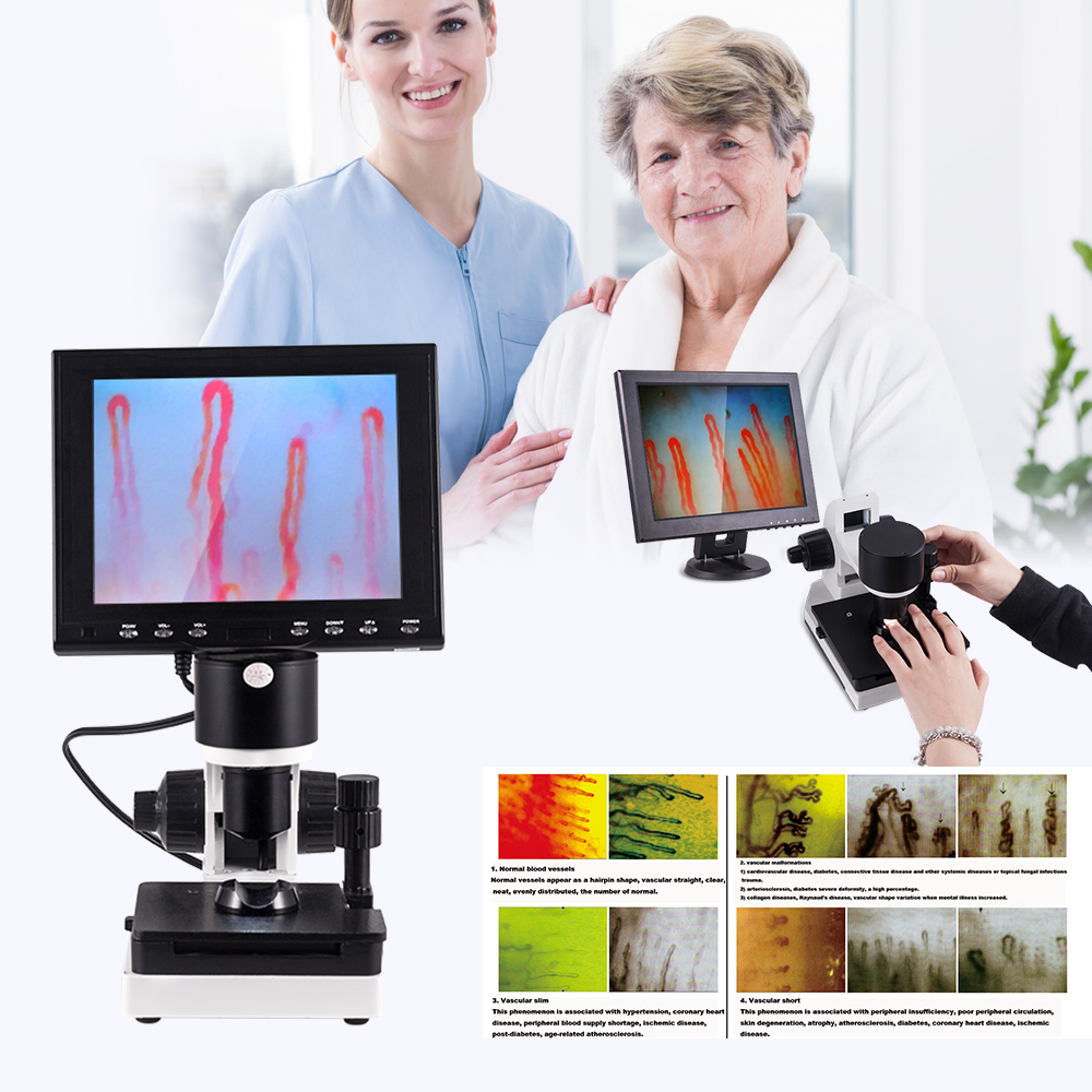
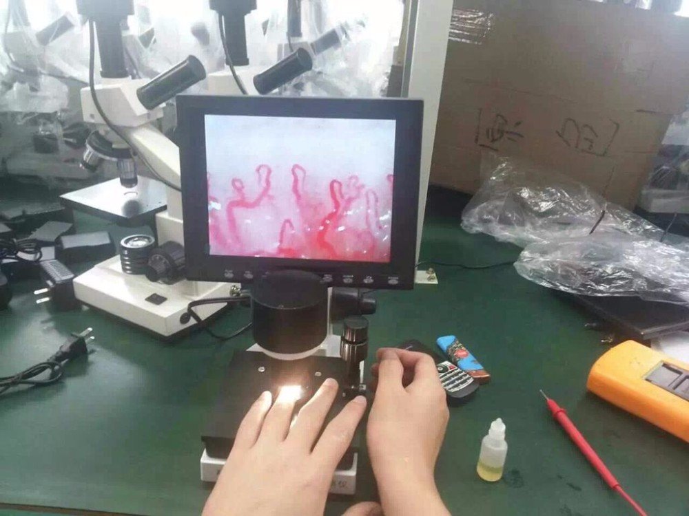
Researchers conducting non-invasive nail capillary microscopy gain valuable field stability by utilizing an integrated finger holder that isolates involuntary tremor during magnification.
Optical Imaging Instrumentation and Benchmarks
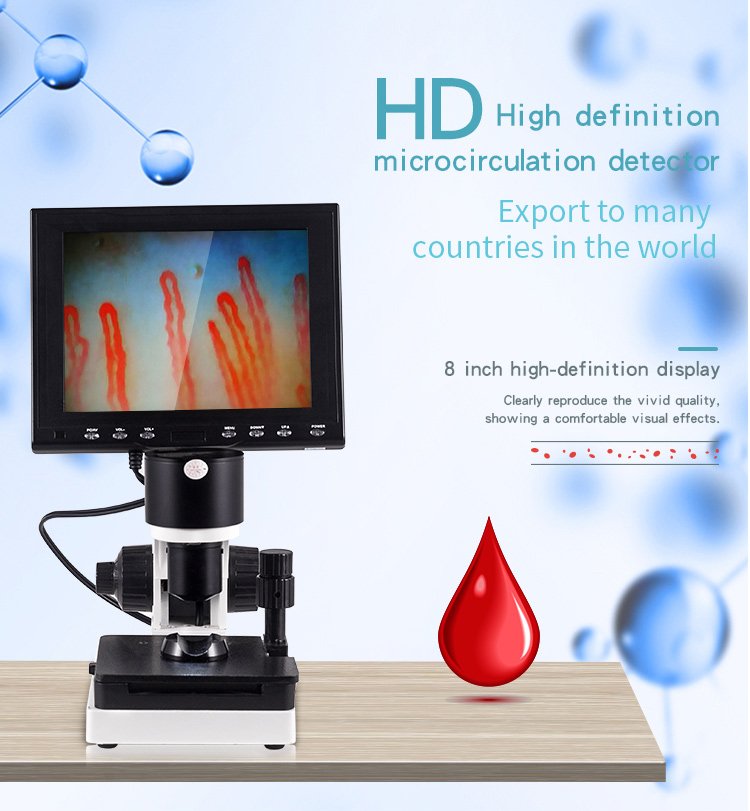
Accurate visual documentation of the medial nail fold requires rigid mechanical stages and dedicated magnification lenses capable of resolving microcirculatory architecture without image distortion. MAIKONG manufactures professional optoelectronic instruments engineered for human skin microcirculation research and teaching environments.
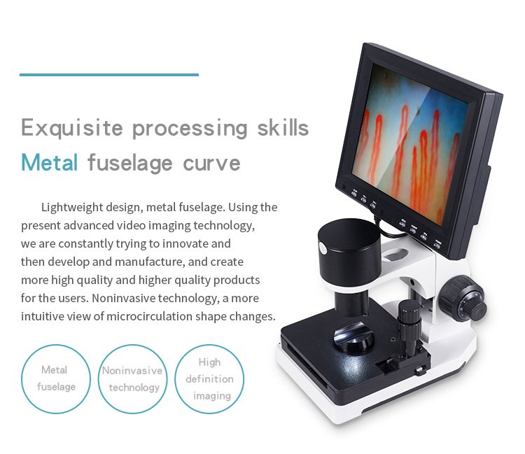
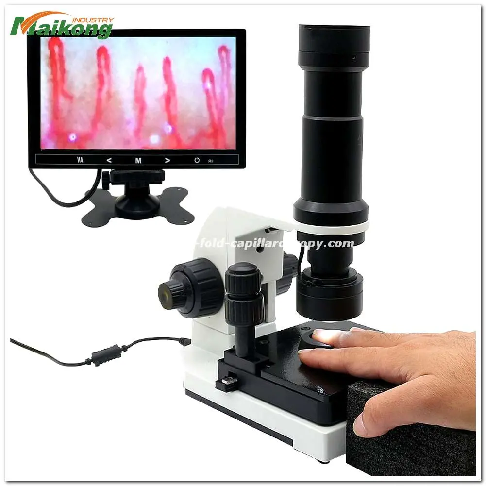
The MK-GY880 platform incorporates a high-stability compound stage, integrated LED cold illumination, and coaxial focusing to simplify boundary alignment across curved periungual tissue.
| Parameter | Specification | Engineering Details |
| Brand Name | MAIKONG | Manufactured in GD, SZ. |
| Magnification | 380X or 240X | Supports 380X, 500X, or 600X optical configurations. |
| Objective Lens | 5X Achromatization | Delivers an effective working range of 25 mm. |
| Stage System | Compound Mechanical | Features double-layer X-Y directional adjustment. |
| Focus System | Coaxial Adjustment | Integrates coarse and ultra-fine mechanical handwheels. |
| Light Source | LED Cold Light | Emits projection lighting without tissue heat transfer. |
| Camera Unit | Integrated Sensor | Transmits live video streams directly to display. |
| Display Panel | Color LCD | Available in 8-inch, 9-inch, 10-inch, and 12-inch sizes. |
Laboratories evaluating specialized nailfold capillaroscopy equipment benefit from the mechanical stability offered by dual-axis stage mobility, allowing researchers to track along the medial margin in seconds.
Package Components and Observation Accessories
Standardized packaging safeguards optical systems against environmental wear during transportation between research laboratories and clinical suites. Every MK-GY880 system ships inside protective reinforced housing designed for secure storage and mobility.
Each component supports direct operational setup, from immersion fluid application to high-contrast video verification on the dedicated monitor.
| Component | Quantity | Function |
| MK-GY880 Host | 1 Unit | Main optical chassis and integrated coaxial focusing body. |
| Color Monitor | 1 Unit | High-resolution LCD screen for real-time video display. |
| Power Adapter | 1 Piece | AC 100-240V 50/60Hz input with DC 12V 2A output. |
| Immersion Oil | 1 Bottle | High-grade cedar oil for eliminating superficial skin refraction. |
| Cotton Swabs | 1 Pack | Lint-free applicators for spreading immersion fluid. |
| Reference Chart | 1 Piece | Visual calibration guide for microcirculation assessments. |
| Storage Case | 1 Piece | Aluminum safety box with high-density memory foam insert. |
At MAIKONG, we manage comprehensive optical manufacturing facilities in GD, SZ, supplying healthcare distributors and academic centers with dependable imaging instruments. Readers seeking detailed information regarding company operations can review the About us resource.
Essential Principles of Nail Fold Anatomy
Mastering periungual anatomy streamlines workflow during both general dermatological reviews and specialized microcirculatory research projects.
- The medial nail fold refers strictly to the soft tissue border facing the anatomical midline of the human body.
- Digit orientation dictates whether the medial border corresponds to the ulnar margin or the radial margin.
- Curved tissue geometry along the medial fold requires precise illumination angles and immersion media to eliminate optical glare.
- Consistent charting across Digits I through V prevents data discrepancy in longitudinal observational studies.
- Stable optical instrumentation with coaxial stage controls simplifies positioning along sloped digital edges.
Frequently Asked Questions
Where exactly is the medial nail fold located?
The medial nail fold is located along the side of the nail plate that faces toward the anatomical midline of the body. On fingers two through five, this corresponds to the inner or ulnar border, whereas on the thumb it represents the margin facing the remaining fingers.
Is the medial nail fold the same as the proximal nail fold?
No, the medial nail fold borders the lateral side of the nail plate, while the proximal nail fold sits at the base of the nail covering the matrix. The two structures meet at the proximal lateral corner of the nail apparatus.
Why does anatomical orientation matter during nail observation?
Standard orientation prevents confusion between the medial and lateral margins during clinical charting. Consistent labeling allows multiple investigators to monitor the exact same microvascular field over time.
Can the medial nail fold be observed with magnification?
Yes, optical magnifiers and microcirculation microscopes can visualize the medial nail fold when paired with immersion oil and proper stage lighting. Stable finger positioning is necessary to maintain focus across the curved digital slope.
What should a professional document when observing this area?
Examiners should record the specific digit number, whether the medial or lateral border was assessed, lighting parameters, and tissue condition. Clear notes on immersion media and focus depth further support reproducibility.
Does the appearance of the medial nail fold vary between individuals?
Yes, skin thickness, cuticle attachment, epidermal hydration, and mechanical wear cause natural structural variation between individuals. Baseline pigmentation and manual labor exposure also influence optical visibility.
Standardizing Periungual Observation
Accurate identification of the medial nail fold strengthens anatomical records and supports reproducible scientific evaluations across clinical and research environments. Maintaining clear terminology bridges the gap between surface dermatological inspection and microvascular imaging.
Establish Distribution or Research Partnerships
Professionals outfitting research laboratories, clinical suites, or distribution networks can source specialized optical microcirculation systems directly from the manufacturer.