
Lateral Nail Fold: Anatomy, Microvascular Context, and Observation Methods
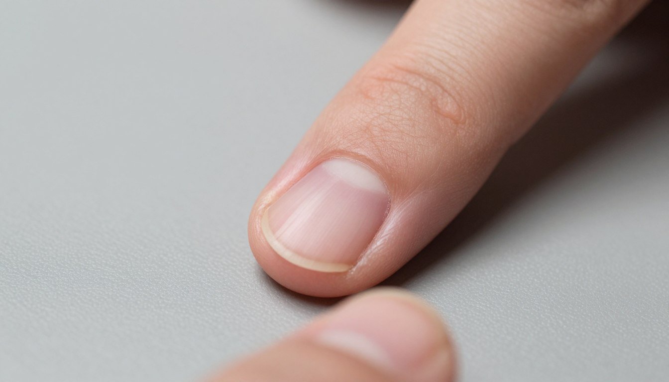
The lateral nail fold is the specialized border of cutaneous tissue that flanks the lateral margins of the nail plate. It forms the lateral protective boundary of the perionychium, anchoring the rigid keratin plate to the digit while shielding the underlying matrix and bed from mechanical shearing and environmental ingress. In microvascular imaging and clinical dermatology, recognizing the distinct architecture of this border helps practitioners differentiate lateral soft tissue patterns from proximal capillary beds.
Selecting appropriate visualization techniques depends on tissue thickness, epidermal pigmentation, and the specific vascular morphology under investigation. Standardized lighting, stable digital magnification, and careful subject positioning help reduce optical artifacts during detailed non-invasive assessments.
Lateral Nail Fold Anatomy and Border Structures
A precise lateral nail fold definition encompasses the soft tissue invagination that runs parallel to the longitudinal axis of the digit along each side of the nail plate. The tissue transitions smoothly from the dorsal epidermis of the distal phalanx into the lateral nail groove, also referred to as the lateral nail sulcus. This anatomic fold stabilizes the hard nail plate as it continuously migrates distally from the germinative nail matrix toward the hyponychium.
Beneath the surface stratum corneum, the lateral folds contain stratified squamous epithelium supported by a dense dermal stroma rich in collagen, elastin, and specialized neurovascular bundles. Because the lateral borders withstand frequent lateral friction, their keratinized layer is generally thicker and more compact than the dorsal skin proximal to the cuticle. This histological difference directly alters surface light reflection and tissue compliance during clinical contact examinations.
The transition between the lateral fold and the adjacent structures defines the mechanical integrity of the entire distal digit.
- Lateral nail sulcus: The deep epithelial groove where the lateral edge of the nail plate rests and glides during growth.
- Dermal papillae: Structural projections providing microvascular nourishment to the periungual epidermis along the digital margins.
- Hyponychial transition: The distal zone where the lateral fold meets the subungual space at the free edge of the plate.
- Proximal junction: The anatomical angle where the lateral fold converges with the proximal nail fold near the eponychium.
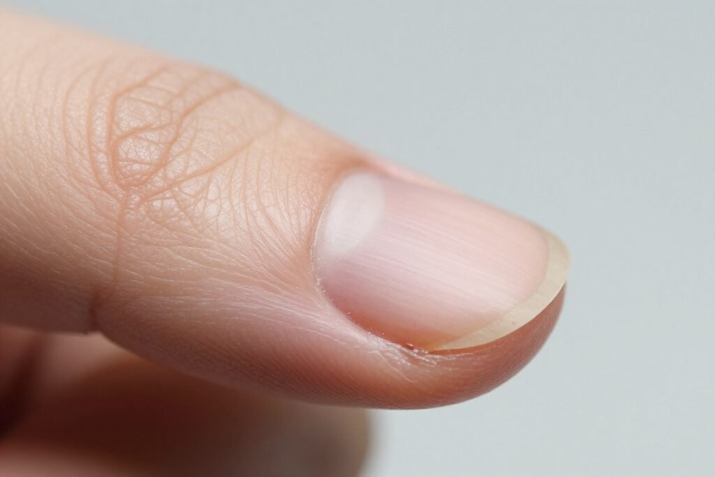
The lateral nail fold anchors the lateral margin of the nail plate within the lateral sulcus.
Structural Differences Across the Perionychium
The perionychium comprises multiple contiguous epithelial structures that support and protect the nail unit. Although the lateral nail fold shares histological characteristics with other periungual folds, its functional role and microvascular orientation differ substantially from the proximal tissue. Differentiating these anatomical zones prevents incorrect interpretations during dermatological evaluations and microvascular research.
In the proximal fold, the capillary loops run horizontal to the skin surface, making them directly visible along the distal edge. Along the lateral folds, the vascular plexus transitions to perpendicular loops similar to standard glabrous skin, which alters loop morphology under direct perpendicular illumination. Furthermore, the lateral fold lacks an extended eponychial seal, relying instead on close mechanical contact with the lateral nail margins to prevent foreign particle entry.
Understanding these anatomical variations clarifies why distinct operational approaches are required when imaging different borders of the nail apparatus.
Lateral Nail Fold Traits
- Vertical or oblique dermal capillary architecture relative to the surface plane.
- Thicker keratinized layer adapted for handling lateral friction and shear forces.
- Continuous lateral sulcus guiding the longitudinal expansion of the nail plate.
- Direct biomechanical stabilization along the lateral sides of the distal phalanx.
Proximal Nail Fold Traits
- Horizontal hairpin capillary loops aligned parallel to the dorsal skin surface.
- Delicate cuticular seal extending over the dorsal aspect of the nail plate.
- Direct overlay above the germinative nail matrix and proximal root.
- Primary site utilized for formal periungual microvascular morphological scoring.
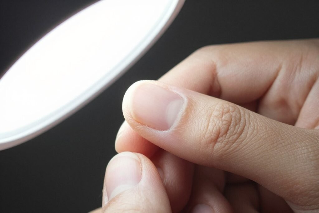
Anatomical differences between proximal and lateral folds require distinct optical positioning strategies.
Microvascular Orientation and Skin Scattering Factors
Optical assessment of microcirculation at the periungual borders requires careful management of light transmission through the stratum corneum. In the lateral nail fold, light scattering is typically elevated due to irregular keratin ridges and hyperkeratotic build-up along the lateral grooves. This optical diffusion can obscure subepidermal microvascular networks unless an appropriate immersion medium is applied.
Applying a high-transparency immersion substance, such as paraffin oil or cedar oil, minimizes surface refraction by matching the refractive index of keratinized skin. This allows optical beams from a specialized nailfold capillaroscopy microscope to penetrate through the superficial epidermis into the papillary dermis. Because the capillary loops in the lateral fold emerge at steeper angles, operators must adjust illumination angles to capture clean microvascular outlines without excessive glare.
Immersion fluids reduce light scattering across the lateral groove, allowing incident rays to penetrate dense keratin layers and expose dermal microstructures with improved optical contrast.
Movement artifacts represent another challenge during lateral tissue evaluation. The curved profile of the lateral digit makes perpendicular focus difficult without stable mechanical support. Utilizing adjustable stage mechanisms and finger stabilizers helps isolate the region of interest while maintaining uniform focal depth across the field of view.
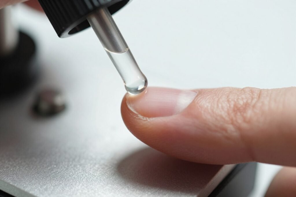
Applying optical immersion oil minimizes surface scattering across dense lateral keratin layers.
Standardized Positioning and Environmental Conditions
Obtaining reliable, reproducible visual records of periungual microcirculation requires strictly controlled operational environments. Cutaneous vascular diameter and blood velocity are sensitive to ambient temperature, emotional stress, and recent physical exertion. Without standardizing these external factors, natural physiological fluctuations may be mistaken for structural variations during visual assessments.
Environmental standards recommend maintaining an ambient room temperature between 22 and 24 degrees Celsius, with relative humidity stabilized near 70 percent. The subject should rest quietly in a seated posture for 15 to 30 minutes prior to observation, allowing digital blood flow to achieve hemodynamic equilibrium. In addition, holding the hand at heart level prevents hydrostatic venous pooling, which can artificially distend peripheral microvascular loops.
- Stabilize the room environment within 22 to 24 degrees Celsius and approximately 70 percent relative humidity before initiating examinations.
- Seat the individual comfortably with the hand supported steadily at approximately heart height to balance hydrostatic pressure.
- Avoid strenuous physical exercise, hot water hand washing, tobacco exposure, and heavy meals for at least one hour before assessment.
- Apply two drops of cedar oil or medical-grade paraffin oil along the lateral sulcus and adjacent tissue folds to optimize optical clarity.
- Align the target digit securely on the stage clamp to eliminate fine tremors during image acquisition.
Positioning the hand at heart level supports hemodynamic stability during non-invasive digital imaging.
Optical Configurations for Periungual Microcirculation Observation
High-resolution periungual evaluation requires specialized optoelectronic instruments designed for superficial cutaneous microscopy. Standard biological microscopes lack the necessary stage depth and illumination angles to accommodate a human digit without causing discomfort. Dedicated digital platforms integrate coaxial focusing mechanisms, compound mechanical stages, and cold LED illumination to deliver stable video streaming of peripheral tissue layers.
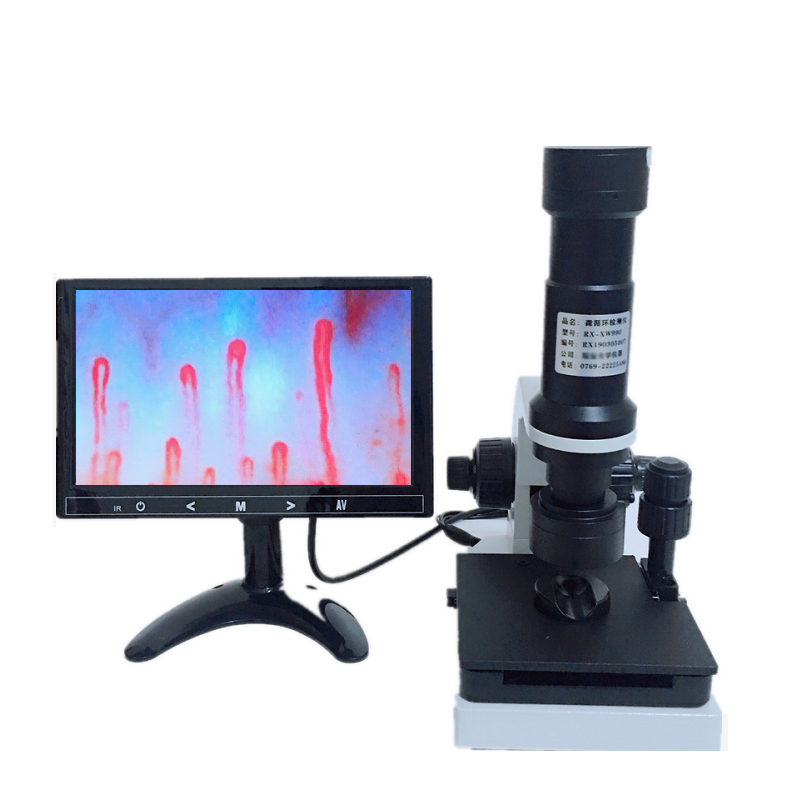
Instruments such as a digital nailfold capillaroscopy microscope utilize achromatic objective lenses paired with high-sensitivity CCD sensors. These sensors output dynamic video feeds directly to integrated LCD screens, allowing operators to locate the primary microvascular rows within seconds. Coaxial coarse and fine adjustment controls enable precise focal depth management across the curved topography of the lateral nail fold.
| Subsystem | Technical Parameter | Functional Contribution |
| Optical Objective | 5X Achromatic | Maintains optical flatness and minimizes chromatic aberration across curved tissue margins. |
| Magnification | 240X to 380X | Resolves structural dermal papillae, capillary loops, and erythrocyte flow dynamics clearly. |
| Illumination | LED Cold Light | Delivers focused target projection without transferring thermal energy to the digital skin surface. |
| Mechanical Stage | X-Y Double Layer | Enables smooth longitudinal and lateral tracking across lateral and proximal nail borders. |
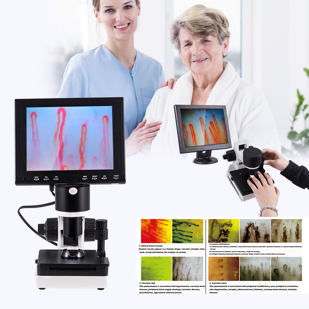
Specialized coaxial stages support steady optical tracking across the curved perionychium.
MAIKONG Optical Inspection Solutions and Specifications
Based in GD, SZ, MAIKONG manufactures professional optoelectronic instruments tailored for academic research institutions, clinical training facilities, and global medical device distributors. With advanced production facilities and strict quality management, we produce high-performance digital observation systems that balance optical precision with operational durability. Our platforms support original equipment manufacturing and private-label customization for specialized healthcare markets worldwide.
The MK-GY880 platform provides rapid microvascular locating, non-invasive observation workflows, and distortion-free video output via high-resolution CCD processing. Below are the reference technical specifications and packaging configurations for the MK-GY880 system.
| Specification Item | System Parameter | Operational Scope |
| Brand & Origin | MAIKONG, GD, SZ | Standardized manufacturing under international production protocols. |
| Magnification Levels | 380X / 500X / 600X | Multiple optical magnification configurations for detailed dermal assessment. |
| Power Supply | 100-240V AC Input | Universal input converting to stable DC 12V 2A output for optoelectronic components. |
| Focusing Drive | Coaxial Mechanism | Coarse and fine dual adjustment knobs for millimeter-level focal accuracy. |
| Display Screen | 8 to 12 Inch LCD | High-definition color monitor providing real-time uncompressed visual streaming. |
| Gross Weight | 7.0 Kilograms | Solid frame architecture housed inside reinforced protective packaging. |
Each observation platform is shipped in a shock-absorbing aluminum password case lined with protective memory foam to prevent mechanical displacement during transit.
| Package Item | Quantity | Item Role |
| MK-GY880 Host | 1 Unit | Main optical base with integrated stage and objective assembly. |
| LCD Monitor | 1 Unit | Dedicated color display screen mounted on top of the optical mast. |
| Power Adapter | 1 Unit | Regulated power supply module compatible with regional electrical outlets. |
| Cedar Oil | 1 Bottle | Optical immersion fluid for reducing superficial epidermal light scattering. |
| Accessories | 1 Set | Cotton swabs, calibration reference chart, and protective operational cover. |

The MK-GY880 platform is secured in a custom aluminum transit case for safe laboratory deployment.
Frequently Asked Questions About the Lateral Nail Fold
What is the lateral nail fold?
The lateral nail fold is the soft tissue structure that flanks the outer lateral edges of the nail plate. It forms the lateral sulcus, guiding longitudinal nail plate growth while shielding subungual compartments from microbial invasion and mechanical damage.
Where is the lateral nail fold located?
The lateral nail fold is located along the medial and lateral margins of every fingernail and toenail. It borders the lateral margins of the nail plate, connecting the dorsal skin of the distal phalanx directly to the lateral nail bed.
How is the lateral nail fold different from the proximal nail fold?
The lateral nail fold runs parallel to the direction of nail growth and contains predominantly vertical or oblique microvascular loops. In contrast, the proximal nail fold sits horizontally above the nail matrix and contains horizontal hairpin capillary loops visible parallel to the surface plane.
Why is the lateral nail fold relevant to nailfold observation?
The lateral nail fold provides a comparative baseline for regional periungual tissue evaluations and structural barrier assessments. Because its keratinized surface is thicker, observing its borders requires specialized optical lighting, stage stabilization, and immersion media to minimize light scattering.
Can a lateral nail fold image be used for diagnosis by itself?
A lateral nail fold image cannot be used for independent clinical diagnosis. High-magnification imaging serves strictly as an observational and documentation tool, and all clinical interpretations must be performed by qualified healthcare practitioners within a comprehensive medical evaluation.
What should a clinic consider when documenting lateral nail fold observations?
Clinics should standardize room temperature, relative humidity, subject rest times, and hand elevation before capturing images. Maintaining consistent optical magnification, stable immersion fluid application, and rigid mechanical stage tracking helps ensure reliable visual documentation over sequential sessions.
Summary of Lateral Nail Fold Evaluation
Understanding the anatomical borders and optical properties of the lateral nail fold supports rigorous clinical research and accurate periungual assessments. Standardizing environmental conditions, controlling skin scattering with immersion media, and utilizing stable coaxial optical stages allows operators to document peripheral microvascular structures with high clarity. These methodological steps help research centers maintain consistent documentation protocols across longitudinal studies.
Equip Your Facility with Professional Observation Systems
Qualified clinical laboratories, university research teams, and healthcare equipment distributors seeking standardized optical platforms can partner directly with MAIKONG for professional hardware configurations.