
Digital Nailfold Capillaroscopy Microscope for Professional Imaging and Research
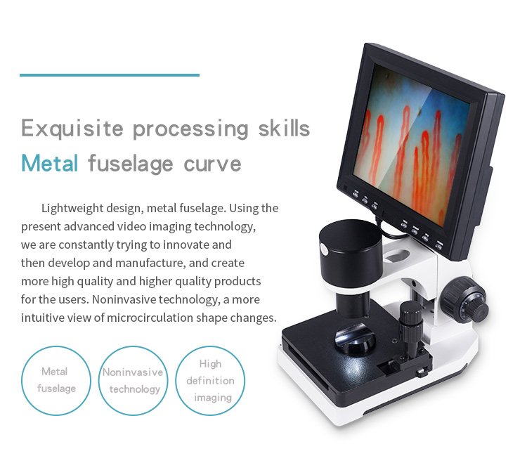
A digital nailfold capillaroscopy microscope is an optoelectronic instrument engineered for the non-invasive visual observation of human periungual microcirculation. The system combines high-magnification optical objectives, projected illumination, and digital video processing to display live capillaries on an integrated monitor. When assessing capillary architecture, clinicians and microvascular researchers rely on this specialized tool to examine morphology and cellular flow without invasive procedures.
Selecting an appropriate instrument depends on optical magnification requirements, mechanical stage precision, and workflow integration within clinical or research environments. Healthcare facilities, academic laboratories, and equipment distributors evaluate hardware stability, ease of adjustment, and customization potential when sourcing microscopy platforms.
Optical and Mechanical Architecture of the Microscope
Modern microcirculation observation instruments combine dedicated optics with integrated digital processing to deliver real-time visual output. An achromatic objective coupled with a high-performance imaging sensor transmits undistorted dynamic video streams directly to an attached color LCD monitor screen.
The system integrates projected LED cold lighting to illuminate the examination area without generating unwanted surface heat. A coaxial coarse and fine focusing mechanism permits smooth vertical travel, while a double-layer mechanical stage provides precise lateral and longitudinal movement for accurate vessel tracking.
Illumination System
An internal LED cold light source directs focused illumination onto the nailfold bed, preserving tissue comfort during observation.
Coaxial Focusing
Independent coarse and fine adjustment wheels allow rapid image location followed by micrometer-level focus refinement.
Compound Stage
The dual-axis X-Y mechanical stage moves smoothly across horizontal and longitudinal axes to position capillary rows.
Professional Examination and Focusing Workflow
Achieving stable microvascular visualization requires methodical setup and careful physical adjustment. Operators place the apparatus on a level workspace, align the monitor, and verify cable connections prior to subject evaluation.
- Mount the color LCD display securely onto the top frame of the mechanical assembly.
- Connect the dedicated power cable and video transmission line to the base interface.
- Power on the unit, allowing the imaging circuit to initialize before placing the subject’s hand.
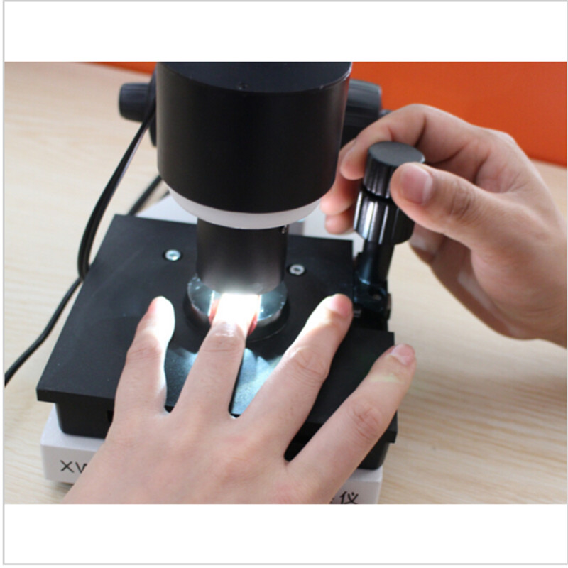
- Apply a thin layer of immersion medium, such as cedar oil or paraffin oil, to the cuticular junction of the left ring finger to minimize light scattering.
- Position the prepared digit comfortably into the finger press beneath the achromatic objective lens.
- Rotate the coarse handwheel to locate the optical plane, then refine the fine adjustment handwheel until capillary loops resolve clearly.
- Translate the compound X-Y stage laterally and longitudinally to scan across the distal capillary line.
Clinical Setting and Subject Preparation Considerations
Physiological variables significantly influence peripheral blood vessel behavior and cutaneous microcirculation appearance. Standardized observational protocols help maintain consistency across serial examinations and multi-subject research projects.
Clinics and research facilities often establish controlled testing suites to limit sympathetic nervous system stimulation before imaging. When preparing subjects for a nailfold capillary test, practitioners verify baseline environmental factors and subject compliance with departmental protocols.
Observation protocols frequently suggest maintaining ambient temperatures between 22 and 24 degrees Celsius with stable humidity near 70 percent. Allowing subjects to rest seated with hands positioned near heart level for several minutes prior to observation helps stabilize local microvascular tone.
Technical Specifications and Configuration Options
Hardware specifications dictate the operational envelope of each digital nailfold capillaroscopy microscope system. The platform balances optical magnification with mechanical stability for rigorous clinical and institutional use.
Configurations support standard magnification profiles alongside extended laboratory viewing options. Buyers assess chassis dimensions, power inputs, and display dimensions to match procurement criteria.
| Parameter | Standard Range | Optional Configuration | Operational Scope |
| Magnification | 380X or 240X | 500X or 600X | Achromatic optical magnification formats |
| Input Voltage | AC 100-240V | 50/60Hz DC | Wide-range power compatibility |
| Output Rating | DC 12V, 2A | Standard supply | Dedicated device power output |
| Objective Lens | 5X Achromat | 25mm range | Achromatic objective optical path |
| Mechanical Stage | Double-layer | Compound X-Y | Dual-axis platform displacement |
| Focus System | Coaxial unit | Coarse and fine | Micrometer precision focus control |
| Illumination | Internal LED | Cold lighting | Target projection cold illumination |
| Integrated Monitor | 8-inch LCD | 9, 10, or 12-inch | Mounted color video display |
| Camera Sensor | CCD sensor | 80,000 pixels | Distortion-free live video feed |
| Net Weight | 6 kg | Base instrument | Sturdy non-jittering chassis foundation |
Standard Package Contents and Accessories
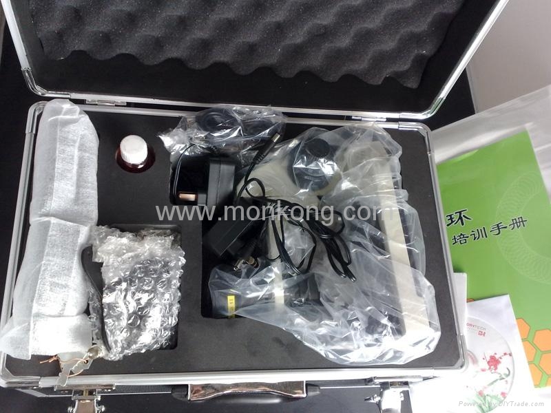
Standardized packaging protects optical assemblies and mechanical components during transport to commercial facilities and teaching hospitals. High-density custom memory foam within an aluminum password case secures the primary chassis against physical shocks.
Each shipment includes operational accessories required for immediate system setup and routine optical maintenance. Procurement teams verify itemized contents against packing lists before departmental deployment.
| Component | Model ID | Included Quantity | Package Role |
| Microscope Chassis | MK-GY880 host | 1 pc | Main optical and stage assembly |
| Display Monitor | LCD color unit | 1 pc | Integrated video observation screen |
| Power Adapter | 12V DC unit | 1 pc | Regulated electrical supply cable |
| Immersion Medium | Cedar oil bottle | 1 pc | Surface index-matching liquid |
| Applicator Swabs | Cotton swabs | 1 pc | Hygienic immersion application tool |
| Reference Chart | Morphology chart | 1 pc | Observational reference document |
Research and Observational Applications
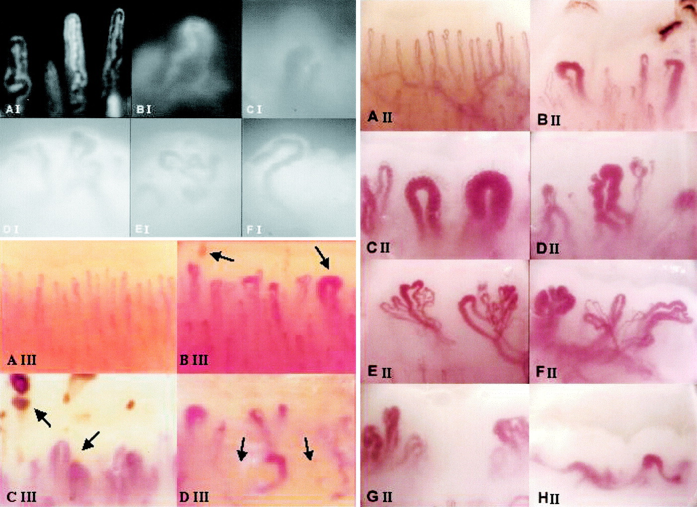
Digital microcirculation microscopes provide essential visualization capabilities across diverse medical research disciplines. Clinicians study cuticular capillary loops to document vascular structural changes, cell grouping, and dynamic microcirculatory patterns.
In specialized settings, the digital nail fold capillaroscopy microscope facilitates systematic imaging across multiple anatomic sites. Clinicians investigating rheumatologic phenomena or microvascular anomalies utilize live feeds to evaluate capillary density, loop width, and hemorrhages.
- Periungual distal nailfold capillary morphology observation and microvascular mapping.
- Dermatological research into peripheral dermal microcirculation, sweat gland structures, and hair follicles.
- Rheumatology research workflows investigating connective tissue vascular patterns alongside standard laboratory metrics.
- Vascular physiology education and teaching hospital training programs for non-invasive microcirculation assessment.
- Longitudinal monitoring of local skin microcirculation in clinical research trials and vascular biology labs.
Investigational and Material Microcirculation Testing
Beyond human clinical research, researchers employ microcirculation microscopy to assess experimental materials and non-clinical consumer goods. Controlled laboratory protocols utilize peripheral vascular observation to track biological responses to external stimuli.
Specialized product testing protocols investigate how specialized fabrics, far-infrared materials, or mineral compositions interact with dermal microvascular beds. These observational projects require stable magnification and dependable lighting to document microvascular velocity changes under controlled conditions.
Investigational studies on mineral products, functional textiles, or far-infrared goods represent exploratory research applications. These evaluations require independent laboratory protocols and do not constitute validated clinical diagnostic procedures.
Operational Considerations and Procedural Boundaries
A digital capillaroscopy microscope functions strictly as an optoelectronic visual observation and recording platform. The instrument does not generate automated diagnostic verdicts, disease classifications, or therapeutic recommendations.
Visual findings require professional interpretation by trained physicians or vascular specialists using established clinical criteria. Image resolution depends on subject preparation, optical cleanliness, operator technique, and proper immersion medium application.
Operational Advantages
- Non-invasive observation of periungual capillary architecture in real time.
- Stable mechanical stage with coaxial fine control reduces operator fatigue.
- Direct monitor transmission allows group viewing during medical training.
Methodological Boundaries
- Image sharpness relies entirely on operator focus and immersion oil quality.
- Microscope optics do not replace formal laboratory testing or clinical exams.
- Local vascular response varies with subject anxiety, temperature, and caffeine intake.
Selection Factors for Equipment Procurement
Procurement managers evaluate multiple mechanical and operational parameters when purchasing capillaroscopy platforms. Mechanical durability, optical stability, and ergonomic stage adjustments dictate daily clinical efficiency.
Distributors compare integrated benchtop consoles against handheld alternatives based on clinical volume and portability needs. Reviewing technical parameters before ordering helps departments match equipment capabilities to institutional documentation standards.
Integrated Stand Systems
- Stable 6 kg base chassis prevents table vibration from blurring images.
- Coaxial coarse and fine handwheels enable micrometer-level focus control.
- Dedicated 8 to 12-inch color LCD display mounts directly to the frame.
- Dual-axis compound mechanical stage supports precise vessel scanning.
Handheld Inspection Tools
- Compact footprint suited for mobile screening and bedside evaluations.
- Flexible positioning across varied peripheral anatomical observation sites.
- USB connection routes video directly into compatible computer software.
- Lower mechanical mass requires steady hand coordination during viewing.
MAIKONG Manufacturing and OEM Capabilities
We produce professional optical inspection platforms at our manufacturing facilities located in SZ, GD. Leveraging a mature regional supply chain and advanced production tooling, MAIKONG delivers both high-end clinical setups and cost-effective microscopy solutions.
Our engineering team supports custom OEM and private-label manufacturing projects for healthcare distributors and brand owners worldwide. We configure display dimensions, optical magnifications, and chassis finishes to align with client branding requirements.
Custom Configuration
We offer tailored hardware adjustments, including optional 500X and 600X magnifications, multiple monitor dimensions, and custom chassis badging.
Supply Chain Reliability
Our GD/SZ production facility maintains strict component inspection standards to deliver consistent mechanical durability across commercial batch orders.
Frequently Asked Questions
What is a digital nailfold capillaroscopy microscope?
A digital nailfold capillaroscopy microscope is an optoelectronic instrument that magnifies the cuticular microcirculation and displays real-time video on a connected screen. It enables clinicians and researchers to observe capillary loop morphology and blood flow dynamics non-invasively.
How does the digital imaging workflow work?
The operator applies immersion oil to the subject’s cuticle, inserts the finger into the stage holder, and illuminates the site with integrated LED cold light. Optical magnification relays live dynamic video through a CCD imaging sensor directly to the integrated LCD monitor.
What magnification and monitor options are available?
Standard systems feature 380X or 240X optical magnification with an 8-inch color LCD monitor. Optional configurations support 500X or 600X magnification alongside 9-inch, 10-inch, or 12-inch displays.
Is the system suitable for research and teaching?
Yes, the integrated monitor and stable coaxial stage make the microscope well suited for medical school lectures, microvascular research labs, and clinical demonstrations. Multiple observers can examine real-time microvascular structures simultaneously without looking through an ocular eyepiece.
What should buyers check before placing a bulk order?
Procurement teams should verify required monitor sizes, local voltage specifications, preferred packaging, and institutional regulatory requirements. Buyers should also evaluate warranty coverage, spare parts availability, and operator training protocols before final purchase confirmation.
Can MAIKONG support OEM or private-label projects?
Yes, MAIKONG provides comprehensive OEM and ODM manufacturing services from our SZ, GD facility. We support custom logo printing, chassis color modifications, tailored packaging, and custom hardware configurations for medical equipment distributors.
What is included in the package?
The standard MK-GY880 package includes the main host chassis, color LCD monitor, 12V power adapter, cedar immersion oil, cotton application swabs, and a morphology reference chart. All items are housed securely in an aluminum case lined with shock-absorbent memory foam.
How can a U.S. distributor request a quotation?
Distributors and clinic purchasing managers can contact MAIKONG directly via email or WhatsApp to discuss order quantities, custom requirements, and wholesale pricing tiers. Our commercial sales team provides formal quotations and international shipment schedules upon request.
Summary and Distribution Partnerships
Digital nailfold microscopy provides an indispensable visual window into peripheral microvascular dynamics for healthcare practitioners and microcirculation scientists. Organizations seeking reliable, cost-effective capillaroscopy equipment can partner directly with MAIKONG for customized manufacturing, OEM branding, and regional distribution agreements.
Request Product Specifications and Wholesale Quotation
Connect with our sales engineering team in SZ, GD to discuss bulk orders, custom configurations, or becoming a local distribution partner.