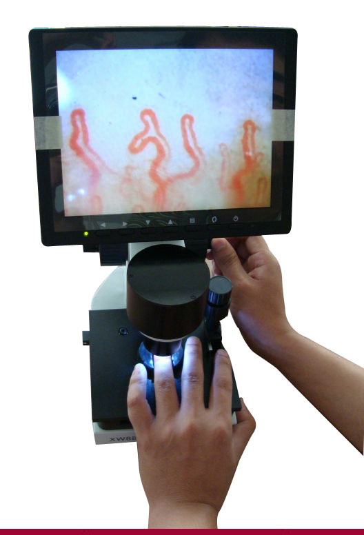
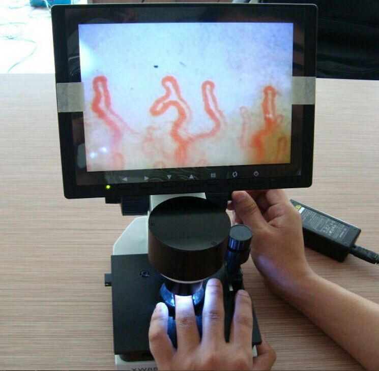
nailfold capillary microscopy application
What is nailfold capillary microscopy and nailfold capillary microscopy application?
nailfold capillary microscopy Widefield capillary microscopy A technique used to evaluate finger nailfold capillaries, using a wide-angle stereomicroscope or ophthalmoscope; enlargement, ↑ tortuosity, or ↓ number of capillaries occur in connective tissue diseases–eg, Raynaud’s phenomenon, scleroderma–to evaluate extent of visceral involvement, MCTD, dermatomyositis, SLE.
Abstract. Nailfold capillaroscopy is a method of great diagnostic value in the differential diagnosis of primary versus secondary Raynaud´s phenomenon, of systemic sclerosis versus other so called connective tissue diseases and of additional diagnostic value in other entities. Rheumatologists, dermatologists, and angiologists in Germany have convened in an interdisciplinary working group in which they synergistically combined their expertise to develop a common nomenclature and standards for the technical performance of nailfold capillary microscopy. The article gives an overview of historical and technical aspects of capillaroscopy, morphologic findings, and disease-specific patterns. It also provides a critical appraisal of its significance in the diagnosis and sequelae of these interdisciplinarily-managed diseases including its performance in children and gives an excursion in the potential perspectives of capillaroscopy in less common indications.
WHAT ARE ITS USES?
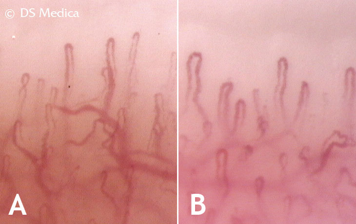
The capillaroscopy allows us to know the extent of the distal vascularisation, which is very important in systemic sclerosis and other connective pathologies, as well as to rule out systemic involvement in patients with Raynaud’s phenomenon with no other associated clinical involvement.
RAYNAUD IS PHENOMENON
The capillaroscopy of the nail bed is a simple, bloodless, economical method which is very useful for studying Raynaud’s phenomenon and other rheumatological conditions. Raynaud’s phenomenon can be defined as the change in the colouring of the fingers and/or toes in response to cold or stress. It traditionally progresses through three stages: paleness (vasospasm), cyanosis (due to increased carboxyhemoglobin) and erythema (reactive hiperemia).
Raynaud’s phenomenon can be primary (Raynaud’s disease) or secondary, associated with a connective tissue disease. Primary Raynaud’s phenomenon is responsible for around 60% of all new cases. 15-20% of cases of Raynaud’s phenomenon are due to a series of non- immunological processes, such as drugs, occupational diseases, neoplasms, etc. The remaining 15-20% are associated with connective tissue diseases.
Raynaud’s is present in over 90% of patients with scleroderma and in 70% of cases it is the first symptom. Although it seems impossible to predict that a patient with Raynaud’s will develop scleroderma, the presence of antinuclear antibodies (ANA) indicates a greater risk of onset.
SCLERODERMA
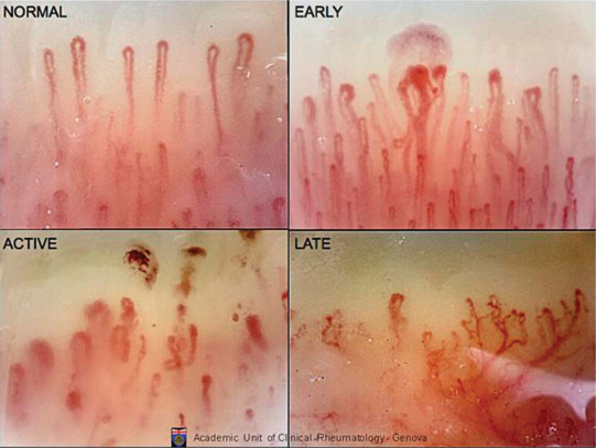
The nail bed capillaroscopy shows morphological alterations at an early stage in some connective tissue diseases of maximum rheumatological interest, particularly capillaroscopy. In these cases, the capillaroscopy traditionally shows the “sclerodermic pattern” characterised by: reduction or absence of capillaries in patches, capillary dilation, and sometimes mega-capillaries and splinter haemorrhages. This “sclerodermic pattern” appears early and when it is observed in patients with Raynaud’s phenomenon, even if it is not very obvious, it should lead to the search for sclerodermic manifestations in internal organs, which can be present without causing any symptoms. The combination of Raynaud’s phenomenon and a “sclerodermic pattern” in a capillaroscopy can precede and therefore predict the onset of scleroderma.
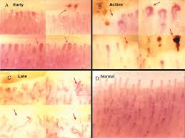
nailfold capillary microscopy chart
CAPILLAROSCOPY AND RHEUMATOLOGICAL CONDITIONS
In dermatomyositis, the capillaroscopy is similar and sometimes indistinguishable from that found in scleroderma. These patients generally present the other clinical, enzymatic or electromyographic manifestations of dermatomyositis that enable its recognition and diagnosis.
In mixed connective tissue disease (MCTD) or overlap syndromes that a sclerodermic component, the findings of the capillaroscopy can be similar although rarely are large capillary dilations and mega-capillaries observed. These capillaroscopies must be analysed by an expert.
In systemic lupus erythematosus, the alterations in the capillaroscopy are non-specific, and it is possible to find focal capillary reduction, albeit not very strong. The capillaries can be somewhat dilated and tortuous, sometimes with criss-crossing of the arterial and venous components (ringlets), the latter being the most characteristic finding in the capillaroscopy.
In primary Raynaud’s phenomenon, the most striking characteristic is the elongated capillaries with undulations throughout the arterial and venous components. Little or no reduction or dilation of the capillaries can be observed. Splinter haemorrhages are scarce and small.
Ultimately, this is an auxiliary diagnostic technique with great value in rheumatology and vascular disease.
Related Items