My Top 10 Peeves of tasly microcirculation test

Where and who need the tasly microcirculation test?
Where and who need the tasly microcirculation test?
The microcirculation microscope is widely used in the field of hospitals, school laboratories, clinics, etc., as the best auxiliary testing equipment. And it is also used in health food industry, as a communication platform for product promotion when, making money equipment, beauty industry.
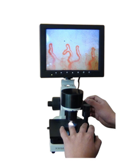
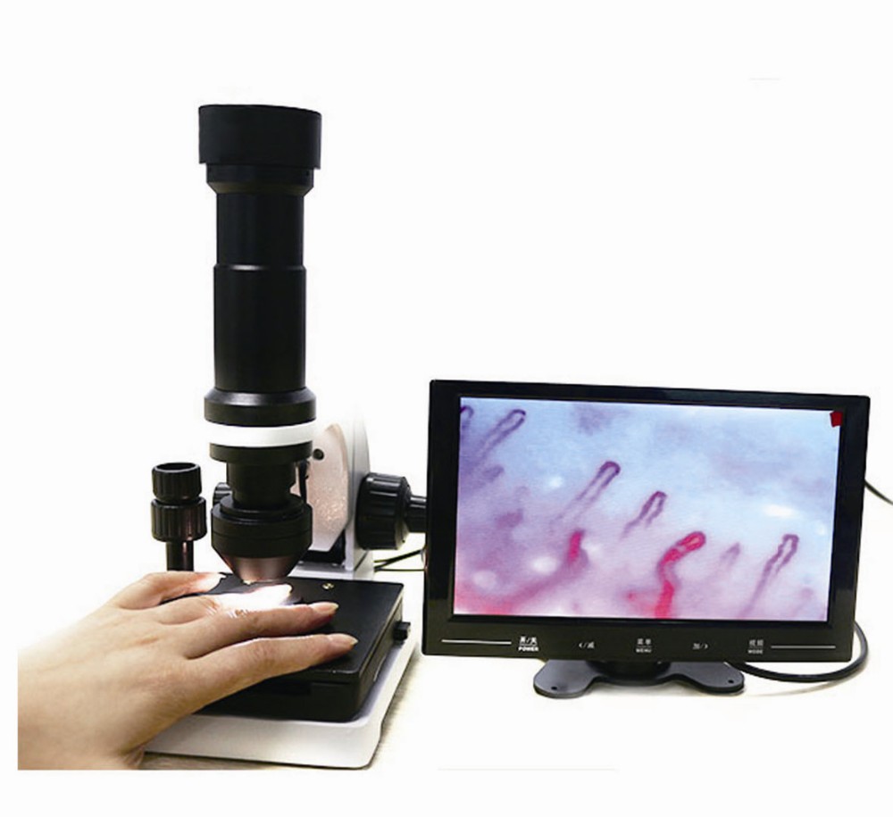
For it is non-invasive, only to observe the end of the finger armor section, you can do the blood microcirculation review. You can observe microcirculation pipe loop patterns, the number of pipe loop, blood flow, bleeding, etc., help determine the sub-health status and blood stasis and brain / cardiovascular related diseases.

How to use tasly microcirculation test?
How to use tasly microcirculation test?
Step 1: connect one side of the power with the plug
Step 2: connect other side of the power with the sub-second line
Step 3 connect one end of the sub-second line with the extend line
Step 4: connect the two end of sub-second line with the machine and the monitor blood microcirculation test machine
Step 5 : connect the video line to the machine and the monitor
Step 6: connect the power. Now the machine is ready for testing

tasly microcirculation test can suggest pulmonary disease activity in systemic sclerosis
tasly microcirculation test can suggest pulmonary disease activity in systemic sclerosis.
OBJECTIVE: To evaluate the association of capillaroscopic alterations with pulmonary disease activity in systemic sclerosis (SSc). METHODS: Ninety-one patients with SSc were studied by means of interview, physical examination, nailfold capillary microscopy (NCM), serology, pulmonary function tests, esophageal transit scintigraphy, Doppler echocardiography, and pulmonary high resolution computed tomography (HRCT). Pulmonary disease activity was diagnosed by the observation of ground-glass opacities on pulmonary HRCT. Capillary loss on NCM was evaluated using the avascular score: patients with mean score > or = 1 or mean number of megacapillaries per finger > or = 1 were considered to have severe capillaroscopic alterations. RESULTS: Patients with higher skin scores, longer disease duration, signs of peripheral ischemia, esophageal dysfunction, antitopoisomerase I antibodies, and ground-glass opacities had higher mean avascular scores (p < or = 0.05 in all tests). The association between ground-glass opacities and higher avascular scores was particularly strong in patients with disease duration < or = 5 years. Among these patients, ground-glass opacities were present in 14 of 19 patients with severe NCM alterations, but were absent in all patients (n = 8) with mild or no NCM alterations (p < 0.001). ROC curves confirmed the ability of NCM to discriminate between patients with and without ground-glass opacities among those with disease duration < or = 5 years. However, NCM could not predict the presence of reduced pulmonary diffusing capacity. CONCLUSION: The severity of NCM abnormalities is associated with lung disease activity in SSc, particularly when the disease duration is relatively short.

What is tasly microcirculation testn?
What is tasly microcirculation testn?
The main functions of the microcirculation are the delivery of oxygen and nutrients and the removal of carbon dioxide (CO2). It also serves to regulate blood flow and tissue perfusion thereby affecting blood pressure and responses to inflammation which can include edema (swelling).
The blood capillaries are where the important functions of the microcirculation take place: the exchange of material between circulation and cells. Capillaries are the smallest of the body’s blood vessels. They are only one cell thick, and they are the sites of the transfer of oxygen and other nutrients from the bloodstream to other tissues in the body; they also collect carbon dioxide waste materials and fluids for return to the veins.

tasly microcirculation test changes in systemic lupus erythematosus: correlations with disease activity and autoantibody profile
tasly microcirculation test changes in systemic lupus erythematosus: correlations with disease activity and autoantibody profile
In systemic lupus erythematosus (SLE) nailfold capillaroscopy (NC) studies have described many different nonspecific patterns. We decided to evaluate NC changes in 44 SLE patients, comparing them with the main clinical, demographic and laboratory parameters, thus to define the real role for NC and its abnormalities in the management of this disease. Fifteen patients (34%) complained of Raynaud’s phenomenon; nine of them (20%) showed relevant capillaroscopic changes (capillaroscopic score >1). In details: three patients (6.8%) had loss of capillaries, while 18 (41%) had a capillary length variability, 16 (36.5%) showing shorter and two (4.5%) longer capillaries; tortuous, meandering, bizarre, ramified and/or bushy capillaries were found in 26 (59%), seven (16%), two (4.5%), three (7%) cases, respectively. An irregular distribution of the capillary array was present in six cases (14%) while microhaemorrhages were found in four cases (9%). 4 patients (9%) showed enlarged capillaries and changes of blood flow. A capillaroscopic score >1 was more frequently associated with higher ECLAM (P < 0.005) and SLEDAI (P < 0.01) activity scores, with the presence of anti-cardiolipin (P < 0.04) and anti-Sm (P < 0.04) antibodies, and also with the presence (P < 0.04) and higher titer (P < 0.001) of anti-dsDNA antibodies. No statistically significant correlation was found among the different capillaroscopy findings, age, disease duration, or treatment, nor with any clinical manifestation of the disease, such as cutaneous, renal or neurological. Our findings confirm the importance of the microvascular involvement in SLE. The NC abnormalities seem to be related to the disease activity and to the presence of many different antibodies, highly involved in the expression of SLE. NC proved to be an easy-to-perform noninvasive technique, able to achieve useful data to better evaluate such a pleomorphic disease as SLE.

Related Items