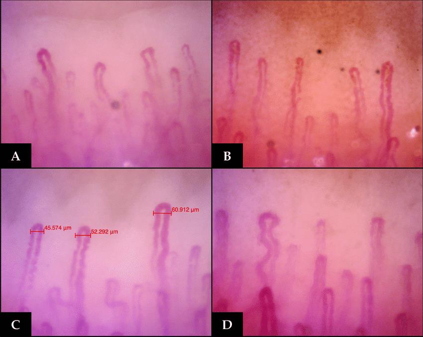
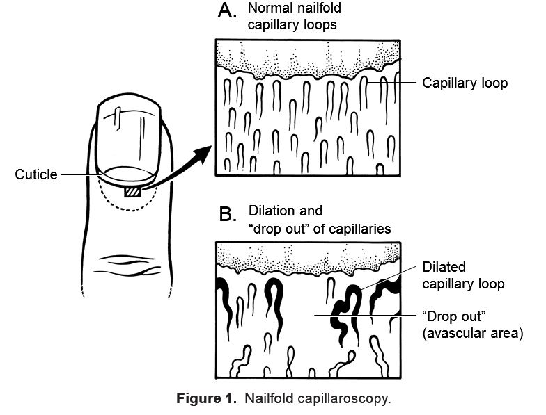
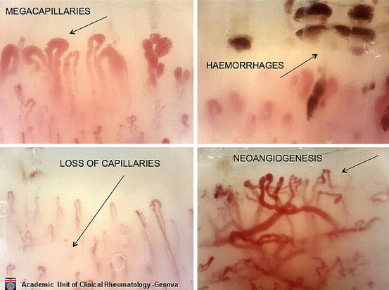
nail fold microscopy
What is nail fold microscopy?
nail fold microscopy
nail fold microscopy
nail fold microscopy
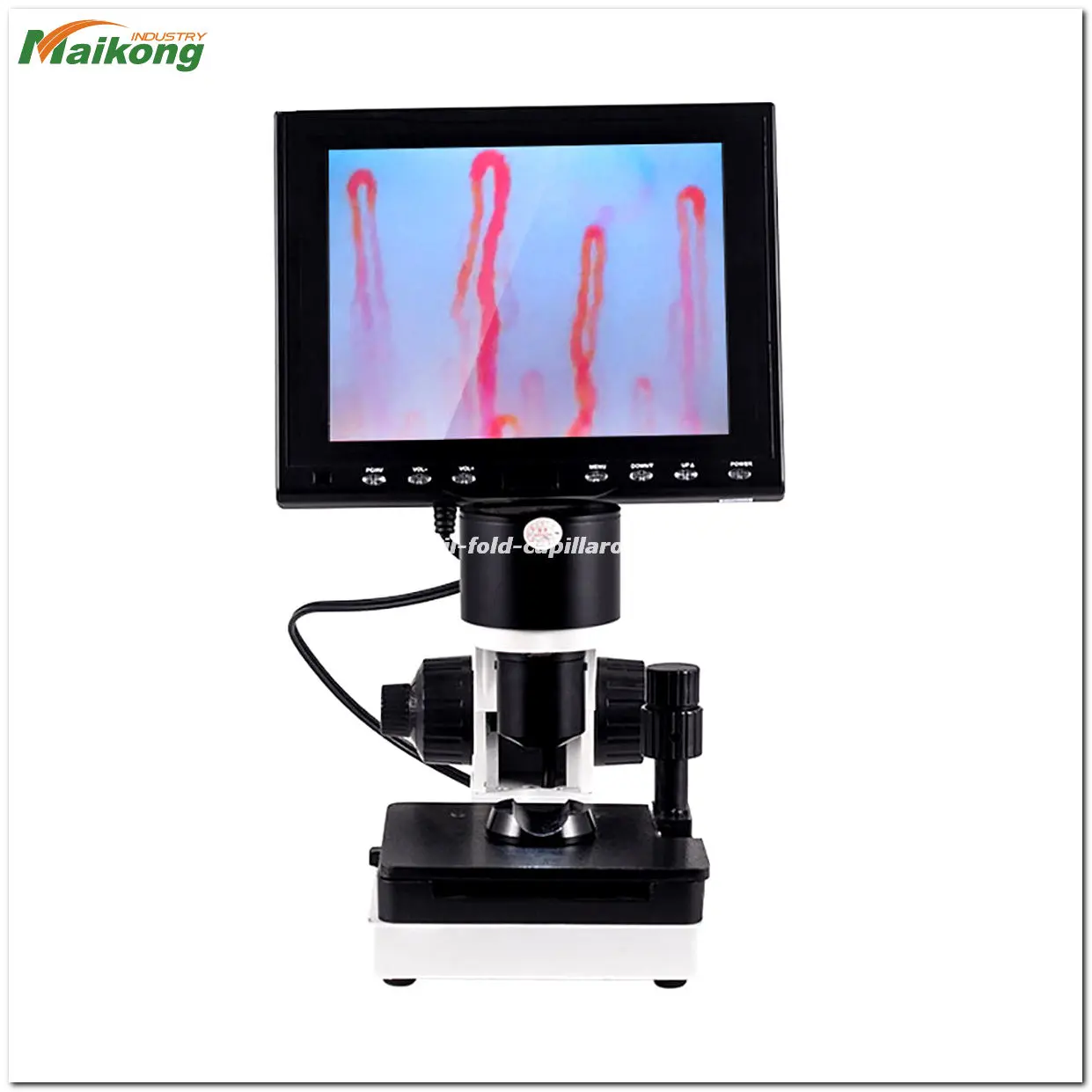
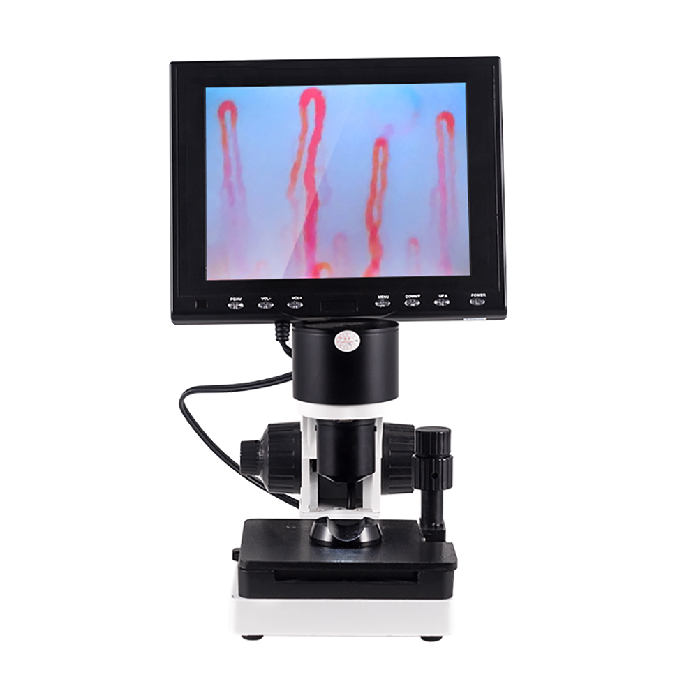
Technical parameters
1.magnification over 400 times
2. built-in camera380 000 pixels
3. light source LED
4.brightness of the light source over 600cd/m2
5. Stage X-Y double layer compound mechanical stage
6. Color LCD monitor 8.5inch (4:3) Color LCD monitor 7 inch (16:9)
7.AC input 100-240V50/60Hz DC output DC12V,2A
8. Package aluminum box (Size 40x29x36cm)
9. Gross weight 6.8kg
Testing conditions
1) Check the room temperature and humidity. Room temperature and humidity
should be kept relatively constant. room temperature should be maintained in the 22-24 degree , relative humidity about 70%
2) the patient generally take seats, keep the height of the hand the same with heart
3) Preparation of paraffin oil or cedar oil.Drop 2 drops in the nailfold skin. (Purpose prove light
transmission, and reduce skin scattering)
4)Testing generally be in the morning or afternoon, and review should at the same time every day .
5) the patient should
a.Avoiding intense activity or manual labor within one hour before testing
b. Take a rest about 15-30 minutes before testing
c.Can not take any drug which will affect the cardiovascular vessels before testing
d. Does not smoke , wash hands or eat within an hour before testing
e.Pay attention to the influence of the Female menstrual
nail fold microscopy
How to use nail fold microscopy?
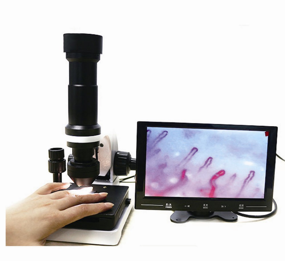
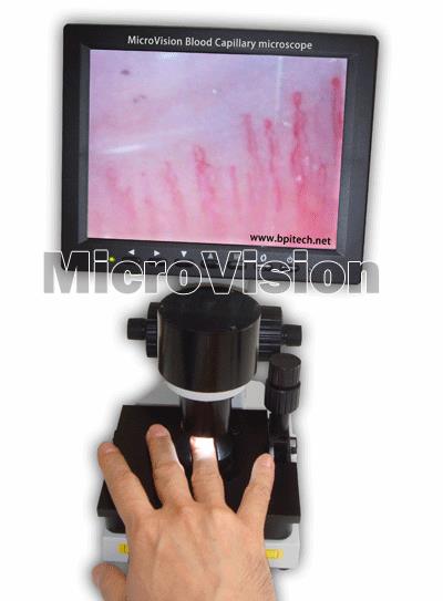
Usage of latest medical microcirculation nailfold capillaroscopy machine
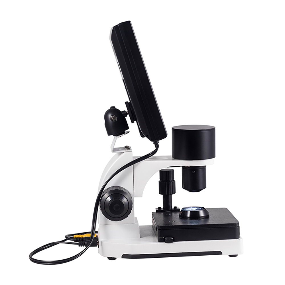
1. Fix the screen on the top of the mechanical machines.
2. Plug in the power line and vidao line to the interface of the base.
3. Connect the plug, the plug shows green lights. Press the “Power” button of the screen, wait 3 seconds, the screnn will light.
4. Paint three drops of oil at the position of the intersection of the ring finger nails and skin. Put the oil coated finger in finger hold.
5. Adjuet the coarse hand wheel, when the screen displays images, then adjust fine handwheel. Picture becomes clearer. The bigger one is the coarse adjustable wheel and the small wheel is the fine handwheel. When the machine is about 2-3cm away from the finger, there will be a transparent image on screen.
6. Then, adjuet platform lateral movement handwheel to et a correct position of the first line of the vessel( capillary tube ) and adjuet the fine coarse handwheel to get a clear picture of the vessel. Your fingers will move from left to right on the double loading platform. So try to find the first line of vessels by rotating the platform lateral movement handwheel to change the position of the finger. And finally rotate the fine handwheel ( the position is showed in the structure ) to get a clear picture of the vessels.
7. If there is spot on the screen, rotate the carmera right and left to eliminate it.

nail fold microscopy
nail fold microscopy
nail fold microscopy
What is nail fold microscopy Benefit?
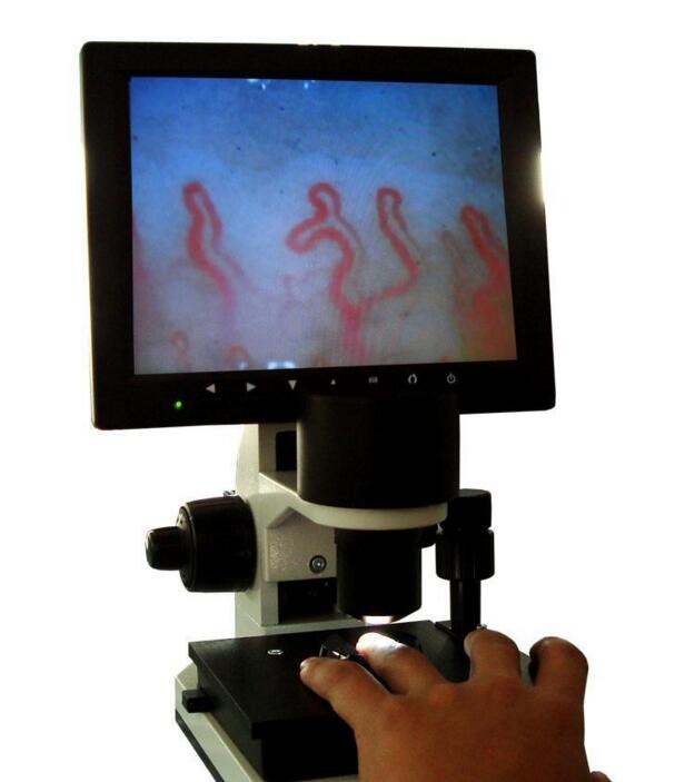
1.in 5 seconds can be observed in the microvascular circulation, simple operation and fast;
2.the microscopic image is stable, clear, do not shake;
3.400 times the microcirculation of a dedicated high-resolution lens of the microscope;
4.handheld ultra-light design, so that the observation of more mobility;
5.with micro-focus wheel, free to adjust the focus length, the use of more convenience
6.lens built-in LED cold light source, accurate projection illumination on an object;
7.using non-invasive way, to observe the microcirculation of human body in time;
8.observing the microcirculation for any part of human body.
What is nail fold microscopy Main Spec?
| Amplification |
400X |
| Ourput device |
480 type wire of TV |
| Objective |
5X,effective range 25mm |
| Objective type |
Achromatization |
| Mechanical stage |
Single |
| Focusing mechanism |
Coaxial coarse and fine adjustment |
| Inner dimming |
LED cold light |
| Power |
12V 1A DC power source |
| Inner dimming |
12V 1A LED cold light |
| Net weight |
4.5kg |
| Gross weight |
6kg |
| Inner package |
Aluminum password box or wooden box, memory foam |
| Out package |
Corrugated box |
| Software |
English software |
| Certificate |
CE approved |
nail fold microscopy
nail fold microscopy
How to Maintenance nail fold microscopy?
1. Place in a cool, dry, no dust, no acid, no-place.
2. The air relative humidity less than 60%.
3. When moving, before installation if two temperature difference, room temperature should be adapted to the use of the instrument, in order to prevent the optical lens fogging, mildew.
4.The objective has been parity, can not disassemble. Dust on the mirror ball blow hair available, or gently wipe with a clean brush (or lens cleaning paper).
5. The camera target surface is dusty, slightly soiled cotton xylene gently wipe test, turn gently rub outward from the center of the target surface wipe.
6. The objective should be taken to avoid being hit.
7. Keep the instrument surface clean.
Related Items