how does dark field microscopy work?
What is dark field microscopy?
how does dark field microscopy work
how does dark field microscopy work
how does dark field microscopy work
how does dark field microscopy work
how does dark field microscopy work

how does dark field microscopy work
All of us are quite familiar with the appearance and visibility of stars on a dark night, this despite their enormous distances from the Earth. Stars can be readily observed at night primarily because of the stark contrast between their faint light and the black sky.
how does dark field microscopy work
how do dark field microscopy work
Yet stars are shining both night and day, but they are invisible during the day because the overwhelming brightness of the sun “blots out” the faint light from the stars, rendering them invisible. During a total solar eclipse, the moon moves between the Earth and the sun blocking out the light of the sun and the stars can now be seen even though it is daytime. In short, the visibility of the faint star light is enormously enhanced against a dark background.
This principle is applied in darkfield (also called darkground) microscopy, a simple and popular method for making unstained transparent specimens clearly visible. Such objects often have refractive indices very close in value to that of their surroundings and are difficult to image in conventional brightfield microscopy. For instance, many small aquatic organisms have a refractive index ranging from 1.2 to 1.4, resulting in a negligible optical difference from the surrounding aqueous medium. These are ideal candidates for dark field microscopy.
dark field microscopy requires blocking out of the central light which ordinarily passes through and around (surrounding) the specimen, allowing only oblique rays from every azimuth to “strike” the specimen mounted on the microscope slide. The top lens of a simple Abbe darkfield condenser is spherically concave, allowing light rays emerging from the surface in all azimuths to form an inverted hollow cone of light with an apex centered in the specimen plane. If no specimen is present and the numerical aperture of the condenser is greater than that of the objective, the oblique rays cross and all such rays will miss entering the objective because of their obliquity. The field of view will appear dark.
The darkfield condenser/objective pair illustrated in Figure 1 is a high-numerical aperture arrangement that represents darkfield microscopy in its most sophisticated configuration, which will be discussed in detail below. The objective contains an internal iris diaphragm that serves to reduce the numerical aperture of the objective to a value below that of the inverted hollow light cone emitted by the condenser. The cardioid condenser is a reflecting darkfield design that relies on internal mirrors to project an aberration-free cone of light onto the specimen plane.
When a specimen is placed on the slide, especially an unstained, non-light absorbing specimen, the oblique rays cross the specimen and are diffracted, reflected, and/or refracted by optical discontinuities (such as the cell membrane, nucleus, and internal organelles) allowing these faint rays to enter the objective. The specimen can then be seen bright on an otherwise black background. In terms of Fourier optics, dark field microscopy removes the zeroth order (unscattered light) from the diffraction pattern formed at the rear focal plane of the objective. This results in an image formed exclusively from higher order diffraction intensities scattered by the specimen.
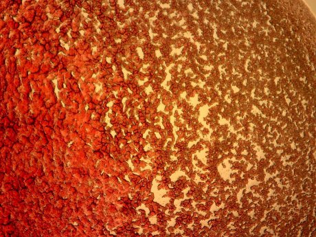
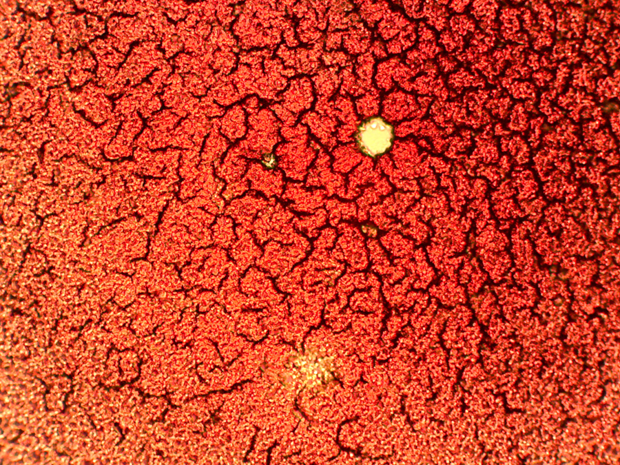
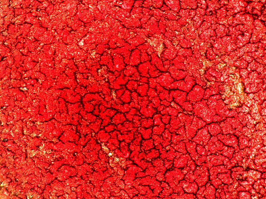
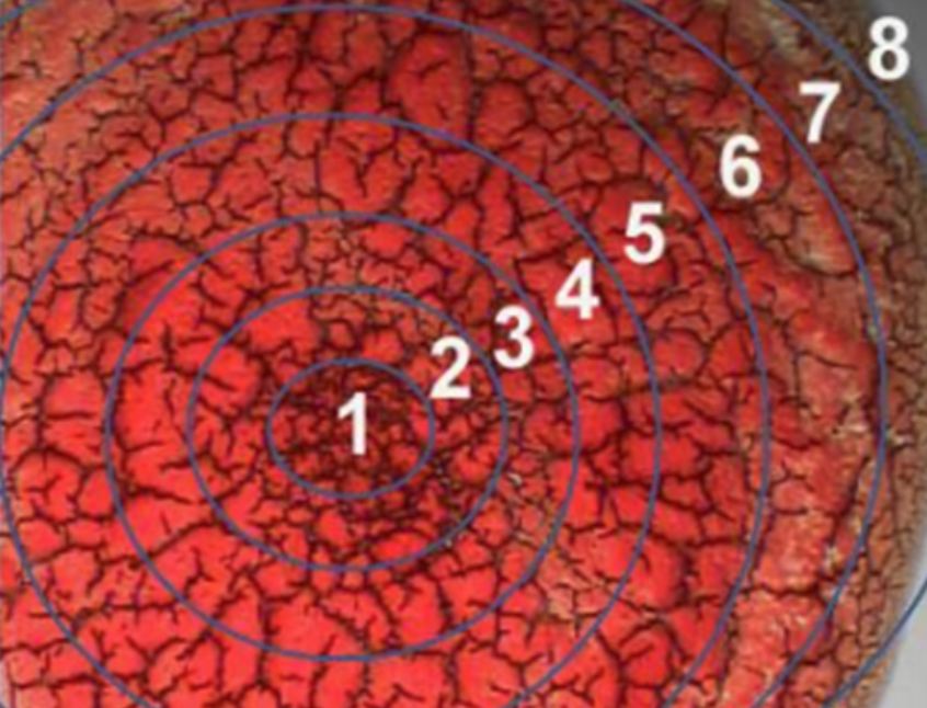
The photomicrographs in Figure 2 illustrate the effects of darkfield and brightfield illumination on silica skeletons from a small marine protozoan (radiolarian) in a whole mount specimen. In ordinary brightfield, skeletal features of the radiolarian are not well defined and tend to be washed out in photomicrographs recorded either with traditional film or digitally captured. was taken in brightfield illumination with the condenser aperture diaphragm closed to a point where diffraction artifacts obscure some of the sample detail. This enhances specimen contrast at the expense of image distortion. Under dark field microscopy, more detail is present, especially in the upper portion of the organism, and the image acquires an apparent three-dimensional appearance. When a red filter is used in conjunction with a darkfield stop , the radiolarian takes on a colorful appearance that is more pleasing, although no additional detail is produced and there is even some reduction in image quality.
Specimens that have smooth reflective surfaces produce images due, in part, to reflection of light into the objective. In situations where the refractive index is different from the surrounding medium or where refractive index gradients occur (as in the edge of a membrane), light is refracted by the specimen. Both instances of reflection and refraction produce relatively small angular changes in the direction of light, allowing some to enter the objective. In contrast, some light striking the specimen is also diffracted, producing a 180-degree arc of light that passes through the entire numerical aperture range of the objective. The resolving power of the
how does dark field microscopy work
how does dark field microscopy work
how does dark field microscopy work
how does dark field microscopy work
dark field microscopy principle?
dark field microscopy is a method which also creates contrast between the object and the surrounding field. As the name implies, the background is dark and the object is bright. A annular stop is also used for dark field, but the stop is now outside the field of view. Only light coming from the outside of the beam passes through the object and it cannot be seen directly. Only when light from the stop is deflected and deviated by the object can it be seen. This method also produces a great deal of glare and therefore the specimen often appears as a bright silhouette rather than as a bright object of which much detail can be determined. The following diagram shows the setup of the dark field light path.
dark field microscopy Conclusion?
A dark field microscope can offer brilliant, light images against a dark background of otherwise difficult to view specimens.Most standard microscopes come with dark field capabilities or accessories to enable this illumination technique.There are many practical applications of dark field, especially in the field of marine biology, in viewing the many specimens you cannot see using alternative techniques.However, a researcher must keep in mind the potential issues and limitations that may arise from dark field illumination.
how does dark field microscopy work?
How dark field microscopy work?-Microscopes are used to magnify objects. Through magnification, an image is made to appear larger than the original object. The magnification of an object can be calculated roughly by multiplying the magnification of the objective lens times the magnification of the ocular lens. Objects are magnified to be able to see small details. There is no limit to the magnification that can be achieved; however, there is a magnification beyond which detail does not become clearer. The result is called empty magnification when objects are made bigger but their details do not become clearer. Therefore, not only magnification but resolution is important to the quality of the information in an image.
The resolving power of the microscope is defined as the ability to distinguish two points apart from each other. The resolution of a microscope is dependent on a number of factors in its construction. There is also an inherent theoretical limit to resolution imposed by the wavelength of visible light (400-600nm). The theoretical limit of resolution (the smallest distance able to be seen between two points) is calculated as:
Resolution = 0.61 l/N.A.
where l represents the wavelength of light used and N.A.is the numerical aperture. The student-grade microscopes generally have much lower resolution than the theoretical limit because of lower quality lenses and illumination systems.
Standard brightfield microscopy relies upon light from the lamp source being gathered by the substage condenser and shaped into a cone whose apex is focused at the plane of the specimen. Specimens are seen because of their ability to change the speed and the path of the light passing through them. This ability is dependent upon the refractive index and the opacity of the specimen. To see a specimen in a brightfield microscope, the light rays passing through it must be changed sufficiently to be able to interfere with each other which produces contrast (differences in light intensities) and, thereby, build an image. If the specimen has a refractive index too similar to the surrounding medium between the microscope stage and the objective lens, it will not be seen. To visualize biological materials well, the materials must have this inherent contrast caused by the proper refractive indices or be artificially stained. These limitations require instructors to find naturally high contrast materials or to enhance contrast by staining them which often requires killing them. Adequately visualizing transparent living materials or thin unstained specimens is not possible with a brightfield microscope.
dark field microscopy relies on a different illumination system. Rather than illuminating the sample with a filled cone of light, the condenser is designed to form a hollow cone of light. The light at the apex of the cone is focused at the plane of the specimen; as this light moves past the specimen plane it spreads again into a hollow cone. The objective lens sits in the dark hollow of this cone; although the light travels around and past the objective lens, no rays enter it (Fig. 1). The entire field appears dark when there is no sample on the microscope stage; thus the name dark field microscopy. When a sample is on the stage, the light at the apex of the cone strikes it. The image is made only by those rays scattered by the sample and captured in the objective lens (note the rays scattered by the specimen in Figure 1). The image appears bright against the dark background. This situation can be compared to the glittery appearance of dust particles in a dark room illuminated by strong shafts of light coming in through a side window. The dust particles are very small, but are easily seen when they scatter the light rays. This is the working principle of dark field microscopy and explains how the image of low contrast material is created: an object will be seen against a dark background if it scatters light which is captured with the proper device such as an objective lens.
The highest quality darkfield microscopes are equipped with specialized costly condensers constructed only for darkfield application. This darkfield effect can be achieved in a brightfield microscope, however, by the addition of a simple “stop”. The stop is a piece of opaque material placed below the substage condenser; it blocks out the center of the beam of light coming from the base of the microscope and forms the hollow cone of light needed for darkfield illumination.

how does dark field microscopy work

how does dark field microscopy work
how does dark field microscopy work

how does dark field microscopy work

how does dark field microscopy work
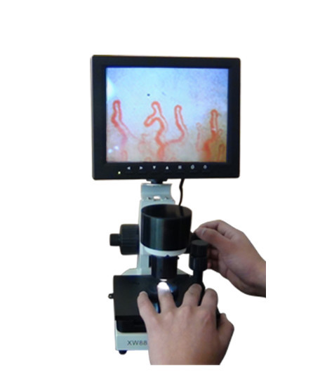
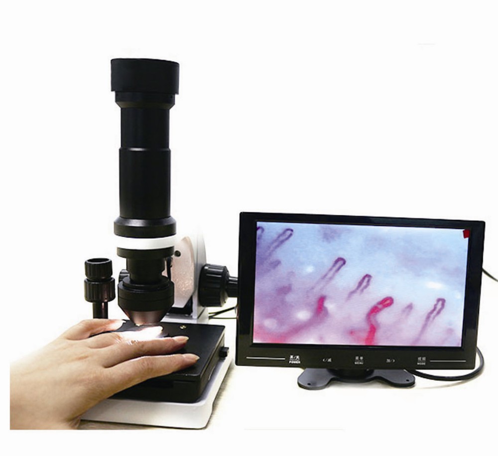
dark field microscopy APPLICATIONS
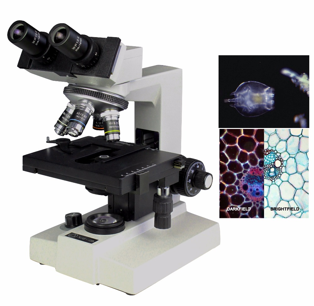
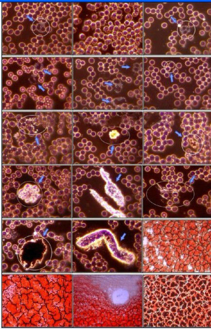
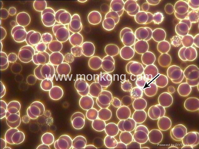
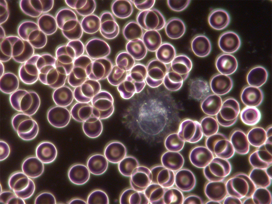
• Viewing blood cells (biological dark field microscope, combined with phase contrast)
• Viewing bacteria (biological dark field microscope, often combined with phase contrast)
• Viewing different types of algae (biological dark field microscope)
• Viewing hairline metal fractures (metallurgical dark field microscope)
• Viewing diamonds and other precious stones (gemological microscope or stereo dark field microscope)
• Viewing shrimp or other invertebrates (stereo dark field microscope)
How to change Dark Field Transformation?
Most stereo and standard compound microscopes have the potential for dark field microscopy.
If a microscope has built-in elements to easily modify for dark field illumination, the manufacturer usually lists this amongst the observation specifications.
You can achieve dark field by using condensers, mirrors and/or a “stop.” Some microscopes come with these accessories or researchers can purchase dark field kits, or even use some common items to adapt a microscope for dark field illumination.
In bright field illumination, the object is lit from below the stage, resulting in a larger, contrasted image that can be studied.
A dark field microscope blocks this central light with a condenser so that only oblique rays hit the object.
An Abbe condenser, for example, contains a concave orb that collects light rays in all azimuths that bounce off a sample to form a cone of illumination.
If there is nothing on the stage, the aperture of the condenser is greater than the objective and the view will be completely black.
A stop is an opaque object that blocks the central light when placed underneath the stage condenser.
This also causes light to scatter in all azimuths, resulting in a cone of light that allows for dark field observation.
Too expensive? What you can do…
If you do not have access to these accessories and cannot afford a dark field kit, there are alternative ways to adapt your microscope for dark field illumination.
The expensive stops are all made of opaque material.
Any possible substitutions cannot have any transparent properties.
One option is to use a circular object, such as a coin; adhere the coin to a larger disk and place below the stage.
You can also cut out a round piece of thick paper, such as construction paper, cardboard or poster-board, and attach to the condenser.
Whatever you use, the trick is to find the right diameter so that the makeshift stop will block the light and only allow the oblique rays to illuminate the specimen.
What is Conclusion about dark field microscopy?
A dark field microscopy can offer brilliant, light images against a dark background of otherwise difficult to view specimens.
Most standard microscopes come with dark field capabilities or accessories to enable this illumination technique.
There are many practical applications of dark field, especially in the field of marine biology, in viewing the many specimens you cannot see using alternative techniques.
However, a researcher must keep in mind the potential issues and limitations that may arise from dark field illumination.
For further information, check out the many microscopy imaging techniques available.
Related Items