Why We Use metatron diagnostic machine review

metatron diagnostic machine review in healthy children and in childhood rheumatic diseases
metatron diagnostic machine review in healthy children and in childhood rheumatic diseases: a prospective single blind observational study Free
Abstract
Objectives: To develop an objective method of nailfold capillaroscopy (NFC), applicable to a wide age range of paediatric patients. To compare the morphological characteristics of the nailfold capillaries in different rheumatology patient groups and controls.
Methods: A colour digital video camera attached to a stereomicroscope was used to capture nailfold capillary images. Computerised image processing was used to analyse and store data. Subsequent quantitative and qualitative morphological analysis was performed in the following paediatric patient and control groups: 18 children with connective tissue diseases (CTD: juvenile dermatomyositis, systemic sclerosis, and undifferentiated connective tissue disease), eight with systemic lupus erythematosus, nine with primary Raynaud’s disease, three with primary vasculitis, 15 with juvenile idiopathic arthritis, 17 healthy children and 20 healthy adults. Images were analysed by a single assessor who was unaware of the patient details.
Results: The NFC technique was simple to perform and gave reproducible results, although some intra- and intersubject variation was noted. Capillary density and width was age related, with younger children having fewer and wider capillaries than older children and adults. Linear capillary density was significantly higher in healthy adults (mean (SD) 8.6 (1.6) capillaries/mm) compared with healthy children (HC 6.9 (0.9) capillaries/mm). The group with CTD had the most abnormal findings, with lower linear density (4.9 (1.7) capillaries/mm) and increased capillary loop width (10.7 (7.3) mm) compared with HC (3.5 (1.7) mm). In addition, 11/18 (61%) patients in the CTD group had more than two definitely abnormal capillaries in at least two nailfolds, an abnormality not seen in other subjects. Two qualitative measures, the degree of avascularity and general disarrangement of capillary pattern, were more commonly observed in the CTD group than in HC. The proportion of tortuous capillaries did not differ significantly between study groups.
Conclusions: This study is unique in measuring objective quantitative and qualitative parameters of the nailfold vasculature across a wide spectrum of age and disease. Differences in capillary morphology and frequency in children with CTD compared with other paediatric diseases and healthy controls were demonstrated. In the clinical situation, an assessment of the general degree of disarrangement may offer a fast tool for assessment of the nailfold vasculature which correlates well with NFC data.

Abnormal metatron diagnostic machine review findings in patients with coronary
Abnormal metatron diagnostic machine review findings in patients with coronary
1. Tambe AA, Demany MA, Zimmerman HA, Mascarenhas E. Angina pectoris and slow flow velocity of dye in coronary arteries – new angiographic finding. Am Heart J. 1972;84:66–71.
2. Beltrame JF, Limaye SB, Horowitz JD. The coronary slow flow phenomenon – a new coronary microvascular disorder. Cardiology. 2002;97:197–202.
3. Goel PK, Gupta SK, Agarwal A, Kapoor A. Slow coronary flow: a distinct angiographic subgroup in syndrome X. Angiology. 2001;52:507–14.
4. Mangieri E, Macchiarelli G, Ciavolella M, Barillà F, Avella A, Martinotti A, Dell’Italia LJ, Scibilia G, Motta P, Campa PP. Slow coronary flow: clinical and histopathological features in patients with otherwise normal epicardial coronary arteries. Cathet Cardiovasc Diagn. 1996;37:375–81.
5. Hawkins BM, Stavrakis S, Rousan TA, Abu-Fadel M, Schechter E. Coronary slow flow–prevalence and clinical correlations. Circ J. 2012;76:936–42.
6. Wozakowska-Kapłon B, Niedziela J, Krzyzak P, Stec S. Clinical manifestations of slow coronary flow from acute coronary syndrome to serious arrhythmias. Cardiol J. 2009;16:462–8.
7. Saya S, Hennebry TA, Lozano P, Lazzara R, Schechter E. Coronary slow flow phenomenon and risk for sudden cardiac death due to ventricular arrhythmias: a case report and review of literature. Clin Cardiol. 2008;31:352.
8. Wang X, Nie SP. The coronary slow flow phenomenon: characteristics, mechanisms and implications. Cardiovasc Diagn Ther. 2011;1:37–43. [PMC free article]
9. Cutolo M, Pizzorni C, Secchi ME, Sulli A. Capillaroscopy. Best Pract Res Clin Rheumatol. 2008;22:1093–108.
10. Cutolo M, Sulli A, Smith V. How to perform and interpret capillaroscopy. Best Pract Res Clin Rheumatol. 2013;2:237–48.
11. Cortes S, Cutolo M. Capillaroscopic patterns in rheumatic diseases. Acta Rheumatol Port. 2007;32:29–36.
12. Dinc A, Melikoglu M, Korkmaz C, Fresko I, Ozdogan H, Yazici H. Nail fold capillary abnormalities in patients with familial Mediterranean fever. Clin Exp Rheumatol. 2001;19:S42–4.
13. Movasat A, Shahram F, Carreira PE, Nadji A, Akhlaghi M, Naderi N, Davatchi F. Nail fold capillaroscopy in Behçet’s disease, analysis of 128 patients. Clin Rheumatol. 2009;28:603–5.
14. Gibson CM, Cannon CP, Daley WL, Dodge JT Jr, Alexander B Jr, Marble SJ, McCabe CH, Raymond L, Fortin T, Poole WK, Braunwald E. TIMI frame count: a quantitative method of assessing coronary artery flow. Circulation. 1996;93:879–88.
15. Bergman R, Sharony L, Schapira D, Nahir MA, Balbir-Gurman A. The handheld dermatoscope as a nail-fold capillaroscopic instrument. Arch Dermatol. 2003;139:1027–30.
16. Yilmaz H, Demir I, Uyar Z. Clinical and coronary angiographic characteristics of patients with coronary slow flow. Acta Cardiol. 2008;63:579–584.
17. Mosseri M, Yarom R, Gotsman MS, Hasin Y. Histologic evidence for small-vessel coronary artery disease in patients with angina pectoris and patent large coronary arteries. Circulation. 1986;74:964–72.
18. Sezgin AT, Sigirci A, Barutcu I, Topal E, Sezgin N, Ozdemir R, Yetkin E, Tandogan I, Kosar F, Ermis N, Yologlu S, Bariskaner E, Cehreli S. Vascular endothelial function in patients with slow coronary flow. Coron Artery Dis. 2003;14:155–61.
19. Cin VG, Pekdemir H, Camsar A, Cicek D, Akkus MN, Parmaksiz T, Katircibas T, Doven O. Diffuse intimal thickening of coronary arteries in slow coronary flow. Jpn Heart J. 2003;44:907–19.
20. Li JJ, Qin XW, Li ZC, Zeng HS, Gao Z, Xu B, Zhang CY, Li J. Increased plasma C-reactive protein and interleukin-6 concentrations in patients with slow coronary flow. Clin Chim Acta. 2007;385:43–7.
21. Turhan H, Saydam GS, Erbay AR, Ayaz S, Yasar AS, Aksoy Y, Basar N, Yetkin E. Increased plasma soluble adhesion molecules; ICAM-1, VCAM-1, and E-selectin levels in patients with slow coronary flow. Int J Cardiol. 2006;108:224–30.
22. Selcuk H, Selcuk MT, Temizhan A, Maden O, Saydam GS, Ulupinar H, Dogan M, Aydin C, Topcu DI, Sasmaz A. Decreased plasma concentrations of adiponectin in patients with slow coronary flow. Heart Vessels. 2009;24:1–7.
23. Camsari A, Pekdemir H, Cicek D, Polat G, Akkus MN, Doven O, Cin VG, Katircibasi T, Parmaksiz T. Endothelin-1 and nitric oxide concentrations and their response to exercise in patients with slow coronary flow. Circ J. 2003;67:1022–1028.
24. Nie SP, Wang X, Geng LL, Liu BQ, Li J, Qiao Y, Liu XM, Luo TY, Dong JZ, Liu XH, Li JJ, Ma CS. Anatomic properties of coronary arteries are correlated to the corrected thrombolysis in myocardial infarction frame count in the coronary slow flow phenomenon. Coron Artery Dis. 2012;3:174–80.
25. Karakaya O, Koçer A, Esen AM, Kargin R, Barutcu I. Impaired cerebral circulation in patients with slow coronary flow. Tohoku J Exp Med. 2011;225:13–6.
26. Camsari A, Ozcan T, Ozer C, Akcay B. Carotid artery intima-media thickness correlates with intravascular ultrasound parameters in patients with slow coronary flow. Atherosclerosis. 2008;200:310–4.
27. Sezgin N, Barutcu I, Sezgin AT, Gullu H, Turkmen M, Esen AM, Karakaya O. Plasma nitric oxide level and its role in slow coronary flow phenomenon. Int Heart J. 2005;46:373–82.
28. Riza Erbay A, Turhan H, Yasar AS, Ayaz S, Sahin O, Senen K, Sasmaz H, Yetkin E. Elevated level of plasma homocysteine in patients with slow coronary flow. Int J Cardiol. 2005;102:419–23.
29. Barutcu I, Sezgin AT, Sezgin N, Gullu H, Esen AM, Topal E, Ozdemir R. Elevated plasma homocysteine level in slow coronary flow. Int J Cardiol. 2005 May;101:143–5.
30. Selcuk MT, Selcuk H, Temizhan A, Maden O, Ulupinar H, Baysal E, Ozeke O, Sasmaz A. Asymmetric dimethylarginine plasma concentrations and L-arginine/asymmetric dimethylarginine ratio in patients with slow coronary flow. Coron Artery Dis. 2007;18:545–51.
31. Kantarci M, Gündogdu F, Doganay S, Duran C, Kalkan ME, Sagsoz ME, Kucuk O, Karakaya A, Kucuk A, Akgün M. Arterial bending angle and wall morphology correlate with slow coronary flow: determination with multidetector CT coronary angiography. Eur J Radiol. 2011;77:111–7.
32. Lambova SN, Müller-Ladner U. Capillaroscopic pattern in systemic sclerosis – an association with dynamics of processes of angio- and vasculogenesis. Microvasc Res. 2010 Dec;80:534–9.
33. Beltrán E, Toll A, Pros A, Carbonel J, Carbonel J, Pujol RM. Assessment of nail fold capillaroscopy by x 30 digital epiluminescence (dermoscopy) in patients with Raynaud phenomenon. Br J Dermatol. 2007;156:892–8.
34. Nagy Z, Czirjak L. Nail fold digital capillaroscopy in 447 patients with connective tissue disease and Raynaud’s disease. J Eur Acad Dermatol Venereol. 2004;18:62–8.
35. Harper FE, Maricq HR, Turner RE, Lidman RW, Leroy EC. A prospective study of Raynaud phenomenon and early connective tissue disease. A five-year report. Am J Med. 1982;72:883–8.
36. Gallucci F, Russo R, Buono R, Acampora R, Madrid E, Uomo G. Indications and results of videocapillaroscopy in clinical practice. Adv Med Sci. 2008;53:149–57.
37. Gasser P, Bühler FR. Nail fold microcirculation in normotensive and essential hypertensive subjects, as assessed by video-microscopy. J Hypertens. 1992;10:83–6.
38. Irving RJ, Walker BR, Noon JP, Watt GC, Webb DJ, Shore AC. Microvascular correlates of blood pressure, plasma glucose, and insulin resistance in health. Cardiovasc Res. 2002;53:271–6.
39. Górska A, Rutkowska-Sak L, Musiej-Nowakowska E, Chlabicz S, Górski S. Nail fold videocapillaroscopy – a useful tool for screening patients with juvenile idiopathic arthritis at the risk of development of premature atherosclerosis. Postepy Hig Med Dosw. 2010;64:296–302.

What is metatron diagnostic machine review ?
What is metatron diagnostic machine review ?
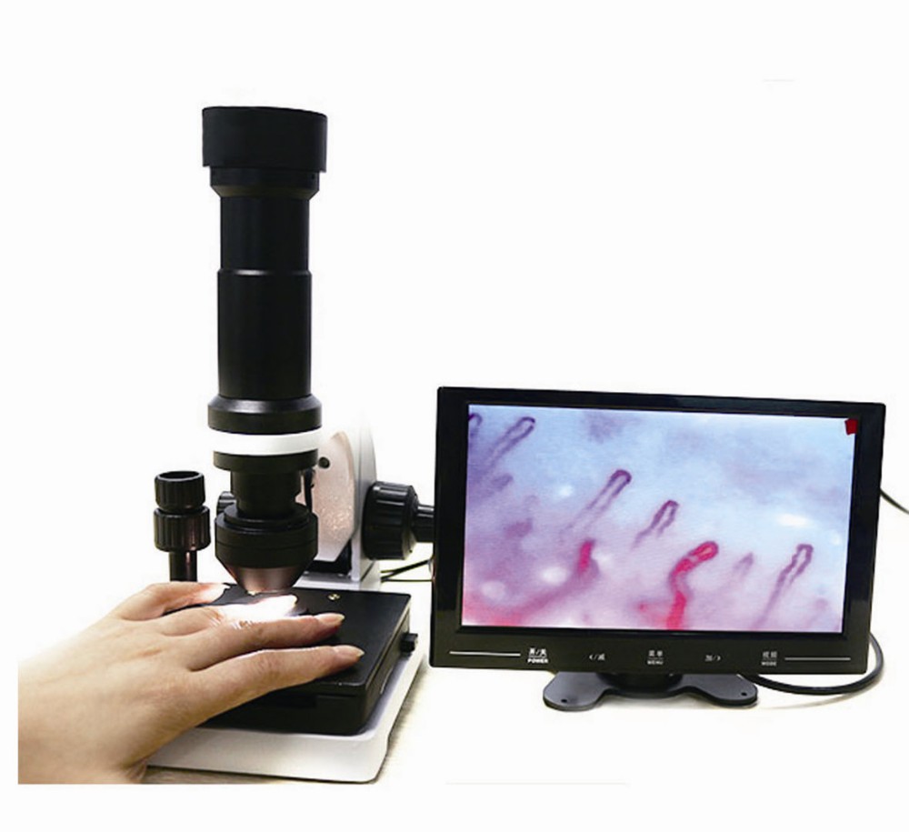
The capillaroscopy is a non-invasive technique at nailfold level, making it possible to assess the characteristics of the nailfold distal capillaries, thanks to a lens and a light that shines on said spot.
The information it provides us with helps to complete the diagnosis of the vasculitic autoimmune process of the patient; it does not permit a diagnosis or specific therapeutic approach on its own.
For its correct visualisation, the patient is recommended:
Not to wear nail varnish and to avoid external harm (bumps, wounds, nail biting)
Not to smoke in the 2 hours prior to the test
To remain in the a room with a temperature of between 22ºC and 25ºC to avoid vasoconstriction episodes due to exogenous factors.
WHAT ARE ITS USES?
The capillaroscopy allows us to know the extent of the distal vascularisation, which is very important in systemic sclerosis and other connective pathologies, as well as to rule out systemic involvement in patients with Raynaud’s phenomenon with no other associated clinical involvement.
RAYNAUD IS PHENOMENON
The capillaroscopy of the nail bed is a simple, bloodless, economical method which is very useful for studying Raynaud’s phenomenon and other rheumatological conditions. Raynaud’s phenomenon can be defined as the change in the colouring of the fingers and/or toes in response to cold or stress. It traditionally progresses through three stages: paleness (vasospasm), cyanosis (due to increased carboxyhemoglobin) and erythema (reactive hiperemia).
Raynaud’s phenomenon can be primary (Raynaud’s disease) or secondary, associated with a connective tissue disease. Primary Raynaud’s phenomenon is responsible for around 60% of all new cases. 15-20% of cases of Raynaud’s phenomenon are due to a series of non- immunological processes, such as drugs, occupational diseases, neoplasms, etc. The remaining 15-20% are associated with connective tissue diseases.
Raynaud’s is present in over 90% of patients with scleroderma and in 70% of cases it is the first symptom. Although it seems impossible to predict that a patient with Raynaud’s will develop scleroderma, the presence of antinuclear antibodies (ANA) indicates a greater risk of onset.
SCLERODERMA
The nail bed capillaroscopy shows morphological alterations at an early stage in some connective tissue diseases of maximum rheumatological interest, particularly capillaroscopy. In these cases, the capillaroscopy traditionally shows the “sclerodermic pattern” characterised by: reduction or absence of capillaries in patches, capillary dilation, and sometimes mega-capillaries and splinter haemorrhages. This “sclerodermic pattern” appears early and when it is observed in patients with Raynaud’s phenomenon, even if it is not very obvious, it should lead to the search for sclerodermic manifestations in internal organs, which can be present without causing any symptoms. The combination of Raynaud’s phenomenon and a “sclerodermic pattern” in a capillaroscopy can precede and therefore predict the onset of scleroderma.
CAPILLAROSCOPY AND RHEUMATOLOGICAL CONDITIONS
In dermatomyositis, the capillaroscopy is similar and sometimes indistinguishable from that found in scleroderma. These patients generally present the other clinical, enzymatic or electromyographic manifestations of dermatomyositis that enable its recognition and diagnosis.
In mixed connective tissue disease (MCTD) or overlap syndromes that a sclerodermic component, the findings of the capillaroscopy can be similar although rarely are large capillary dilations and mega-capillaries observed. These capillaroscopies must be analysed by an expert.
In systemic lupus erythematosus, the alterations in the capillaroscopy are non-specific, and it is possible to find focal capillary reduction, albeit not very strong. The capillaries can be somewhat dilated and tortuous, sometimes with criss-crossing of the arterial and venous components (ringlets), the latter being the most characteristic finding in the capillaroscopy.
In primary Raynaud’s phenomenon, the most striking characteristic is the elongated capillaries with undulations throughout the arterial and venous components. Little or no reduction or dilation of the capillaries can be observed. Splinter haemorrhages are scarce and small.
Ultimately, this is an auxiliary diagnostic technique with great value in rheumatology and vascular disease.

What is metatron diagnostic machine review Structure?
What is metatron diagnostic machine review Structure?
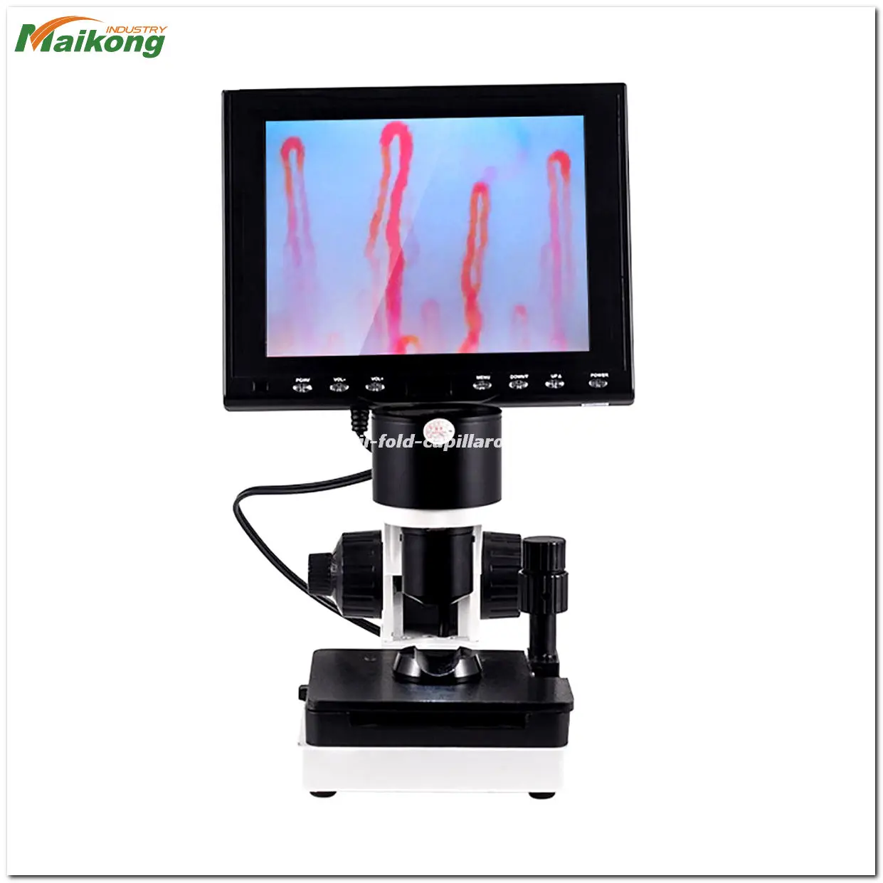
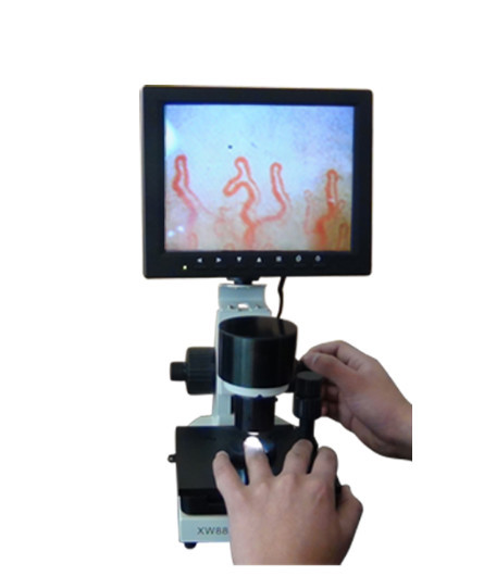
1. 8 inch monitor
2. imported chip CCD
3. fine handwheel
4. coarse handwheel
5. platform lateral movement
6. longitudinal movement of the platform
7. finger hold
8. image spot adjustment ( could totate from left and right )

What is metatron diagnostic machine review Functions?
What is metatron diagnostic machine review Functions?
Functions of latest medical microcirculation nailfold capillaroscopy machine-Latest medical microcirculation nailfold capillaroscopy machine is a new photoelectric instrument of non-invasive and no side effects,whice is used to check the nailfold microcirculation of human body.It’s widely used in clinical for diagnosising the early microcirculation change, Condition forecast, curative effect judgment and prognosis estimation of many diseases (such as heart head blood-vessel, high blood pressure, stroke, diabetes, rheumatoid arthritis, etc.).

Related Items