What (Influencer Name) Gets Wrong About tasly microcirculation test
 Gets Wrong About tasly microcirculation test")
who need tasly microcirculation test?
who need tasly microcirculation test?
People who easily feel tired
People who always burn midnight oil or stay up all nigh long working
Long-term smokers
Insufficient oxygen in blood
People with hypertension
Stroke patient
Drunkard
People who breathe difficulty after light exercises
People who work under exposure of radiation
Cardiac dyfunction
Obesity
After a luxurious meal
 Gets Wrong About tasly microcirculation test")
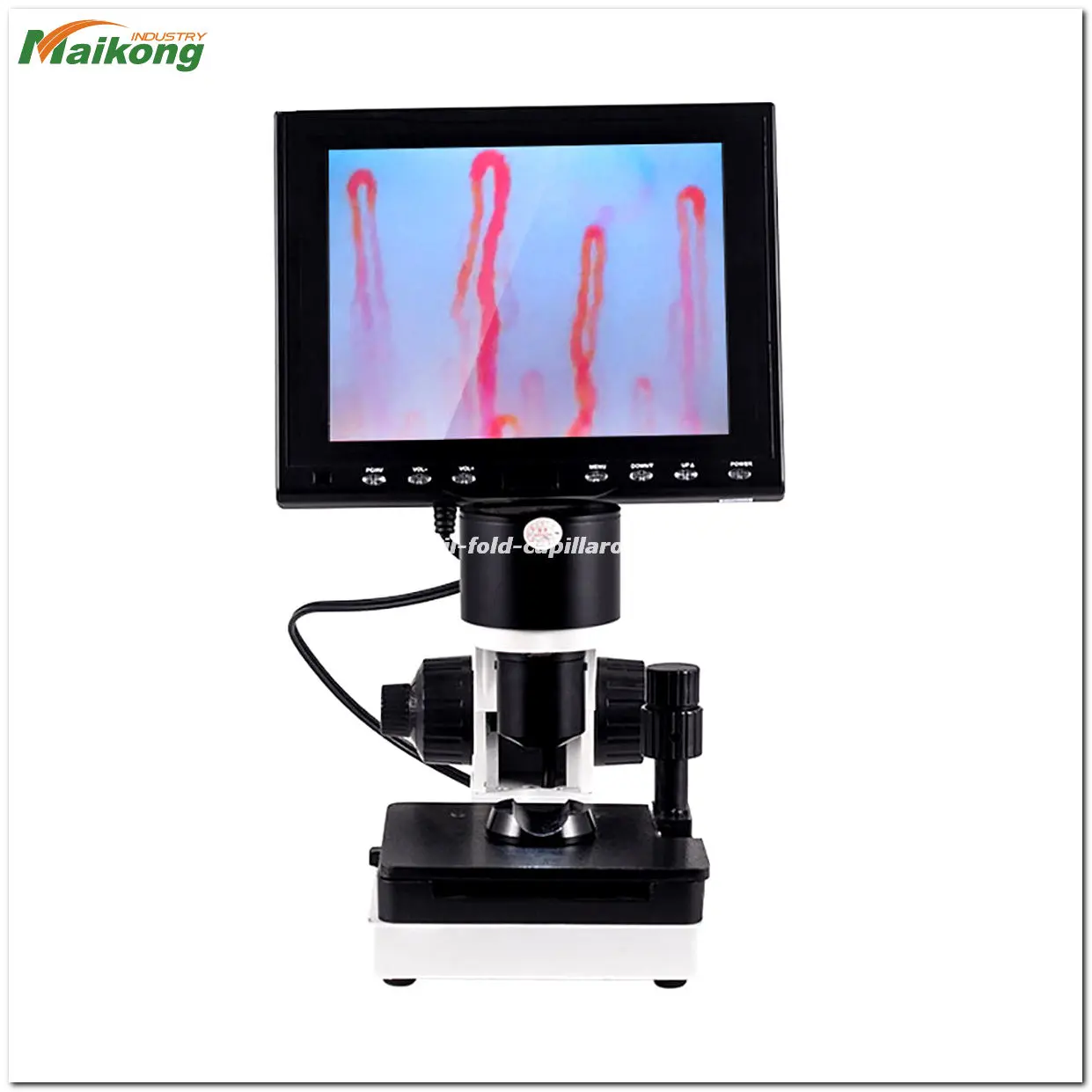
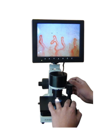
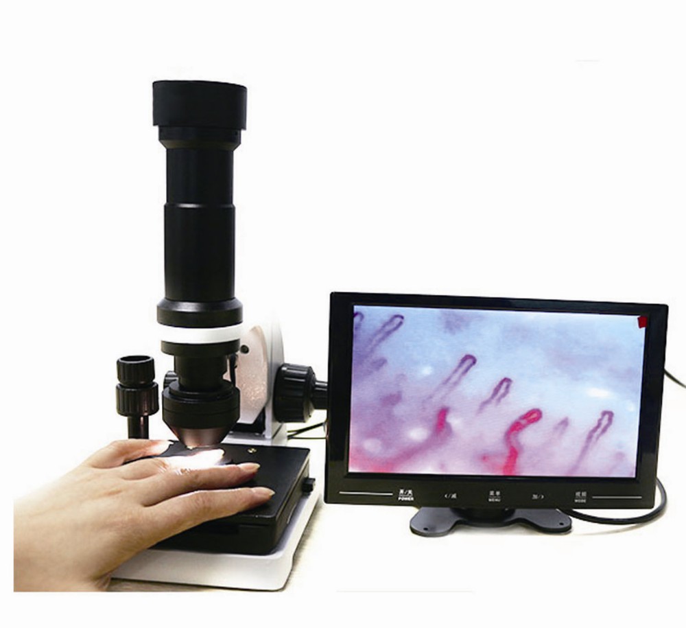
What is tasly microcirculation test ?
What is tasly microcirculation test ?
The capillaroscopy is a non-invasive technique at nailfold level, making it possible to assess the characteristics of the nailfold distal capillaries, thanks to a lens and a light that shines on said spot.
The information it provides us with helps to complete the diagnosis of the vasculitic autoimmune process of the patient; it does not permit a diagnosis or specific therapeutic approach on its own.
For its correct visualisation, the patient is recommended:
Not to wear nail varnish and to avoid external harm (bumps, wounds, nail biting)
Not to smoke in the 2 hours prior to the test
To remain in the a room with a temperature of between 22ºC and 25ºC to avoid vasoconstriction episodes due to exogenous factors.
WHAT ARE ITS USES?
The capillaroscopy allows us to know the extent of the distal vascularisation, which is very important in systemic sclerosis and other connective pathologies, as well as to rule out systemic involvement in patients with Raynaud’s phenomenon with no other associated clinical involvement.
RAYNAUD IS PHENOMENON
The capillaroscopy of the nail bed is a simple, bloodless, economical method which is very useful for studying Raynaud’s phenomenon and other rheumatological conditions. Raynaud’s phenomenon can be defined as the change in the colouring of the fingers and/or toes in response to cold or stress. It traditionally progresses through three stages: paleness (vasospasm), cyanosis (due to increased carboxyhemoglobin) and erythema (reactive hiperemia).
Raynaud’s phenomenon can be primary (Raynaud’s disease) or secondary, associated with a connective tissue disease. Primary Raynaud’s phenomenon is responsible for around 60% of all new cases. 15-20% of cases of Raynaud’s phenomenon are due to a series of non- immunological processes, such as drugs, occupational diseases, neoplasms, etc. The remaining 15-20% are associated with connective tissue diseases.
Raynaud’s is present in over 90% of patients with scleroderma and in 70% of cases it is the first symptom. Although it seems impossible to predict that a patient with Raynaud’s will develop scleroderma, the presence of antinuclear antibodies (ANA) indicates a greater risk of onset.
SCLERODERMA
The nail bed capillaroscopy shows morphological alterations at an early stage in some connective tissue diseases of maximum rheumatological interest, particularly capillaroscopy. In these cases, the capillaroscopy traditionally shows the “sclerodermic pattern” characterised by: reduction or absence of capillaries in patches, capillary dilation, and sometimes mega-capillaries and splinter haemorrhages. This “sclerodermic pattern” appears early and when it is observed in patients with Raynaud’s phenomenon, even if it is not very obvious, it should lead to the search for sclerodermic manifestations in internal organs, which can be present without causing any symptoms. The combination of Raynaud’s phenomenon and a “sclerodermic pattern” in a capillaroscopy can precede and therefore predict the onset of scleroderma.
CAPILLAROSCOPY AND RHEUMATOLOGICAL CONDITIONS
In dermatomyositis, the capillaroscopy is similar and sometimes indistinguishable from that found in scleroderma. These patients generally present the other clinical, enzymatic or electromyographic manifestations of dermatomyositis that enable its recognition and diagnosis.
In mixed connective tissue disease (MCTD) or overlap syndromes that a sclerodermic component, the findings of the capillaroscopy can be similar although rarely are large capillary dilations and mega-capillaries observed. These capillaroscopies must be analysed by an expert.
In systemic lupus erythematosus, the alterations in the capillaroscopy are non-specific, and it is possible to find focal capillary reduction, albeit not very strong. The capillaries can be somewhat dilated and tortuous, sometimes with criss-crossing of the arterial and venous components (ringlets), the latter being the most characteristic finding in the capillaroscopy.
In primary Raynaud’s phenomenon, the most striking characteristic is the elongated capillaries with undulations throughout the arterial and venous components. Little or no reduction or dilation of the capillaries can be observed. Splinter haemorrhages are scarce and small.
Ultimately, this is an auxiliary diagnostic technique with great value in rheumatology and vascular disease.
 Gets Wrong About tasly microcirculation test")
tasly microcirculation test: A Basic Screening Tool in Raynaud’s Phenomenon
tasly microcirculation test: A Basic Screening Tool in Raynaud’s Phenomenon
The term “Raynaud’s phenomenon” refers to a defined triphasic colour change pattern (white, blue, red) of the skin of the fingers. The initial pallor, which may lead to cyanosis (blue coloration) of the digits, is followed by reactive hyperemia and can be accompanied by numbness, paresthesias and pain. Raynaud’s is triggered by exposure to cold, emotional stress and specific drugs. Pathophysiologically it is characterized by an excessive vasoconstrictive response (dominance of vasoconstrisctive over vasodilatory factors) of the small arterioles and the digital arteries.Etiologically it can be classified as primary or secondary, mainly either to systemic sclerosis and related diseases, or to the thoracic outlet syndrome. As such, Raynaud’s phenomenon represents an important clinical manifestation of microvascular involvement. Rare cases of paraneoplastic Raynaud’s have been described, the pathogenesis of which remains unclarified, has however been most likely attributed to neuroendocrine substances secreted by the tumor.Despite the evolution of the technology involved over the decades, nailfold capillaroscopy remains very simple in principle. The term refers to the non-invasive observation of the nailfold capillary bed. Nailfold capillaroscopy can be performed with basic equipment such as a magnifying lens, an ophthalmoscope, a dermatoscope/ stereomicroscope or a conventional wide field microscope; the nailfold video capillaroscope, however, is the gold standard tool, and provides the technology for the visualization and analysis both of morphological as well as rheological parameters of the skin microvasculature. Qualitative and quantitative microangiopathic parameters that can be recorded and analyzed include, among others, microhemorrhages, plexus morphology, capillary density, and morphologic anomalies of the end row loops. In normal conditions, or in primary Raynaud’s, the normal nailfold capillaroscopic pattern shows a regular disposition of the capillary loops along with the nailbed. In subjects suffering from secondary Raynaud’s phenomenon, alterations of the capillaroscopic findings should alert the physician of the possibility of a connective tissue disease or other conditions not yet detected.This review aims to outline the major characteristics of the normal nailfold microvascular network and the healthy variations that may be encountered in relation to age, race, employment etc, and highlight its applicability not only as a powerful tool in the screening of a variety of vascular disorders, in particular of autoimmune origin, but also as a potent tool in the follow-up of patients with established microvascular disorders in response to treatment.As microcirculation involvement is attracting increasing interest in chronic vascular diseases such as diabetes mellitus or hypertension, as a prognostic marker for macrovascular disease and target organ involvement, the acquaintance with and application of techniques assessing microvascular compromise gains particular significance. The advantages of capillaroscopy (low cost, uninvasiveness, repeatability, high sensitivity, good specificity and easy interpretation of the results) render this technique a valuable asset for the clinician.
 Gets Wrong About tasly microcirculation test")
What is tasly microcirculation test?
What is tasly microcirculation test?
tasly microcirculation test examination has been used since late 1950s as a non-invasive in-vivo technique for diagnosing and monitoring connective tissue disease in adults. Disorders such as Raynaud’s phenomenon, progressive systemic sclerosis, and rheumatoid arthritis were detected in more than 80% of adult patients, by analyzing such high resolution images. In this research paper, we propose a framework to classify nailfold capillary microscopy images into SLE (Systemic Lupus Erythematosus) and PSS (Progress Systemic sclerosis) diseases. Based on statistical data collected in Taichung Veteran’s General Hospital (TCVGH), Taiwan, higher percentage of adult patients are find with SLE patients, when compared with PSS. According to ARA (American Rheumatism Association), patients’ outside features are obviously. The PSS patients are the same way to finding the features. And in the same time, we refer from doctor’s idea add other conditions to help judging. In the first step, RP is the most important element. Most of the SLE and PSS features are raised by RP. In the other way, the features can be found in both of SLE and PSS patients. In order to divide the SLE and PSS patients into the correct class, other conditions are existed in necessary.
 Gets Wrong About tasly microcirculation test")
Related Items