What it REALLY means to capillaroscopy
What it REALLY means to capillaroscopy
capillaroscopy Abstract
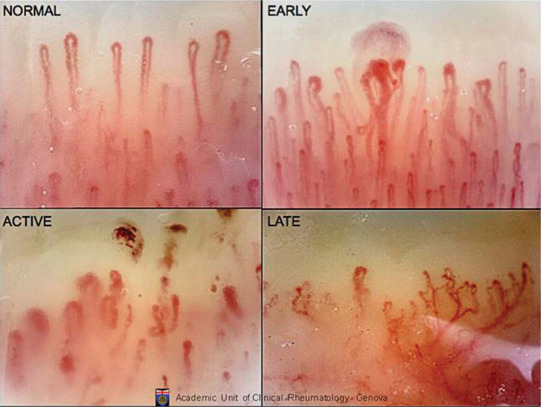
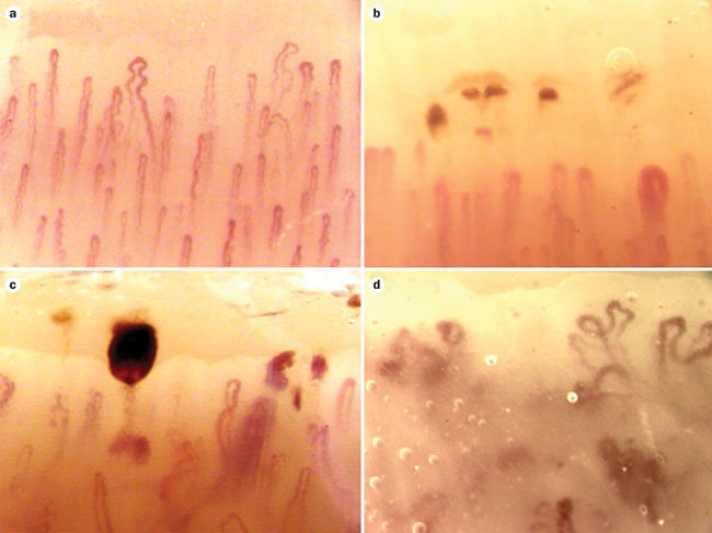
Capillaroscopy is a non-invasive, easy and safe diagnostic technique designed to evaluate small vessels of the microcirculation in the nailfold. It can reveal both the general architecture of capillary rows and fine details of particular vessels. The most important indications for performing capillaroscopy include differential diagnosis of primary and secondary Raynaud’s phenomenon, as well as assessment of scleroderma spectrum disorders. In systemic sclerosis capillary abnormalities appear and evolve in a clearly defined sequence called the scleroderma pattern, which correlates with internal organ involvement. Capillaroscopy is also listed as a systemic sclerosis classification criterion recognized by the European League Against Rheumatism (EULAR). With digitized equipment, capillaroscopy allows for precise qualitative and quantitative evaluation of the microcirculation and is a valuable tool in the rheumatologists’ daily practice.
capillaroscopy Introduction
Capillaroscopy is a non-invasive diagnostic technique designed to evaluate small vessels of the microcirculation . Nailfold capillaries were first observed in the 17th century with primitive magnifying equipment, and in the early 19th century the first associations between inflammation and capillary alterations were made. Beginning with the works of Maurice Raynaud’s , research in the second half of the 19th and first decades of the 20th century established a direct link between capillary abnormalities and certain medical conditions. In the 1930s, interest in capillaroscopy began to decline, to rise again in the 1980s and 1990s. With the advent of modern digital equipment and evidence-based methodology, at the beginning of the 21st century, we can witness a renaissance of the capillaroscopic technique and widespread recognition of its significance.
capillaroscopy Principles Microcirculation
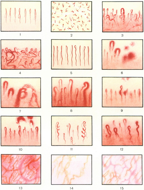
The vasculature of the microcirculation consists of the smallest blood vessels in the human body – arterioles, capillaries and venules. The capillaries, in turn, are formed by an arterial limb, capillary loop and venous limb. This pattern is found in every tissue except liver, spleen and bone marrow, where capillaries are replaced by sinusoids. Microcirculation’s main function is capillary exchange – delivery of oxygen and nutrients to tissues and removal of carbon dioxide and waste products . In a systemic disease in which vascular damage is one of the pathogenetic factors, abnormalities in capillary morphology can be observed long before the onset of clinical symptoms. In patients already diagnosed with a systemic disease, damage to the capillaries may reflect the involvement of internal organs and help determine the stage of the disease .
Microcirculation in capillaries is routinely evaluated within the skin of the nailfold. The entire skin abounds in capillaries; however, they run perpendicular to the skin surface, and only the tip of the loop is visible. In the nailfold, terminal rows of capillaries run parallel to the skin surface and, therefore, all morphological details and the nature of the blood flow can be examined .
Performing capillaroscopy
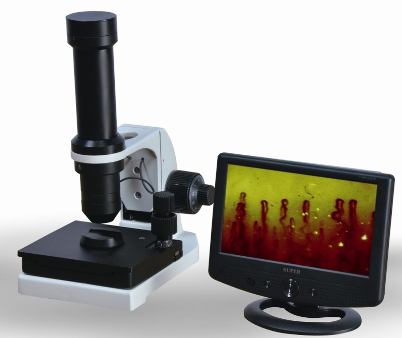
A range of optical devices can be used to perform capillaroscopic examination, for example a dermatoscope, an ophthalmoscope or a traditional microscope. However, it is best to perform the examination with equipment dedicated for capillaroscopy, i.e. a stereomicroscope or a digital videocapillaroscope. Of these two, the latter is preferred, as a hand-held probe can be easily used in every situation, e.g. for bedside examination or in patients with severe flexion contractures.
In order to enhance skin transparency, a drop of immersion oil is applied to the nailfold before capillaroscopy. In a routine examination, all fingers except the thumbs are evaluated. Each finger should be examined in two magnifications: × 50, showing the general architecture of the terminal capillary row, and × 200–300, in which morphological details of a single capillary can be assessed. Digital equipment allows the obtained images to be stored and used for an objective comparison if the patient needs subsequent examinations .
Patient preparation
Environmental factors can cause physiological constriction of capillaries, thus greatly affecting the capillaroscopic image. Before the examination, patients should be acclimatised at a temperature of 20–22°C for 15–20 minutes, and should refrain from smoking and drinking caffeine for 4 hours. Capillaroscopy should not be performed if the patient has recently (in the last 3 weeks) undergone any cosmetic procedure involving the nailfold area, since the consequent micro-traumas can give false-positive results
Related Items
